. 2017 Mar 14:8:193.
doi: 10.3389/fimmu.2017.00193. eCollection 2017.

Canonical and Cross-reactive Binding of NK Cell Inhibitory Receptors to HLA-C Allotypes Is Dictated by Peptides Bound to HLA-C

Affiliations

Canonical and Cross-reactive Binding of NK Cell Inhibitory Receptors to HLA-C Allotypes Is Dictated by Peptides Bound to HLA-C

Malcolm J W Sim et al. Front Immunol. .

Abstract

Background: Human natural killer (NK) cell activity is regulated by a family of killer cell immunoglobulin-like receptors (KIRs) that bind human leukocyte antigen (HLA) class I. Combinations of KIR and HLA genotypes are associated with disease, including susceptibility to viral infection and disorders of pregnancy. KIR2DL1 binds HLA-C alleles of group C2 (Lys80). KIR2DL2 and KIR2DL3 bind HLA-C alleles of group C1 (Asn80). However, this model cannot explain HLA-C allelic effects in disease or the impact of HLA-bound peptides. The goal of this study was to determine the extent to which the endogenous HLA-C peptide repertoire can influence the specific binding of inhibitory KIR to HLA-C allotypes.

Results: The impact of HLA-C bound peptide on inhibitory KIR binding was investigated taking advantage of the fact that HLA-C*05:01 (HLA-C group 2, C2) and HLA-C*08:02 (HLA-C group 1, C1) have identical sequences apart from the key KIR specificity determining epitope at residues 77 and 80. Endogenous peptides were eluted from HLA-C*05:01 and used to test the peptide dependence of KIR2DL1 and KIR2DL2/3 binding to HLA-C*05:01 and HLA-C*08:02 and subsequent impact on NK cell function. Specific binding of KIR2DL1 to the C2 allotype occurred with the majority of peptides tested. In contrast, KIR2DL2/3 binding to the C1 allotype occurred with only a subset of peptides. Cross-reactive binding of KIR2DL2/3 with the C2 allotype was restricted to even fewer peptides. Unexpectedly, two peptides promoted binding of the C2 allotype-specific KIR2DL1 to the C1 allotype. We showed that presentation of endogenous peptides or HIV Gag peptides by HLA-C can promote KIR cross-reactive binding.

Conclusion: KIR2DL2/3 binding to C1 is more peptide selective than that of KIR2DL1 binding to C2, providing an explanation for KIR2DL3-C1 interactions appearing weaker than KIR2DL1-C2. In addition, cross-reactive binding of KIR is characterized by even higher peptide selectivity. We demonstrate a hierarchy of functional peptide selectivity of KIR-HLA-C interactions with relevance to NK cell biology and human disease associations. This selective peptide sequence-driven binding of KIR provides a potential mechanism for pathogen as well as self-peptide to modulate NK cell activation through altering levels of inhibition.

Keywords: human leukocyte antigen; immunogenetics; innate immunity; killer cell Ig-like receptors; natural killer cell.


Figures
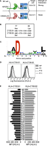
Figure 1
Figure 1
Human leukocyte antigen (HLA)-C*05:01 (group C2) and HLA-C*08:02 (group C1) are almost identical in sequence and HLA-C*05:01-eluted endogenous peptides bind HLA-C*05:01 and HLA-C*08:02. (A) Schematic showing how the specificity of inhibitory KIR for different HLA-C allotypes is defined by an amino acid dimorphism at positions 77 and 80 of HLA-C, where KIR2DL1 binds group C2 allotypes (Asn77Lys80) and KIR2DL2 and KIR2DL3 bind group C1 allotypes (Ser77Asn80). (B) Nucleotide sequence alignment of amino acid positions 77–80 of HLA-C*05:01 and HLA-C*08:02. Bold indicates nucleotide differences. (C) The motif for HLA-C*05:01 9mer peptides. (D) HLA-I expression on 221–C*05:01–ICP47 (left) and RMA-S-C*08:02 (right) cells after overnight incubation at 26°C with 100 μM HLA-C*05:01 peptides P2, P10, and P11 and no peptide (NP). (E) HLA-I expression on 221–C*05:01–ICP47 (left) and RMA-S-C*08:02 (right) cells after overnight incubation at 26°C with 100 μM of 1 of 28 HLA-C*05:01 peptides and NP. Mean MFI and SEM of three independent experiments are shown.
Figure 2
Figure 2
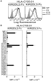
KIR2DL2 and KIR2DL3 binding to human leukocyte antigen (HLA)-C*08:02 is more peptide selective than KIR2DL1 binding to HLA-C*05:01. (A) KIR2DL1-Fc binding to peptide-loaded 221–C*05:01–ICP47 (left), KIR2DL2-Fc (middle) and KIR2DL3-Fc (right) binding to RMA-S-C*08:02 cells. Cells were loaded overnight at 26°C with no peptide (NP) or 100 μM HLA-C*05:01 peptides P2, P4, and P7. (B) Same as in panel (A). Killer cell Ig-like receptor (KIR-Fc) binding was assessed after overnight incubation at 26°C with NP or 100 μM of 28 individual HLA-C*05:01 peptides. Mean MFI and SEM of three independent experiments are shown. KIR-Fc (3.6 μg/ml) was conjugated to protein A-alexa 647.
Figure 3
Figure 3
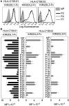
Cross-reactive KIR2DL2 and KIR2DL3 binding to the C2 allotype human leukocyte antigen (HLA)-C*05:01 is highly peptide selective. (A) KIR2DL2-Fc (left) and KIR2DL3-Fc (right) binding to peptide-loaded 221–C*05:01–ICP47, cells. Cells were loaded overnight at 26°C with no peptide (NP) or 100 μM HLA-C*05:01 peptides P2, P10, and P11. (B) Same as in panel (A). Killer cell Ig-like receptors (KIR)-Fc binding was assessed after overnight incubation at 26°C with NP or 100 μM of 28 individual HLA-C*05:01 peptides. Mean MFI and SEM of three independent experiments are shown. KIR-Fc (3.6 μg/ml) was conjugated to protein A-alexa 647.
Figure 4
Figure 4
Canonical and cross-reactive KIR2DL2 and KIR2DL3 binding is controlled by amino acids at positions 7 and 8. (A) KIR2DL1-Fc, KIR2DL2-Fc, and KIR2DL3-Fc binding to peptide-loaded 221–C*05:01–ICP47 (left, black) and KIR2DL2-Fc and KIR2DL3-Fc (right, gray) binding to RMA-S-C*08:02 cells. Cells were loaded overnight at 26°C with no peptide (NP) or 100 μM of peptide P2 (P2-IIDKSGSTV) and amino acid variants of peptide 2 with substitutions at positions 7 and 8. Amino acid sequence substitutions at positions 7 and 8 are shown. Mean MFI and SEM of three independent experiments are shown. Killer cell Ig-like receptors (KIR-Fc, 3.6 μg/ml) were conjugated to protein A-alexa 647. (B) KIR2DL1-Fc, KIR2DL2-Fc, and KIR2DL3-Fc binding to peptide-loaded 221–C*05:01–ICP47 (left, black) and KIR2DL2-Fc and KIR2DL3-Fc (right, gray) binding to RMA-S-C*08:02 cells. Cells were loaded overnight at 26°C with NP or 100 μM of peptides P10 (P10-IVDKSGRTL), P11 (P11-AGDDAPRAV), and amino acid variants of P10 and P11 with substitutions at positions 7 and 8. Amino acid sequence at positions 7 and 8 is shown. Mean MFI and SEM of three independent experiments are shown. KIR-Fc (3.6 μg/ml) was conjugated to protein A-alexa 647.
Figure 5
Figure 5
High peptide selectivity, cross-reactive binding and functional inhibition of KIR2DL3+ natural killer (NK) cells by HLA-C*05:01 (C2). (A) Specific lysis of 221–C*05:01–ICP47 cells at different E:T by YTS-2DL3 NK cells. 221–C*05:01–ICP47 target cells were used after overnight culture at 26°C in the presence of no peptide (NP), P2, P2-AA, P2-EE, P2-GG, and P7. (B) Relative-specific lysis of 221–C*05:01–ICP47 cells by YTS-2DL3 NK cells compared to NP at10:1 (E:T). Mean and SEM of three independent experiments are shown. *** = p < 0.001 by Student’s t-test. (C) Flow cytometry histograms showing degranulation (CD107a expression) of KIR2DL3-SP (left) and KIR3DL1-SP (right) NK cells in response to 221–C*05:01–ICP47 cells loaded with P2, P2-AA, P2-EE, or NP. (D) Box plots showing the fraction of CD107a+ KIR2DL3-SP NK cells in response to 221–C*05:01–ICP47 cells loaded with P2, P2-AA, P2-EE, P2-GG, P2-LA, P11, P11-7A, P10, P10-AA, P10-7E, or NP (n = 3–5). The CD107a+ values were normalized to the fraction of CD107a+ KIR2DL3-SP NK cells that degranulated in the absence of peptide (set at 1.0) for each donor (CD107a+ ranged from 62.8 to 79.0%, n = 5). ***p < 0.001, **p < 0.01, and *p < 0.05 by Kruskal–Wallis test with Dunn’s multiple comparisons test. (E) KIR2DL3-Fc binding to 221–C*05:01–ICP47 cells correlated with degranulation (CD107a+) of KIR2DL3-SP NK cells in response to 221–C*05:01–ICP47 cells in the presence of P2, P2-AA, P2-EE, P2-GG, P2-LA, P11, P11-7A, P10, P10-AA, P10-7E, or NP. Spearman’s correlation r = −0.955, p < 0.0001. CD107a expression was normalized to value obtained in response to NP for each donor.
Figure 6
Figure 6
High peptide selectivity and functional cross-reactive binding of KIR2DL1 to the C1 allotype human leukocyte antigen (HLA)-C*08:02. (A) KIR2DL1-Fc binding to RMA-S-C*08:02 cells after overnight incubation at 26°C with no peptide (NP) or 100 μM of 28 individual HLA-C*05:01 peptides. Mean MFI and SEM of three independent experiments are shown. (B) KIR2DL1-Fc binding to RMA-S-C*08:02 cells after overnight incubation at 26°C with NP or 100 μM of peptides P10, P10-7E, P11, P11-7A, P2, P2-AA, P2-RA, P2-KA, and P2-EE. Mean MFI and SEM of three independent experiments are shown. KIR-Fc (3.6 μg/ml) was conjugated to protein A-alexa 647. (C) Flow cytometry histograms showing degranulation (CD107a expression) of KIR2DL1-SP (left) and KIR3DL1-SP (right) natural killer (NK) cells in response to 221–C*08:02–ICP47 cells pre-incubated overnight with P10, P10-7E, or NP. Expression of CD107a on KIR2DL1-SP NK cells with no target cells is also shown. (D) Box plots showing CD107a expression on KIR2DL1-SP NK cells in response to 221–C*08:02–ICP47 cells pre-incubated overnight with P10, P10-7E, P11, P11-7A, P2, P2-RA, P2-KA, P2-EE, or NP (n = 5–8). The CD107a+ values were normalized to the fraction of CD107a+ KIR2DL1-SP NK cells that degranulated in the absence of peptide (set at 1.0) for each donor (CD107a+ ranged from 23.7 to 68.4%, n = 8). ****p < 0.0001 and **p < 0.01 by Kruskal–Wallis test with Dunn’s multiple comparisons test. (E) KIR2DL1-Fc binding to RMA-S-C*08:02 cells is correlated with degranulation (CD107a+) of KIR2DL1-SP NK cells in response to 221–C*08:02–ICP47 cells loaded with P10, P10-7E, P11, P11-7A, P2, P2-RA, P2-KA, P2-EE, or NP. CD107a expression was normalized to value obtained in response to NP for each donor. Spearman’s correlation, p = 0.0004, r = −0.95.
Figure 7
Figure 7
KIR2DL2 and KIR2DL3 are more peptide selective than KIR2DL1 but less stringent about allotype preference. KIR2DL1, with strong specificity for C2 allotype, exhibits low peptide selectivity in binding C2. KIR2DL2 and KIR2DL3 cross-reactivity with C2 is associated with greater peptide selectivity. KIR2DL1 cross-reacted with C1 in the context of only two peptides. This hierarchy in peptide selectivity of killer cell Ig-like receptors (KIR)–HLA-C interactions is relevant to natural killer (NK) biology and understanding of disease associations. Peptide sequence-driven binding of KIR to HLA-C provides a potential mechanism for pathogen as well as self-peptide to modulate NK cell function through altering levels of KIR binding and KIR-mediated inhibition.

Similar articles

Cited by

References

    1. Cerwenka A, Lanier LL. Natural killer cells, viruses and cancer. Nat Rev Immunol (2001) 1:41–9.10.1038/35095564 - DOI - PubMed
    1. Long EO, Sik Kim H, Liu D, Peterson ME, Rajagopalan S. Controlling natural killer cell responses: integration of signals for activation and inhibition. Annu Rev Immunol (2013) 31:227–58.10.1146/annurev-immunol-020711-075005 - DOI - PMC - PubMed
    1. Parham P. MHC class I molecules and KIRs in human history, health and survival. Nat Rev Immunol (2005) 5:201–14.10.1038/nri1570 - DOI - PubMed
    1. Biassoni R, Falco M, Cambiaggi A, Costa P, Verdiani S, Pende D, et al. Amino acid substitutions can influence the natural killer (NK)-mediated recognition of HLA-C molecules. Role of serine-77 and lysine-80 in the target cell protection from lysis mediated by “group 2” or “group 1” NK clones. J Exp Med (1995) 182:605–9.10.1084/jem.182.2.605 - DOI - PMC - PubMed
    1. Winter CC, Long EO. A single amino acid in the p58 killer cell inhibitory receptor controls the ability of natural killer cells to discriminate between the two groups of HLA-C allotypes. J Immunol (1997) 158:4026–8. - PubMed