. 2017 May 1;113(6):656-670.
doi: 10.1093/cvr/cvx036.

Heterologous desensitization of cardiac β-adrenergic signal via hormone-induced βAR/arrestin/PDE4 complexes

Affiliations

Heterologous desensitization of cardiac β-adrenergic signal via hormone-induced βAR/arrestin/PDE4 complexes

Qian Shi et al. Cardiovasc Res. .

Abstract

Aims: Cardiac β-adrenergic receptor (βAR) signalling is susceptible to heterologous desensitization by different neurohormonal stimuli in clinical conditions associated with heart failure. We aim to examine the underlying mechanism of cross talk between βARs and a set of G-protein coupled receptors (GPCRs) activated by hormones/agonists.

Methods and results: Rat ventricular cardiomyocytes were used to determine heterologous phosphorylation of βARs under a series of GPCR agonists. Activation of Gs-coupled dopamine receptor, adenosine receptor, relaxin receptor and prostaglandin E2 receptor, and Gq-coupled α1 adrenergic receptor and angiotensin II type 1 receptor promotes phosphorylation of β1AR and β2AR at putative protein kinase A (PKA) phosphorylation sites; but activation of Gi-coupled α2 adrenergic receptor and activation of protease-activated receptor does not. The GPCR agonists that promote β2AR phosphorylation effectively inhibit βAR agonist isoproterenol-induced PKA phosphorylation of phospholamban and contractile function in ventricular cardiomyocytes. Heterologous GPCR stimuli have minimal to small effect on isoproterenol-induced β2AR activation and G-protein coupling for cyclic adenosine monophosphate (cAMP) production. However, these GPCR stimuli significantly promote phosphorylation of phosphodiesterase 4D (PDE4D), and recruit PDE4D to the phosphorylated β2AR in a β-arrestin 2 dependent manner without promoting β2AR endocytosis. The increased binding between β2AR and PDE4D effectively hydrolyzes cAMP signal generated by subsequent stimulation with isoproterenol. Mutation of PKA phosphorylation sites in β2AR, inhibition of PDE4, or genetic ablation of PDE4D or β-arrestin 2 abolishes this heterologous inhibitory effect. Ablation of β-arrestin 2 or PDE4D gene also rescues β-adrenergic stimuli-induced myocyte contractile function.

Conclusions: These data reveal essential roles of β-arrestin 2 and PDE4D in a common mechanism for heterologous desensitization of cardiac βARs under hormonal stimulation, which is associated with impaired cardiac function during the development of pathophysiological conditions.

Keywords: Heterologous desensitization; PKA; PKC; Phosphodiesterase 4; β-Arrestin 2.


Figures

Figure 1
Figure 1
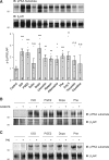
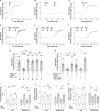
Activation of different GPCRs promotes phosphorylation of β2AR in adult ventricular cardiomyocytes. Adult rat ventricular cardiomyocytes expressing flag-β2AR were cultured for 48 h before stimulation with different GPCR agonists as indicated (1 µM for 5 min, respectively). The flag-β2AR was immunoprecipitated using anti-FLAG beads. The elution was subjected to western blot to detect β2AR phosphorylation. The phosphorylation of β2AR at putative PKA phosphorylation sites is quantified and expressed as fold over non-stimulated group (Control). Data are obtained from 3–7 independent experiments and shown as mean ± S.E.M. n.s, not significant; *P ≤ 0.05; **P ≤ 0.01; ***P ≤ 0.001 (vs. Control group) by student’s unpaired t-test. ISO, isoproterenol; PGE2, Prostaglandin E2; Dobu, Dobutamine; Dopa, Dopamine; Phe, Phenylephrine; Ang II, Angiotensin II. Adult rat ventricular cardiomyocytes expressing flag-β2AR were pretreated with 1 µM of PKC inhibitor GO6976 (B) or 10 µM of PKA peptide inhibitor PKI (C) for 30 min followed by stimulation with different GPCR agonists as indicated (1 µM for 5 min, respectively). Flag-β2AR was immunoprecipitated using anti-FLAG beads. Phosphorylated β2AR at putative PKA phosphorylation sites and total β2AR were detected by western blot. Data represent 3–5 independent experiments.
Figure 2
Figure 2
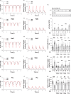
Activation of different GPCRs inhibits β-adrenergic stimulation induced PLB phosphorylation, calcium handling and myocyte contractility. (AE) Representative traces of myocyte sarcomere length fractional shortening and (FJ) representative traces of intracellular calcium release (calcium transient) from adult rat ventricular cardiomyocytes treated with or without 100 nM isoproterenol (ISO, A and F); 1 µM PGE2 before 100 nM ISO (B and G); 1 µM angiotensin II (Ang II) before 100 nM ISO (C and H); 1 µM dopamine (Dopa) before 100 nM ISO (D and I); 1 µM phenylephrine (Phe) before 100 nM ISO (E and J). (K) Adult rat ventricular cardiomyocytes were pretreated with indicated GPCR agonists (1 µM for 5 min, respectively), then stimulated by 100 nM ISO for 5 min. Phosphorylation of PLB at Serine 16 site was detected by western blot and quantified as fold over non-stimulated group (Control group, without ISO stimulation condition), and shown in bar graph with scatter plots. Data are collected from 7 to 9 independent experiments. The percent shortening in adult rat ventricular myocyte sarcomere length (L), the amplitude of calcium release (calcium transient) (M), and the rate of calcium decay (τ) are shown in bar graph with scatter plots. Data are obtained from four independent experiments and shown as mean ± S.E.M. In (K)—(N), *P ≤ 0.05; **P ≤ 0.01; ****P ≤ 0.0001 (vs. ISO alone group) by one-way ANOVA with Dunnett’s multiple comparison test. Between paired groups, n.s, not significant; #P ≤ 0.05; ###P ≤ 0.001; ####P ≤ 0.0001 by student’s unpaired t- test.
Figure 3
Figure 3
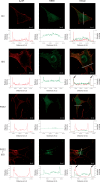
Heterologous phosphorylation of β2AR does not affect β-adrenergic stimulation-induced receptor active conformation change. WT neonatal cardiomyocytes expressing flag-β2AR and Nb80-GFP were stimulated with or without isoproterenol (ISO, 1 μM) for 5 min or in the presence of PGE2 (1 μM) for 5 min then stimulated with or without ISO (1 μM) for 5 min. The cells were then fixed and labelled with anti-flag antibody, followed by a red fluorescent secondary antibody. Data represents five independent experiments. The representative dual colour confocal immunofluorescence images show Nb80-GFP (green) and flag-β2AR (red) distribution and their localization (shown as merge) (Scale bar = 10 µm). The relative fluorescence intensity of Nb80-GFP and flag-β2AR along the white solid line are measured and plotted in the graphs below each individual image, respectively. The arrowheads indicate the positions where fluorescence peaks of Nb80-GFP and flag-β2AR are overlapped.
Figure 4
Figure 4
Heterologous phosphorylation of β2AR promotes β-arrestin 2 dependent recruitment and activation of PDE4D to β2AR. (A) Adult rat ventricular cardiomyocytes were stimulated with different GPCR agonists as indicated (1 µM for 5 min, respectively). Cells were then lysed, and phosphorylation of PDE4D at Serine 190 site was detected in total lysate by western blot using anti-phospho-specific PDE4D antibody. The levels of phosphorylation of PDE4D are quantified and expressed as fold over non-stimulated group (Control). Data are obtained from three independent experiments and shown as mean ± S.E.M. *P < 0.05; ***P < 0.001; ****P < 0.0001 by one-way ANOVA with Dunnett’s multiple comparison test. (B) WT or β-arrestin-2 KO neonatal cardiomyocytes were infected with flag-β2AR together GFP-N-terminal-PDE4D5 adenoviruses. After stimulation with 1 µM PGE2 for 5 min, flag-β2AR was immunoprecipitated using anti-FLAG M2 beads. The elution was subjected to western blot to detect β2AR and PDE4D5, respectively. The PDE4D levels are quantified and expressed as fold over β2AR levels in corresponding non-stimulated groups. (C) Adult rat ventricular cardiomyocytes were infected with mCherry-PDE4D5 together with flag-β2AR or flag-β2AR-PKA4A (lacking four PKA phosphorylation sites) adenovirus and cultured for 48 h. After stimulation with 1 µM PGE2 or 1 µM Angiotensin II (Ang II) for 5 min, cardiomyocytes were lysed and β2AR was immunoprecipitated using anti-FLAG M2 beads. The elution was subjected to western blot to detect β2AR and PDE4D5, respectively. The PDE4D5 levels are quantified and expressed as fold over β2AR levels in corresponding non-stimulated groups. In (B) and (C), data are obtained from four independent experiments and shown as mean ± S.E.M. Between paired groups, n.s, not significant; *P < 0.05 by student’s unpaired t-test.
Figure 5
Figure 5
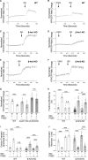
Inhibition of PDE4 or deletion of PDE4D rescues desensitized β-adrenergic signal after PGE2 treatment. WT mouse neonatal cardiomyocytes expressing PM-ICUE3 FRET biosensor were stimulated with 1 µM ISO without (A) or with 1 µM selective PDE4 inhibitor rolipram (Roli) (E); or cardiomyocytes were pretreated with 1 µM PGE2 for 5 min followed by stimulation with 1 µM ISO in the absence (C) or presence of 1 µM Roli addition at different times (B, D, and F). Representative traces of normalized YFP/CFP (FRET) ratio changes with different stimulations as indicated are shown. (G) The normalized FRET ratio changes corresponding to (AF) are shown in bar graph with scatter plots. Data are obtained from five independent experiments and shown as mean ± S.E.M. ****P ≤ 0.0001 (vs. ISO + PGE2 group); Between paired groups, n.s, not significant; #P ≤ 0.05; ##P ≤ 0.05; ####P ≤ 0.0001 by one-way ANOVA with Dunnett’s multiple comparison test. (H) WT mouse neonatal cardiomyocytes expressing PM- ICUE3 FRET biosensor were stimulated with 10 nM ISO followed by Forskolin (50 µM) and IBMX (100 µM); or 10 nM ISO together with 100 nM selective PDE4 inhibitor rolipram (Roli). Alternatively cardiomyocytes were pretreated with 1 µM PGE2 for 5 min followed by stimulation with 10 nM ISO in the absence or presence of 100 nM Roli. The normalized FRET ratio changes are shown in bar graph with scatter plots. Data are obtained from three experiments and shown as mean ± S.E.M. ****P ≤ 0.0001 (6 vs. ISO + PGE2 group); Between paired groups, n.s, not significant; #P ≤ 0.05; ##P ≤ 0.05 by one-way ANOVA with Dunnett’s multiple comparison test. The percent shortening in adult WT or PDE4D KO mouse ventricular cardiomyocyte sarcomere length (I), the amplitude of calcium release (calcium transient) (J) and the rate of calcium decay (τ) (K) are shown in bar graph with scatter plots. In (IK), data are obtained from three independent experiments and shown as mean ± S.E.M. Between paired groups, n.s, not significant; *P ≤ 0.05; **P ≤ 0.01; ***P ≤ 0.001; ****P ≤ 0.0001 by student’s unpaired t-test.
Figure 6
Figure 6
Deletion of β-arrestin 2 rescues desensitized β-adrenergic signal after PGE2 treatment. WT (A, B), β-arrestin 1 KO (C, D) or β-arrestin 2 KO (E, F) neonatal cardiomyocytes expressing SR-AKAR3 FRET biosensor were stimulated with 1 µM ISO directly or pretreated with 1 µM PGE2 for 5 min followed by stimulation with 1 µM ISO. Representative traces of normalized YFP/CFP (FRET) ratio changes with different stimulations as indicated are shown. (G) The normalized FRET ratio changes corresponding to (AF) are shown in bar graph with scatter plots. Data are obtained from four independent experiments and shown as mean ± S.E.M. Between paired groups, n.s, not significant; ***P ≤ 0.001; ****P ≤ 0.0001 by student’s unpaired t-test. The percent shortening in adult WT or β-arrestin 2 KO mouse ventricular cardiomyocyte sarcomere length (H), the amplitude of calcium release (calcium transient) (I) and the rate of calcium decay (τ) ( J) are shown in bar graph with scatter plots. In (HJ), data are obtained from three independent experiments and shown as mean ± S.E.M. Between paired groups, *P ≤ 0.05; **P ≤ 0.01; ****P ≤ 0.0001 by student’s unpaired t-test.
Figure 7
Figure 7
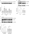
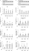
Deletion of β-arrestin 2 abolishes the inhibitory effect of different GPCR agonists on β-adrenergic signal in cardiomyocytes. Adult mouse ventricular cardiomyocytes were pretreated with indicated GPCR agonists (1 µM for 5 min, respectively), then stimulated by 100 nM ISO for 5 min. Phosphorylation of PLB at Serine 16 site was detected by western blot. PLB Phosphorylation levels in WT (A) or β-arrestin 2 KO (E) cardiomyocytes are quantified and expressed as fold over non-stimulated group (Control group, without ISO stimulation condition). Data are obtained from 3 to 4 independent experiments and shown as mean ± S.E.M. *P ≤ 0.05; ***P ≤ 0.001; ****P ≤ 0.0001 (vs. ISO alone group) by one-way ANOVA with Dunnett’s multiple comparison test. The percent shortening in adult WT (B) or β-arrestin 2 KO (F) mouse ventricular cardiomyocyte sarcomere length, the amplitude of calcium release (calcium transient) in WT (C) or β-arrestin 2 KO myocytes (G), and the rate of calcium decay (τ) in WT (D) or β-arrestin 2 KO myocytes (H) are shown in bar graph with scatter plots. Data are obtained from three independent experiments and shown as mean ± S.E.M. **P ≤ 0.01; ****P ≤ 0.0001 (vs. ISO alone group) by one-way ANOVA with Dunnett’s multiple comparison test.
Figure 8
Figure 8
Working model of cross talk between different GPCRs and β2AR resulting in decreased cardiac contractile response. Schematic diagram depicts key signalling components responsible for cross talk between Gs- or Gq-coupled receptors and β2AR. Gs- and Gq-coupled receptors promote PKA- and PKC-mediated phosphorylation of β2AR. The phosphorylated β2AR recruits PDE4D to the cell surface in a β-arrestin 2-dependent manner, which enhances hydrolysis of cAMP induced by β-adrenergic stimulation, effectively blocking β-adrenergic signal transduction to the inner cell organelle such as the SR.

Similar articles

Cited by

References

    1. Gainetdinov RR, Premont RT, Bohn LM, Lefkowitz RJ, Caron MG.. Desensitization of G protein-coupled receptors and neuronal functions. Annu Rev Neurosci 2004;27:107–144. - PubMed
    1. Chuang TT, Iacovelli L, Sallese M, De Blasi A. . G protein-coupled receptors: heterologous regulation of homologous desensitization and its implications. Trends Pharmacol Sci 1996;17:416–421. - PubMed
    1. Rockman HA, Koch WJ, Lefkowitz RJ.. Seven-transmembrane-spanning receptors and heart function. Nature 2002;415:206–212. - PubMed
    1. Moulédous L, Froment C, Dauvillier S, Burlet-Schiltz O, Zajac J-M, Mollereau C.. GRK2 protein-mediated transphosphorylation contributes to loss of function of μ-opioid receptors induced by neuropeptide FF (NPFF2) receptors. J Biol Chem 2012;287:12736–12749. - PMC - PubMed
    1. Cheng Y, Tao Y, Sun J, Wang Y, Xu X, Chen J, Chi Z, Liu J.. Adenosine A(1) receptor agonist N(6)-cyclohexyl-adenosine induced phosphorylation of delta opioid receptor and desensitization of its signaling. Acta Pharmacol Sin 2010;31:784–790. - PMC - PubMed

MeSH terms