. 2017 Feb 28:4:9.
doi: 10.3389/fmolb.2017.00009. eCollection 2017.

The Copper Efflux Regulator CueR Is Subject to ATP-Dependent Proteolysis in Escherichia coli

Affiliations

The Copper Efflux Regulator CueR Is Subject to ATP-Dependent Proteolysis in Escherichia coli

Lisa-Marie Bittner et al. Front Mol Biosci. .

Abstract

The trace element copper serves as cofactor for many enzymes but is toxic at elevated concentrations. In bacteria, the intracellular copper level is maintained by copper efflux systems including the Cue system controlled by the transcription factor CueR. CueR, a member of the MerR family, forms homodimers, and binds monovalent copper ions with high affinity. It activates transcription of the copper tolerance genes copA and cueO via a conserved DNA-distortion mechanism. The mechanism how CueR-induced transcription is turned off is not fully understood. Here, we report that Escherichia coli CueR is prone to proteolysis by the AAA+ proteases Lon, ClpXP, and ClpAP. Using a set of CueR variants, we show that CueR degradation is not altered by mutations affecting copper binding, dimerization or DNA binding of CueR, but requires an accessible C terminus. Except for a twofold stabilization shortly after a copper pulse, proteolysis of CueR is largely copper-independent. Our results suggest that ATP-dependent proteolysis contributes to copper homeostasis in E. coli by turnover of CueR, probably to allow steady monitoring of changes of the intracellular copper level and shut-off of CueR-dependent transcription.

Keywords: AAA+ proteases; ClpAP; ClpXP; CueR; Lon; MerR family; copper homoeostasis; proteolysis.


Figures
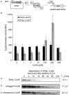
Figure 1
Figure 1
Activity and stability of CueR in E. coli. Schematic presentation of the in vivo CueR activity assay (A). E. coli ΔcueR, Φ(copA-lacZ) cells were transformed with the empty vector pASK-IBA5(+) or the inducible plasmid encoding Strep_CueR and grown to exponential growth phase (M9 minimal medium; with the addition of 30 ng/ml AHT; 30°C). Cells were stressed with increasing CuSO4 concentrations for 1 h and β-galactosidase activity was measured in Miller Units (MU). Standard deviations were calculated from at least two independent experiments (B). Plasmid-encoded Strep_CueR was expressed for 20 min in exponential growth phase (M9 minimal medium; 30°C) in E. coli (MC4100). Translation was blocked by addition of Cm. Samples were taken at indicated time points, subjected to SDS-PAGE, Western transfer, and immunodetection. Half-lives (T1/2) and standard deviations were calculated from 10 independent experiments (C). In vivo degradation experiments with plasmid-encoded untagged CueR were performed as described above. Half-lives (T1/2) and standard deviations were calculated from five independent experiments (D). Stability of endogenous CueR was determined in E. coli MC4100 as described above. Half-lives (T1/2) and standard deviations were calculated from two independent experiments (E).
Figure 2
Figure 2
Strep_CueR is degraded by Lon, ClpXP, and ClpAP protease. Plasmid-encoded Strep_CueR was expressed for 20 min in exponential growth phase (M9 minimal medium; 30°C) in different protease-deficient E. coli strains and their corresponding wild-type (Wt) strains. Translation was blocked by addition of Cm or with spectinomycin for the strain lacking all three proteases (ΔclpXP, Δlon, ΔhslUV) and its parental strain (MG1655) since the triple knockout strain is resistant to Cm. Samples were taken at indicated time points, subjected to SDS-PAGE, Western transfer, and immunodetection. Half-lives (T1/2) and standard deviations were calculated from at least two or three independent experiments.
Figure 3
Figure 3
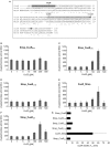
Activity and stability of various CueR variants in E. coli. Comparison of the amino acid sequence of CueR, ZntR, and MerR. DNA-binding domain and dimerization domain of CueR are marked in dark gray and light gray, respectively. Amino acids, which were substituted in different variants used in this study, are highlighted with arrows and the two copper-binding cysteines of CueR are indicated with gray circles. (* = identical amino acid; : = conserved substitution;. = semi conserved substitution; − = lacking amino acid). Alignment was performed by using the align tool of the uniprot database (http://www.uniprot.org/) (A). E. coli ΔcueR, Φ(copA-lacZ) cells harboring inducible plasmids encoding Strep_CueRR18A (B), Strep_CueRA78C (C), Strep_CueRC112S (D), CueR_Strep (E), or Strep_CueRΔC5 (F) were grown in M9 minimal medium with 30 ng/ml AHT at 30°C to log phase. Cells were then treated with increasing CuSO4 concentrations for 1 h. β-galactosidase activity and standard deviations were calculated from at least two independent experiments (B–F). Plasmid-encoded CueR variants were expressed for 20 min in exponential growth phase (M9 minimal medium; 30°C). Translation was blocked by addition of Cm. Samples were taken at indicated time points, subjected to SDS-PAGE, Western transfer, and immunodetection. Half-lives (T1/2) and standard deviations were calculated from at least three independent experiments. For comparison half-life of Strep_CueR is presented (G).
Figure 4
Figure 4
Strep_CueR is not degraded by Lon in vitro. Lon_His6, His6_CspD, and Strep_CueR were purified and used for in vitro degradation experiments (A,B). Degradation experiments were initialized by addition of 20 mM ATP (+ATP). An approach without ATP addition (-ATP) served as control. Samples were taken at indicated time points, subjected to SDS-PAGE and Coomassie staining for His6_CspD (A) or were subjected to Western transfer, and immunodetection for Strep_CueR (B). Data are representative of five independent experiments.
Figure 5
Figure 5
Stability of Strep_CueR in response to increasing CuSO4 concentrations. E. coli MC4100 (Wt) cells harboring a plasmid encoding for Strep_CueR were grown to exponential phase (M9 minimal medium at 30°C). Cultures were supplemented with varying CuSO4 concentrations for 1 h followed by in vivo degradation experiments. Translation was blocked by addition of Cm. Samples were taken at indicated time points, subjected to SDS-PAGE, Western transfer, and immunodetection. Half-lives (T1/2) and standard deviations were calculated from at least two independent experiments.
Figure 6
Figure 6
Degradation of Strep_CueR in different growth phases under varying CuSO4 concentrations. Stability of Strep_CueR was determined in LB medium at 37°C in different growth phases (I–VI) (A) in E. coli MC4100 (Wt) under varying CuSO4 concentrations (B-E). Defined CuSO4 concentrations (0-200 μM) were added right from inoculation of the main culture. In vivo degradation experiments were performed after 20 min of Strep_CueR induction in every growth phase. Translation was blocked by addition of Cm. Samples were taken at indicated time points, subjected to SDS-PAGE, Western transfer, and immunodetection. Half-lives (T1/2) and standard deviations were calculated from at least two independent experiments. Strep_CueR was not detectable in in vivo degradation experiments (V) and (VI).
Figure 7
Figure 7
Stability of Strep_CueR in different growth phases before and after a CuSO4 pulse. Stability of Strep_CueR was determined in different growth phases in E. coli MC4100 (Wt) before and after a copper pulse (arrow) with different CuSO4 concentrations added to the main culture. Cells were grown to different growth phases (I-VI) in LB medium at 37°C (A). The first two in vivo degradation experiments were performed without CuSO4 treatment (I and II) (B–D). After 20 min of Strep_CueR induction in every growth phase, translation was blocked by addition of Cm. Samples were taken at indicated time points, subjected to SDS-PAGE, Western transfer, and immunodetection. Approx. 2.5 h after inoculation a CuSO4 pulse (10–200 μM CuSO4) was given to the main cultures and in vivo degradation experiments in further growth phases (III–VI) followed like described before. Half-lives (T1/2) and standard deviations were calculated from at least two independent experiments. Strep_CueR was not detectable in in vivo degradation experiments (V) and (VI).

Similar articles

Cited by

References

    1. Baba T., Ara T., Hasegawa M., Takai Y., Okumura Y., Baba M., et al. . (2006). Construction of Escherichia coli K-12 in-frame, single-gene knockout mutants: the Keio collection. Mol. Syst. Biol. 2:2006.0008. 10.1038/msb4100050 - DOI - PMC - PubMed
    1. Bachmann B. J. (1972). Pedigrees of some mutant strains of Escherichia coli K-12. Bacteriol. Rev. 36, 525–557. - PMC - PubMed
    1. Baker T. A., Sauer R. T. (2006). ATP-dependent proteases of bacteria: recognition logic and operating principles. Trends Biochem. Sci. 31, 647–653. 10.1016/j.tibs.2006.10.006 - DOI - PMC - PubMed
    1. Barembruch C., Hengge R. (2007). Cellular levels and activity of the flagellar sigma factor FliA of Escherichia coli are controlled by FlgM-modulated proteolysis. Mol. Microbiol. 65, 76–89. 10.1111/j.1365-2958.2007.05770.x - DOI - PubMed
    1. Battesti A., Gottesman S. (2013). Roles of adaptor proteins in regulation of bacterial proteolysis. Curr. Opin. Microbiol. 16, 140–147. 10.1016/j.mib.2013.01.002 - DOI - PMC - PubMed

LinkOut - more resources