. 2017 Mar 8:7:43295.
doi: 10.1038/srep43295.

Post-translational modifications in PrP expand the conformational diversity of prions in vivo

Affiliations

Post-translational modifications in PrP expand the conformational diversity of prions in vivo

Patricia Aguilar-Calvo et al. Sci Rep. .

Abstract

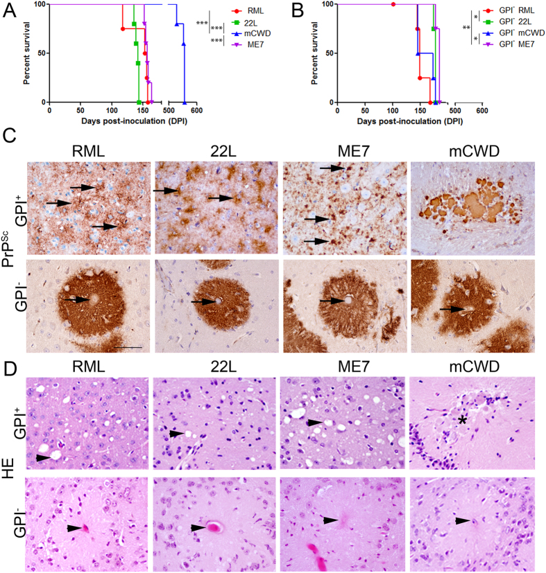
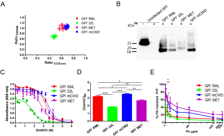
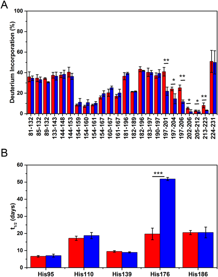
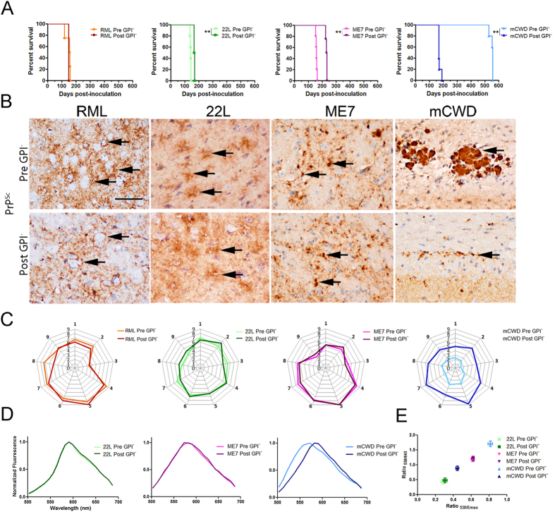
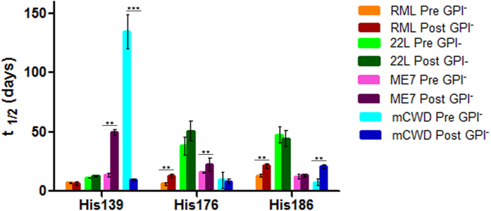
Misfolded prion protein aggregates (PrPSc) show remarkable structural diversity and are associated with highly variable disease phenotypes. Similarly, other proteins, including amyloid-β, tau, α-synuclein, and serum amyloid A, misfold into distinct conformers linked to different clinical diseases through poorly understood mechanisms. Here we use mice expressing glycophosphatidylinositol (GPI)-anchorless prion protein, PrPC, together with hydrogen-deuterium exchange coupled with mass spectrometry (HXMS) and a battery of biochemical and biophysical tools to investigate how post-translational modifications impact the aggregated prion protein properties and disease phenotype. Four GPI-anchorless prion strains caused a nearly identical clinical and pathological disease phenotype, yet maintained their structural diversity in the anchorless state. HXMS studies revealed that GPI-anchorless PrPSc is characterized by substantially higher protection against hydrogen/deuterium exchange in the C-terminal region near the N-glycan sites, suggesting this region had become more ordered in the anchorless state. For one strain, passage of GPI-anchorless prions into wild type mice led to the emergence of a novel strain with a unique biochemical and phenotypic signature. For the new strain, histidine hydrogen-deuterium mass spectrometry revealed altered packing arrangements of β-sheets that encompass residues 139 and 186 of PrPSc. These findings show how variation in post-translational modifications may explain the emergence of new protein conformations in vivo and also provide a basis for understanding how the misfolded protein structure impacts the disease.


Conflict of interest statement

The authors declare no competing financial interests.

Figures

Figure 1
Figure 1. Tg(GPI-PrP) mice infected with four prion strains show similar incubation periods and pathology in brain.
(A,B) Survival curves of WT (A) and Tg(GPI-PrP) (B) mice inoculated with RML, 22 L, mCWD and ME7 prion strains show variable incubation periods in the WT mice, but similar incubation periods in the Tg(GPI-PrP) mice after 4–5 passages. (C) PrPSc aggregates in the brain of prion-infected WT or Tg(GPI-PrP) mice. In WT mice (GPI+), RML forms diffuse, patchy aggregates (arrows), 22 L forms fine aggregates on cell membranes (arrows), ME7 forms dense, punctate aggregates (arrows), and mCWD forms large, dense plaques. Corresponding anchorless prions (GPI) all form dense plaques that infiltrate and extend beyond a central vessel (arrows) and are present in all brain regions. Brain regions shown: RML: thalamus; 22 L: dorsal striatum; ME7: dorsal striatum; mCWD: corpus callosum; GPI-anchorless strains: cerebral cortex. (D) Prion-infected WT mice show spongiform degeneration in the brain after infection with 3 of 4 strains (arrow heads). mCWD plaques (asterisk) lead to very little spongiform change. Note the central vessel and lack of spongiform degeneration in prion-infected Tg(GPI-PrP) mice (arrow head). Scale bar = 100 μm. N = 4–5 mice/group. One-way ANOVA followed by Tukey’s multiple comparison test for survival times, *P < 0.05; **P < 0.05; ***P < 0.001.
Figure 2
Figure 2. Biochemical properties of the GPI-anchorless prions.
(A) PTAA labelling of GPI-anchorless prions. GPI 22 L shows significant differences in the ratios of emitted light intensity at ratios of 533 nm/640 nm and 533 nm/emission maximum compared with the 3 other anchorless prion strains. (B) Electrophoretic mobility of GPI RML, 22 L, mCWD and ME7 prions reveals that the GPI ME7 PrPSc has a higher molecular weight than the other three strains, indicative of a longer PK-resistant PrPSc core fragment. (C) GPI RML, 22 L, mCWD and ME7 exposed to different concentrations of GdnHCl prior to PK digestion show significant differences in the aggregate stability among the strains. Plotted are the mean and standard error (SE) of the remaining PrPSc as measured by ELISA from 3–4 mice per strain, each run in triplicate. (D) The [GdnHCl1/2] values for each strain reveal significant differences between the strains (one-way ANOVA followed by Tukey’s multiple comparison test). (E) The resistance of GPI-anchorless PrPSc to proteinase K shows a significant differences between the strains. (two-way ANOVA followed by Bonferroni post-tests of GPI- anchorless prions revealed significant differences in GPI RML vs GPI mCWD at 10 μg/ml PK**, GPI RML vs GPI ME7 at 10 μg/ml PK*, and GPI 22 L vs GPI mCWD at 10 and 50 μg/ml PK*). *P < 0.05; **P < 0.01; ***P < 0.001.
Figure 3
Figure 3. Structural comparison of GPI-anchorless RML prions at different passages by mass spectrometry based methods.
(A) Backbone amide H/D exchange for PrPSc purified from GPI-anchorless PrP mice after the first (red) and fourth (blue) passage of RML prions. The samples were incubated in D2O for 24 hours at 37 °C, and deuterium incorporation for each peptic fragment derived from these samples was assessed by mass spectrometry. (B) Histidine H/D exchange for first (red) and fourth (blue) passage PrPSc. The parameter t1/2 represents the half-time of exchange reaction for individual His residues. Error bars indicate standard deviation based on three experiments. Student’s t-test: *P < 0.05; **P < 0.01; ***P < 0.001.
Figure 4
Figure 4. mCWD prions form a new prion strain following passage through the GPI-anchorless mice.
(A) Survival curves of WT mice inoculated with RML, 22 L, ME7 and mCWD (Pre GPI) and GPI-anchorless RML, 22 L, ME7 and mCWD (Post GPI) prions. mCWD shows a significantly shortened survival time after passage through the anchorless state, whereas ME7 prions show a modest lengthening in the survival time. (B) Morphology of PrPSc aggregates in the brains of prion-infected WT mice inoculated with the original strains and GPI-anchorless prions. GPI RML, 22 L, mCWD and ME7 passaged into WT mice show diffuse to punctate PrPSc aggregates (arrows) similar to those induced by the original RML, 22 L, and ME7 strains. In contrast, GPI-mCWD led to small clusters of plaques in WT mice, differing markedly from the large, discrete dense plaques of original GPI-anchored mCWD, although both plaque types were enriched in the corpus callosum (arrows). (C) Lesion profile of WT mice infected with Pre and Post GPI prion strains. For the Pre and Post GPI RML, 22 L and ME7 infected WT mice, the severity of spongiosis, astrogliosis, and PrPSc deposition were scored for nine brain regions (see Methods) and were nearly superimposable. For Post GPI- mCWD, the regions and severity of spongiosis, astrogliosis, and PrPSc distribution were more widespread and severe as compared to Pre GPI mCWD. Radial plots show the mean from 4–7 mice per strain. (D) Emission spectra of PTAA-labelled Pre and Post GPI prions in the brain of WT mice (for 22 L and ME7) and in the brain of tga20 mice (for mCWD prions) measured at wavelengths from 500–700 nm. (E) Ratios at 538 nm/643 nm and at 538 nm/emission maximum are shown. Brain regions shown in (B): RML, Pre: thalamus, Post: dorsal striatum; 22 L, Pre and Post: cerebral cortex; ME7, Pre and Post: dorsal striatum; mCWD, Pre and Post: corpus callosum. N = 4–5 mice per group. Scale bar = 100 μm. Logrank test for survival times, *P < 0.05; **P < 0.01; ***P < 0.001.
Figure 5
Figure 5. mCWD prions post-GPI-passage differ biochemically from the original mCWD strain.
(A) The Post GPI RML, 22 L, ME7 and mCWD strains show no differences in electrophoretic mobility as compared to the original strains. (B) [GdnHCl1/2] of Pre and Post GPI strains show that RML and 22 L have not changed following passage through an anchorless state whereas ME7 and mCWD prions are less stable than the original strain. (C) The di-, mono-, and unglycosylated PrP bands of PK-digested Pre and Post GPI RML, 22 L, ME7 and mCWD strains strains were measured following western blotting. Triplots show similar glycoform profiles for all strains except mCWD, which varied significantly from the original strain (2-tailed, unpaired Student’s t test). (D) PK-resistance of Post GPI -RML and −22L were similar to their respective Pre GPI strains, while Post GPI-ME7 show lower PK resistance than -ME7 and Post GPI-mCWD prions show higher PK resistance than–mCWD. Two-way ANOVA followed by Bonferroni post-tests of Pre and Post GPI prions revealed significant differences in RML at 10 and 50 μg/ml PK, and mCWD at 10, 50, and 100 μg/ml and at 250 μg/ml PK. (E) Western blots show the solubility of Pre and Post GPI RML, 22 L, ME7 and mCWD strains. S: supernatant and P: pellet. (F) Quantification of the pellet fraction for all strains (3 animals per strain). Two-way ANOVA followed by Bonferroni post-tests of Pre and Post GPI prions revealed significant differences in the PrPSc solubility of Pre- and Post GPI-mCWD prions. *P < 0.05; **P < 0.01; ***P < 0.001.
Figure 6
Figure 6. H/D exchange for GPI-anchored prion RML, 22 L, ME7 and mCWD before and after passage through GPI-anchorless mice.
Data for His95 and His110 was incomplete due to low abundance of peptide fragments containing these residues and is not shown. The parameter t1/2 represents the half-time of exchange reaction for individual His residues. Error bars indicate standard deviation based on three experiments. **P < 0.01; ***P < 0.005.

Similar articles

Cited by

References

    1. Prusiner S. B. Novel proteinaceous infectious particles cause scrapie. Science 216, 136–144 (1982). - PubMed
    1. Aguzzi A., Sigurdson C. & Heikenwaelder M. Molecular mechanisms of prion pathogenesis. Annu. Rev. Pathol. 3, 11–40 (2008). - PubMed
    1. Boluda S. et al.. Differential induction and spread of tau pathology in young PS19 tau transgenic mice following intracerebral injections of pathological tau from Alzheimer’s disease or corticobasal degeneration brains. Acta Neuropathol. 129, 221–237 (2015). - PMC - PubMed
    1. Bousset L. et al.. Structural and functional characterization of two alpha-synuclein strains. Nat. Commun. 4, 2575 (2013). - PMC - PubMed
    1. Peelaerts W. et al.. alpha-Synuclein strains cause distinct synucleinopathies after local and systemic administration. Nature 522, 340–344 (2015). - PubMed

Publication types