. 2017 Apr 21;292(16):6633-6643.
doi: 10.1074/jbc.M116.770057. Epub 2017 Mar 7.

Discoidin domain receptor 2 mediates collagen-induced activation of membrane-type 1 matrix metalloproteinase in human fibroblasts

Affiliations

Discoidin domain receptor 2 mediates collagen-induced activation of membrane-type 1 matrix metalloproteinase in human fibroblasts

Iwona Majkowska et al. J Biol Chem. .

Abstract

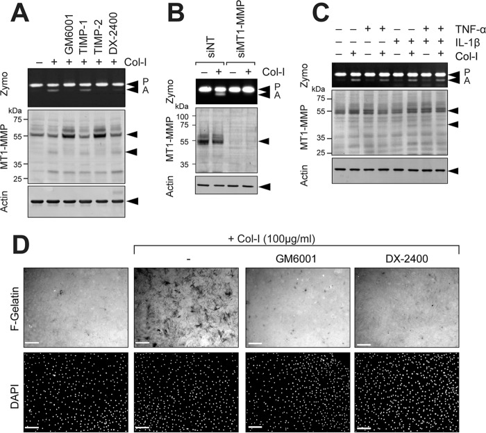
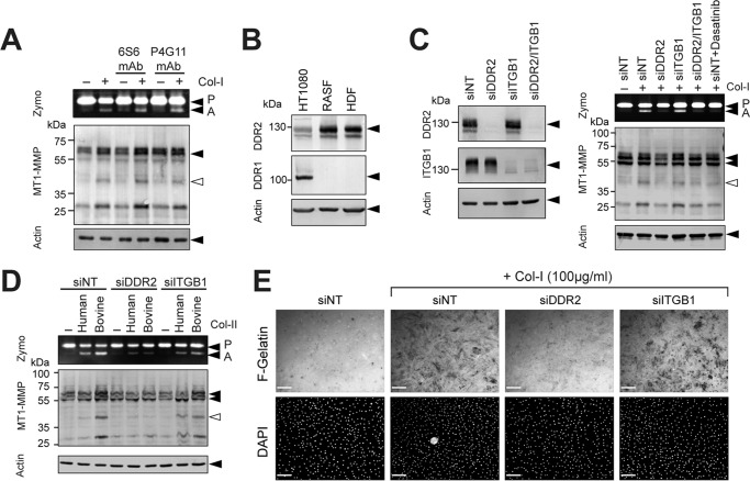
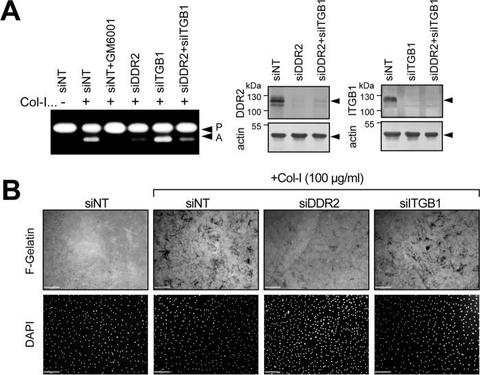
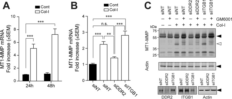
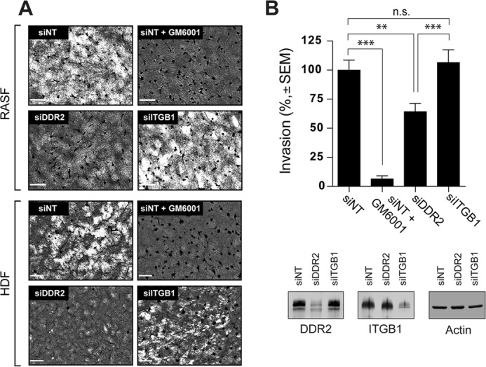
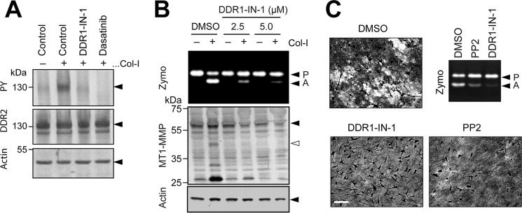
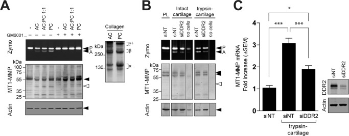
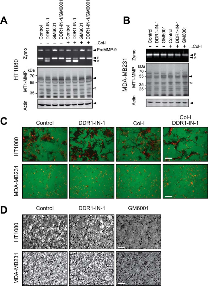
Membrane-type 1 matrix metalloproteinase (MT1-MMP) is a membrane-bound MMP that is highly expressed in cells with invading capacity, including fibroblasts and invasive cancer cells. However, pathways of MT1-MMP up-regulation are not clearly understood. A potential physiological stimulus for MT1-MMP expression is fibrillar collagen, and it has been shown that it up-regulates both MT1-MMP gene and functions in various cell types. However, the mechanisms of collagen-mediated MT1-MMP activation and its physiological relevance are not known. In this study, we identified discoidin domain receptor 2 (DDR2) as a crucial receptor that mediates this process in human fibroblasts. Knocking down DDR2, but not the β1 integrin subunit, a common subunit for all collagen-binding integrins, inhibited the collagen-induced MT1-MMP-dependent activation of pro-MMP-2 and up-regulation of MT1-MMP at the gene and protein levels. Interestingly, DDR2 knockdown or pharmacological inhibition of DDR2 also inhibited the MT1-MMP-dependent cellular degradation of collagen film, suggesting that cell-surface collagen degradation by MT1-MMP involves DDR2-mediated collagen signaling. This DDR2-mediated mechanism is only present in non-transformed mesenchymal cells as collagen-induced MT1-MMP activation in HT1080 fibrosarcoma cells and MT1-MMP function in MDA-MB231 breast cancer cells were not affected by DDR kinase inhibition. DDR2 activation was found to be noticeably more effective when cells were stimulated by collagen without the non-helical telopeptide region compared with intact collagen fibrils. Furthermore, DDR2-dependent MT1-MMP activation by cartilage was found to be more efficient when the tissue was partially damaged. These data suggest that DDR2 is a microenvironment sensor that regulates fibroblast migration in a collagen-rich environment.

Keywords: DDR2; MMP-2; MT1-MMP; arthritis; cancer; collagen; fibroblast; matrix metalloproteinase (MMP).


Conflict of interest statement

The authors declare that they have no conflicts of interest with the contents of this article

Figures

Figure 1.
Figure 1.
Collagen activates MT1-MMP functions in RASF. A, human RASF were cultured on a plastic dish and stimulated with or without collagen I (Col-I; 100 μg/ml) for 24 h in the presence or absence of GM6001 (10 μm), TIMP-1 (100 nm), TIMP-2 (100 nm), or DX-2400 (500 nm). Media were analyzed by zymography for pro-MMP-2 activation (Zymo), and cells were subjected to Western blotting analyses for MT1-MMP and actin. P, pro-MMP-2; A, active MMP-2. B, human RASF transfected with siNT or MT1-MMP siRNA (siMT1-MMP) were cultured and stimulated with collagen I (100 μg/ml) for 24 h. Conditioned media and cell lysates were analyzed as in A. C, human RASF were stimulated with collagen I, TNF-α (10 ng/ml), and/or IL-1β (10 ng/ml) for 24 h as indicated, and the media and cell lysates were analyzed as in A. D, human RASF were subjected to F-gelatin film degradation assay. Cells were cultured on F-gelatin-coated coverslips for 48 h in the presence or absence of collagen I (100 μg/ml) with or without GM6001 (10 μm) or DX-2400 (500 nm). Cells were fixed, nuclei were stained with DAPI, and images were captured with a fluorescence microscope. Scale bars, 270 μm.
Figure 2.
Figure 2.
DDR2 but not integrins mediates collagen signaling to activate MT1-MMP functions in RASF. A, human RASF were stimulated with collagen I (Col-I) in the presence or absence of β1 integrin-inhibitory antibody (6S6) or -activating antibody (P4G11) for 24 h in serum-free medium. Conditioned media and cell lysates were analyzed as in A. B, cell lysates from HT1080 human fibrosarcoma cells, RASF, and HDF were analyzed for DDR1 and DDR2 expression by Western blotting. RASF and HDF express DDR2 but not DDR1, whereas HT1080 expresses both DDRs. C, RASF were transfected with siRNA for DDR2 (siDDR2), β1 integrin (siITGB1), and/or siNT as indicated. 48 h later, cell lysates were subjected to Western blotting analyses for DDR2, ITGB1, and actin (left panel). 48 h after siRNA transfection, cells were also stimulated with collagen I (100 μg/ml) and cultured for a further 72 h. siNT-transfected cells were also treated with dasatinib (Dasa; 100 nm). Conditioned media and cell lysates were analyzed as in A (right panel). D, RASF transfected with siNT, siRNA for DDR2, or siRNA for ITGB1 were stimulated with collagen II (Col-II) of human origin (100 μg/ml) and bovine origin (100 μg/ml) for 48 h. Conditioned media and cell lysates were analyzed as in A. E, RASF transfected with siNT, siRNA for DDR2, or siRNA for ITGB1 were subjected to F-gelatin film degradation assay as in D. Scale bars, 270 μm. P, pro-MMP-2; A, active MMP-2; Zymo, zymography.
Figure 3.
Figure 3.
DDR2 mediates collagen signaling to activate MT1-MMP functions in HDF. A, HDF were transfected with siRNA for DDR2 (siDDR2), siRNA for β1 integrin (siITGB1), and/or siNT as indicated. 48 h later, cell lysates were subjected to Western blotting analyses for DDR2, ITGB1, and actin (right panel).48 h after siRNA transfection, cells were further stimulated with collagen I (Col-I) (100 μg/ml) and cultured for a further 48 h. Conditioned media were analyzed by zymography. B, HDF transfected with siNT, siRNA for DDR2, or siRNA for ITGB1 were subjected to F-gelatin film degradation assay as in Fig. 2E. Scale bars, 270 μm. P, pro-MMP-2; A, active MMP-2.
Figure 4.
Figure 4.
Role of DDR2 in collagen-induced MT1-MMP gene expression. A, human RASF were stimulated with or without collagen I (Col-I) (open bars or closed bars, respectively) for 24 or 48 h, and the level of MT1-MMP gene expression was examined by qPCR (n = 6). ***, p > 0.001. B, RASF transfected with siNT, siRNA for DDR2 (siDDR2), or siRNA for β1 integrin (siITGB1) were stimulated with collagen (open bars). Error bars represent S.E. (n = 6). ***, p > 0.001; **, p > 0.01; n.s., not significant. C, RASF transfected with siNT, siRNA for DDR2, or siRNA for ITGB1 were stimulated with collagen I (100 μg/ml) in the presence or absence of GM6001for 72 h. Cell lysates were subjected to Western blotting analyses for MT1-MMP, actin, DDR2, and ITGB1. Cont, control.
Figure 5.
Figure 5.
Role of DDR2 in MT1-MMP-dependent collagen film degradation and collagen invasion. A, human RASF or HDF transfected with siNT, siRNA for DDR2 (siDDR2), or siRNA for β1 integrin (siITGB1) were subjected to collagen film degradation assay as described under “Experimental Procedures” (left panel). Conditioned media from this assay were analyzed by zymography. P, pro-MMP-2; A, active MMP-2. Scale bars, 270 μm. B, human RASF transfected with siNT, siRNA for DDR2, or siRNA for ITGB1 were subjected to Transwell collagen invasion assay as described under “Experimental Procedures” (upper panel). Levels of DDR2, ITGB1, and actin in transfected cells were analyzed by Western blotting (bottom panel). These are combined data of three independent experiments (n = 6 for each experiments). Error bars represent S.E. ***, p > 0.001; n.s., not significant.
Figure 6.
Figure 6.
Effect of pharmacological inhibition of DDR kinase on MT1-MMP functions. A, HEK293 cells were transfected with DDR2 expression plasmid and stimulated with collagen I (Col-I) (100 μg/ml) in the presence or absence of DDR1-IN-1 (5 μm) or dasatinib (100 nm) for 4 h. Cell lysates were analyzed by Western blotting for phosphotyrosine (PY) (4G10), DDR2, and actin. B, human RASF were stimulated with collagen I (100 μg/ml) in the presence or absence of DDR1-IN-1 at 2.5 or 5.0 μm for 48 h. Conditioned media and cell lysates were analyzed as in Fig. 2A. C, RASF were subjected to collagen film degradation assay in the presence or absence of DDR1-IN-1 (5 μm) or the Src tyrosine kinase inhibitor PP2 (10 μm). Culture media from the assay were analyzed by zymography (Zymo). P, pro-MMP-2; A, active MMP-2.
Figure 7.
Figure 7.
Effect of partial damage on collagen and cartilage for activation of MT1-MMP function. A, human RASF were treated with AC and/or PC (total of 100 μg/ml each) in the presence or absence of GM6001 (10 μm) as indicated. Conditioned media and cell lysates were analyzed as in Fig. 2A. AC and PC used in this experiment were analyzed by SDS-PAGE under reducing conditions (50 μg/lane). The collagen bands were visualized by staining with Coomassie Brilliant Blue (right panel). B, disks of bovine nasal cartilage were treated with or without trypsin (100 μg/ml) for 18 h. Cartilage was washed and treated with soybean trypsin inhibitor (200 μg/ml) for 24 h to inhibit trypsin activity followed by extensive washing with PBS. RASF transfected with siNT or siRNA for DDR2 (siDDR2) were cultured on the cartilage disks for 96 h. Conditioned media and cell/cartilage disk extracts were analyzed as in Fig. 2A. The gel/blot lanes from cartilage alone without cells (no cells) were spliced and placed next to the samples of the cells transfected with siRNA for DDR2. Note that the no-cells sample from intact cartilage has pro-MMP-2 most likely derived from chondrocytes in the tissue. C, human RASF transfected with siNT or siRNA for DDR2 were cultured on cartilage treated with trypsin and analyzed for MT1-MMP gene expression by qPCR (left panel). Levels of DDR2 in the cells were analyzed by Western blotting (right panel). Error bars represent S.E. (n = 6). ***, p > 0.001; *, p > 0.1. Zymo, zymography, P, pro-MMP-2; A, active MMP-2.
Figure 8.
Figure 8.
Effect of pharmacological inhibition of DDRs in cancer cells on collagen-induced MT1-MMP activation. A, HT1080 cells were treated with collagen I (Col-I) in the presence or absence of DDR1-IN-1 (5 μm) and/or GM6001 (10 μm) as indicated. Conditioned media and cell lysates were analyzed as in Fig. 2A. B, MDA-MB231 cells were treated with collagen in the presence or absence of DDR1-IN-1 (5 μm) and/or GM6001 (10 μm) as indicated. Exogenous pro-MMP-2-containing conditioned medium from HEK293 cells stably expressing pro-MMP-2 was added in the medium as MDA-MB231 does not produce pro-MMP-2. Conditioned media and cell lysates were analyzed as in A. C, HT1080 or MDA-MB231 cells were subjected to gelatin film degradation assay in the presence or absence of DDR1-IN-1 (5 μm) and/or collagen I (100 μg/ml) as indicated. Scale bars, 270 μm. D, HT1080 or MDA-MB231 cells were subjected to collagen film degradation assay in the presence or absence of DDR1-IN-1 (5 μm) or collagen I (100 μg/ml) as indicated. Scale bar, 270 μm. Zymo, zymography, P, pro-MMP-2; A, active MMP-2.

Similar articles

Cited by

References

    1. Itoh Y. (2015) Membrane-type matrix metalloproteinases: their functions and regulations. Matrix Biol. 44-46, 207–223 - PubMed
    1. Sabeh F., Ota I., Holmbeck K., Birkedal-Hansen H., Soloway P., Balbin M., Lopez-Otin C., Shapiro S., Inada M., Krane S., Allen E., Chung D., and Weiss S. J. (2004) Tumor cell traffic through the extracellular matrix is controlled by the membrane-anchored collagenase MT1-MMP. J. Cell Biol. 167, 769–781 - PMC - PubMed
    1. Sakamoto T., and Seiki M. (2010) A membrane protease regulates energy production in macrophages by activating hypoxia-inducible factor-1 via a non-proteolytic mechanism. J. Biol. Chem. 285, 29951–29964 - PMC - PubMed
    1. Sakamoto T., Niiya D., and Seiki M. (2011) Targeting the Warburg effect that arises in tumor cells expressing membrane type-1 matrix metalloproteinase. J. Biol. Chem. 286, 14691–14704 - PMC - PubMed
    1. Overall C. M., and Sodek J. (1990) Concanavalin A produces a matrix-degradative phenotype in human fibroblasts. Induction and endogenous activation of collagenase, 72-kDa gelatinase, and Pump-1 is accompanied by the suppression of the tissue inhibitor of matrix metalloproteinases. J. Biol. Chem. 265, 21141–21151 - PubMed

Publication types

MeSH terms

LinkOut - more resources