. 2017 Jul;189(1):21-35.
doi: 10.1111/cei.12959. Epub 2017 Apr 5.

Immunomodulatory glc/man-directed Dolichos lablab lectin (DLL) evokes anti-tumour response in vivo by counteracting angiogenic gene expressions

Affiliations

Immunomodulatory glc/man-directed Dolichos lablab lectin (DLL) evokes anti-tumour response in vivo by counteracting angiogenic gene expressions

V Vigneshwaran et al. Clin Exp Immunol. 2017 Jul.

Abstract

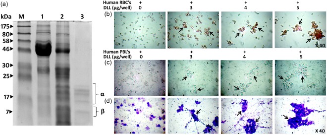
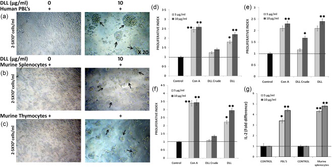
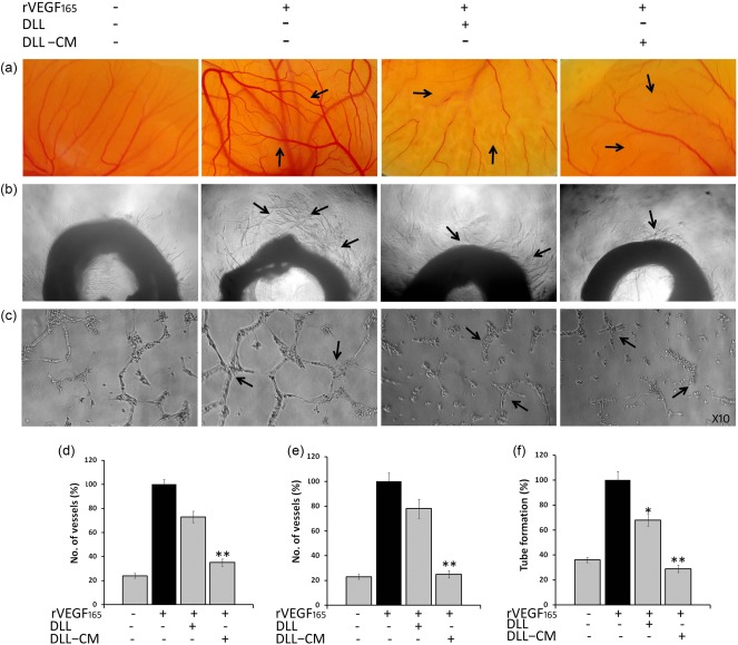
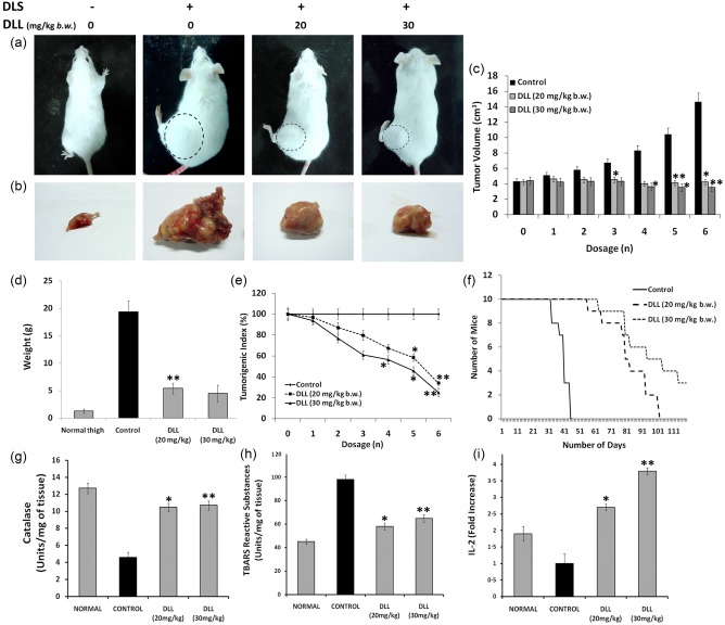
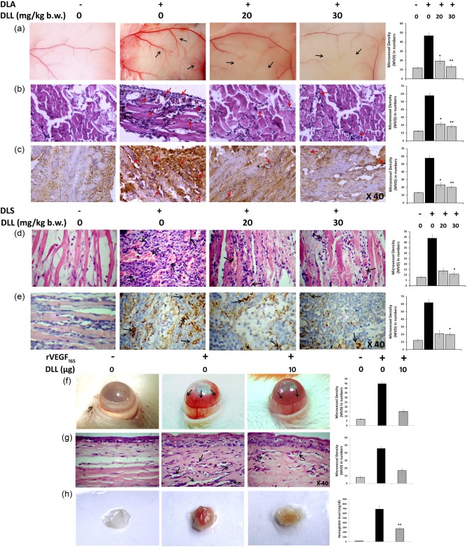
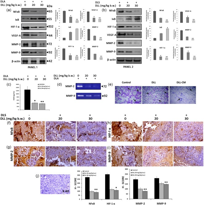
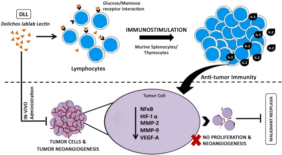
Neovascularization and jeopardized immunity has been critically emphasized for the establishment of malignant progression. Lectins are the diverse class of carbohydrate interacting proteins, having great potential as immunopotentiating and anti-cancer agents. The present investigation sought to demonstrate the anti-proliferative activity of Dolichos lablab lectin (DLL) encompassing immunomodulatory attributes. DLL specific to glucose and mannose carbohydrate moieties has been purified to homogeneity from the common dietary legume D. lablab. Results elucidated that DLL agglutinated blood cells non-specifically and displayed striking mitogenicity to human and murine lymphocytes in vitro with interleukin (IL)-2 production. The DLL-conditioned medium exerted cytotoxicity towards malignant cells and neoangiogenesis in vitro. Similarly, in-vivo anti-tumour investigation of DLL elucidated the regressed proliferation of ascitic and solid tumour cells, which was paralleled with blockade of tumour neovasculature. DLL-treated mice showed an up-regulated immunoregulatory cytokine IL-2 in contrast to severely declined levels in control mice. Mechanistic validation revealed that DLL has abrogated the microvessel formation by weakening the proangiogenic signals, specifically nuclear factor kappa B (NF-κB), hypoxia inducible factor 1α (HIF-1 α), matrix metalloproteinase (MMP)-2 and 9 and vascular endothelial growth factor (VEGF) in malignant cells leading to tumour regression. In summary, it is evident that the dietary lectin DLL potentially dampens the malignant establishment by mitigating neoangiogenesis and immune shutdown. For the first time, to our knowledge, this study illustrates the critical role of DLL as an immunostimulatory and anti-angiogenic molecule in cancer therapeutics.

Keywords: Dolichos lablab lectin; anti-tumour; immunomodulation; mitogenecity; tumour neovasculature.


Figures

Figure 1
Figure 1
Purified Dolichos lablab lectin (DLL) exerts potent erythro‐ and leucoagglutinating activity. (a) Molecular mass determination of purified D. lablab lectin by sodium dodecyl sulphate‐polyacrylamide gel electrophoresis (SDS‐PAGE) under reducing conditions. Lane profile: M = marker, 1 = crude D. lablab extracts (3%), 2 = ammonium sulphate precipitated (20–60%) DLL extracts, 3 = purified Glc/Man‐specific DLL. Lane 3 shows purified protein fractions with five subunits with apparent masses ranging from 10 to 22 kDa. The lectin activity of the purified DLL was examined by its cell agglutinating characteristics employing human red blood cells (RBCs) and leucocytes. (b) DLL potently agglutinates 2% trypsinized human RBCs in sharp contrast to the standard untreated with distinguished separate RBCs. (c) DLL displays leucocyte agglutinating behaviour in a concentration‐dependent manner. The leucocytes were separated through density gradient centrifugation, as described in the Materials and methods section. The illustrations are true‐colour microscopic photographs at ×40 optical resolution. (d) Leucocytes visualized post‐DLL treatment and leucocyte staining at ×40 resolution exhibit significant agglutinating pattern that is conspicuously distinctive to that of control. The dark arrows indicate the agglutinated cells. The results are representative of three independent experiments. [Colour figure can be viewed at wileyonlinelibrary.com].
Figure 2
Figure 2
Immunostimulatory activity of Dolichos lablab lectin (DLL) on human and murine lymphocytes in vitro. To investigate the mitogenic attribute of DLL, in‐vitro cultured lymphocytes were treated with varying lectin concentrations and subjected to 3‐(4,5‐dimethylthiazol‐2‐yl)‐2,5‐diphenyltetrazoliumbromide (MTT) proliferation assay, as told in the Methodology section. (a) Representative visual depictions of the purified DLL‐induced mitogenicity on human peripheral blood lymphocytes (2·5 × 106 cells/ml) at 10 μg/ml concentration. The cell cluster formation, a prerequisite event in the cellular proliferation, could be observed in DLL, whereas untreated cells appear distinctively separated. Similar cell clusters were also observed in (b) murine splenocytes (c) and thymocytes. The true‐coloured and unstained images were observed through brightfield inverted microscope (Olympus) at ×20 optical magnification. The proliferative effect of DLL was expressed in terms of proliferative index. The index for control (non‐stimulated) cells was taken as 1·0 and for others it is calculated as a ratio of absorbance at 570 nm of the test sample to that of the control. Graphs indicating the proliferative index of DLL on human peripheral blood lymphocytes (PBLs) (d), splenocytes (e) and thymocytes (f) in vitro. (g) Effect of DLL on ex‐vivo interleukin (IL)‐2 production by human PBL and murine splenocytes. Results are the means of three determinations, each conducted in triplicate. Statistically significant values are *P < 0·05; ** P < 0·01. [Colour figure can be viewed at wileyonlinelibrary.com].
Figure 3
Figure 3
Anti‐angiogenic activity of Dolichos lablab lectin‐conditioned medium (DLL‐CM) in in‐vitro angiogenic assay models. Indirect angioinhibitory effect of DLL on various angiogenic systems in vitro. DLL‐CM was the supernatant of DLL exposed to murine splenocytes after 24 h incubation. DLL‐CM exhibits regressed angiogenesis in (a) ex‐vivo chorioallontoic membrane (CAM) and (b) rat aortic angiogenesis assay. DLL directly exhibited only a minimal angiolytic effect. (c) DLL‐CM exposed to human umbilical vein endothelial cells (HUVEC) cells inhibits the tube‐forming efficiency. Pictographical representation of (d) ex‐vivo CAM, (e) aortic ring angiogenesis and (f) tube formation assay. Data were represented as mean ± standard error of the mean (s.e.m.) of three independent experiments. Statistically significant values are expressed as *P < 0·05 and **P < 0·01. [Colour figure can be viewed at wileyonlinelibrary.com].
Figure 4
Figure 4
Dolichos lablab lectin (DLL) mitigates the proliferation of murine Dalton's lymphoma solid tumour in‐vivo with up‐regulation of IL‐2 levels. Solid tumour was induced by injecting Dalton's lymphoma cells (1 × 106 cells/mouse) in hindlimbs of Swiss albino mice. After the palpable development of tumour, six doses of DLL (20 and 30 mg/kg body weight b.w.) were administered intraperitoneally (i.p.) to tumour‐bearing mice on every alternative day. (a) Tumour morphology of the DLS‐induced mice with and without DLL treatment. The circular dotted lines represent the extent of tumour growth. (b) Gross anatomical appearance of the excised DLS tumours. Note the extensive vascularization surrounding the tumour tissue of the untreated control. (c) The effect of DLL on DLS tumour progression at different time‐points. (d) Weight of the excised normal thigh, control and DLL‐treated tumours (g). (e) Tumorigenic index elucidating the dose‐dependent decrease of tumour progression post‐DLL regimen. (f) Kaplan–Meier survival curve of the animals treated with DLL. Effect of DLL in the endogenous hepatic anti‐oxidant system (d) pronounced catalase enzymes levels in DLL‐treated mice. Note control with severely declined enzyme content. (e) Decreased lipid peroxidation in DLL‐treated mice, as assessed by the thiobarbituric reactive substances (TBARS) method. (f) Fold difference in the serum interleukin (IL)‐2 levels of DLL‐treated animals compared to control. The control animal shows declined levels of IL‐2, whereas DLL at 20 and 30 mg/kg b.w. via i.p. administration shows pronounced IL‐2 levels. Results are the means of three determinations, each conducted in triplicate. Statistically significant values are *P < 0·05; ** P < 0·01. [Colour figure can be viewed at wileyonlinelibrary.com].
Figure 5
Figure 5
Dolichos lablab lectin (DLL) abrogates tumoral and non‐tumoral angiogenesis in vivo. The effect of DLL in the tumour‐induced angiogenesis was evaluated by murine ascites and solid tumour models in vivo, treated with DLL 20 and 30 mg/kg body weight intraperitoneally (i.p.). (a) The peritoneal lining of mice showing the visible suppression of peritoneal microvessel in DLL‐treated mice. (b) Haematoxylin and eosin (H&E)‐stained peritoneum sections showing the decreased vascular count (c). CD31 immunohistochemistry (IHC) of the peritoneal sections depicting hypervascularization (intensive brownish staining) in the control section in sharp contrast to the DLL. The reticence of tumour angiogenesis was confirmed by (d) H&E and (e) IHC (CD31) photomicrographs of solid tumour sections depicting a significant reduction in vessel density. The effect of DLL on corneal angiogenesis and matrigel plug model systems was studied by inducing angiogenesis with recombinant vascular endothelial factor (rVEGF)165 and treatment with DLL. (f) DLL regresses rat corneal angiogenesis. (g) Micrographs of H&E‐stained rat corneal sections confirming the reduced microvascular density/high‐power field (MVD/HPF) upon DLL treatment. The black arrows indicate the extent of vascularization. The representative MVD counts were provided adjacent to every corresponding image. (h) Matrigel plug assay depicting control with aberrant vascularization in contrast to the DLL‐treated mice. Pictograph depicting the level of haemoglobin content in matrigel implants is provided adjacently. Results are the means of three independent experiments. Statistically significant values are *P < 0·05; ** P < 0·01. [Colour figure can be viewed at wileyonlinelibrary.com].
Figure 6
Figure 6
Translational down‐regulation of tumoral angiogenic gene expression by Dolichos lablab lectin (DLL). Immunoblots, gelatin zymography and immunohistochemistry (IHC) were carried out using in‐vivo tumour cells of both Dalton's ascites lymphoma (DLA) and Dalton's lymphoma solid tumour (DLS) with or without DLL treatment in two different concentrations. (a,b) Immunoblots showing altered translational expression of proangiogenic genes such as nuclear factor kappa B (NF‐κB), inhibitory kappa B (I‐κB), hypoxia inducible factor 1α (HIF‐1α), vascular endothelial growth factor (VEGF)‐A, matrix metalloproteinase (MMP)‐2 and 9 in ascitic (panel 1) and solid tumour (panel 2). The graphs indicate the densitometric values of corresponding Western blot data. (c) Reduction of secreted serum VEGF levels in DLL‐treated DLA mice in vivo. (d) Gelatin zymography showing reduced gelatinolytic activity in DLL‐treated mice. (e) DLL‐conditioned medium (DLL‐CM) inhibits the migration of A549 cell in vitro. IHV should be replaced with IHC detection of DLL‐induced altered gene expression. (f,i) IHC detection of proangiogenic gene expression in solid tumours in vivo after DLL treatment. The intense brown staining indicates the respective protein expression. Representative graphs demonstrating the gene expression corresponding to the IHC data are given below. (j) Representative counter haematoxylin‐stained section. Data are represented as mean ± standard error of the mean (s.e.m.) of three independent experiments. Statistically significant values are expressed as *P < 0·05 and **P < 0·01. [Colour figure can be viewed at wileyonlinelibrary.com].
Figure 7
Figure 7
Schematic demonstration of glc/man interacting Dolichos lablab lectin (DLL) induced anti‐tumour effect. Hypothetical model diagram depicting the possible mechanism of action of immunostimulatory DLL in eliciting the potent anti‐neoplastic and anti‐angiogenic response in in‐vivo murine Dalton's lymphoma tumour. [Colour figure can be viewed at wileyonlinelibrary.com].

Similar articles

Cited by

References

    1. Rabinovich GA, Gabrilovich D, Sotomayor EM. Immunosuppressive strategies that are mediated by tumor cells. Annu Rev Immunol 2007; 25:267–96. - PMC - PubMed
    1. Ghirelli C, Hagemann T. Targeting immunosuppression for cancer therapy. J Clin Invest 2013; 123:2355–7. - PMC - PubMed
    1. Deepak P, Kumar S Jr, Kishore D, Acharya A. IL‐13 from Th2‐type cells suppresses induction of antigen‐specific Th1 immunity in a T‐cell lymphoma. Int Immunol 2010; 22:53–63. - PubMed
    1. Rosenberg SA. IL‐2: the first effective immunotherapy for human cancer. J Immunol 2014; 192:5451–8. - PMC - PubMed
    1. Douglas H, Weinberg AR. Hallmarks of cancer: the next generation. Cell 2011; 144:646–74. - PubMed

Publication types

MeSH terms