. 2017 Apr 28;91(10):e00175-17.
doi: 10.1128/JVI.00175-17. Print 2017 May 15.

Conformational States of a Soluble, Uncleaved HIV-1 Envelope Trimer

Affiliations

Conformational States of a Soluble, Uncleaved HIV-1 Envelope Trimer

Yuhang Liu et al. J Virol. .

Abstract

The HIV-1 envelope spike [Env; trimeric (gp160)3 cleaved to (gp120/gp41)3] induces membrane fusion, leading to viral entry. It is also the viral component targeted by neutralizing antibodies. Vaccine development requires production, in quantities suitable for clinical studies, of a recombinant form that resembles functional Env. HIV-1 gp140 trimers-the uncleaved ectodomains of (gp160)3-from a few selected viral isolates adopt a compact conformation with many antigenic properties of native Env spikes. One is currently being evaluated in a clinical trial. We report here low-resolution (20 Å) electron cryomicroscopy (cryoEM) structures of this gp140 trimer, which adopts two principal conformations, one closed and the other slightly open. The former is indistinguishable at this resolution from those adopted by a stabilized, cleaved trimer (SOSIP) or by a membrane-bound Env trimer with a truncated cytoplasmic tail (EnvΔCT). The latter conformation is closer to a partially open Env trimer than to the fully open conformation induced by CD4. These results show that a stable, uncleaved HIV-1 gp140 trimer has a compact structure close to that of native Env.IMPORTANCE Development of any HIV vaccine with a protein component (for either priming or boosting) requires production of a recombinant form to mimic the trimeric, functional HIV-1 envelope spike in quantities suitable for clinical studies. Our understanding of the envelope structure has depended in part on a cleaved, soluble trimer, known as SOSIP.664, stabilized by several modifications, including an engineered disulfide. This construct, which is difficult to produce in large quantities, has yet to induce better antibody responses than those to other envelope-based immunogens, even in animal models. The uncleaved ectodomain of the envelope protein, called gp140, has also been made as a soluble form to mimic the native Env present on the virion surface. Most HIV-1 gp140 preparations are not stable, however, and have an inhomogeneous conformation. The results presented here show that gp140 preparations from suitable isolates can adopt a compact, native-like structure, supporting its use as a vaccine candidate.

Keywords: cryoEM; envelope; human immunodeficiency virus; immunogen.


Figures

FIG 1
FIG 1
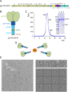
Production and characterization of the C97ZA012 gp140 trimer for cryoEM analysis. (A) Schematic representation of the gp140 construct used in the EM study. The gp140 region includes the following segments: C1 to C5, conserved regions 1 to 5; V1 to V5, variable regions 1 to 5; F, fusion peptide; HR1, heptad repeat 1; C-C loop, immunodominant loop with a conserved disulfide bond; HR2, heptad repeat 2; and MPER, membrane-proximal external region. The C-terminal end of gp140 was fused directly to a GCN4 trimer sequence, followed by a GSGG linker, a foldon sequence, a GSG linker, and another GCN4 sequence ending with a histidine tag. (B) 3D organization of the gp140 trimer. The length of the added stem region can be ∼100 Å in an extended conformation. (C) The His6-tagged gp140-GFG protein was purified from supernatants of 293T cells stably transfected with the expression construct. Purified gp140 was resolved by gel filtration chromatography on a Superose 6 column. The molecular mass standards included thyroglobulin (670 kDa), ferritin (440 kDa), γ-globulin (158 kDa), and ovalbumin (44 kDa). Peak fractions were pooled and analyzed by Coomassie-stained SDS-PAGE (inset; lane 1, gp140 described in reference ; lane 2, gp140-GFG; and lane 3, purified complex of gp140-GFG and 4-domain CD4). (D) Diagram of rosette formation when the gp140-GFG protein was incubated with Ni-NTA (in red)-conjugated gold beads (in orange). (E) Representative area of a micrograph of gp140 rosettes under cryo conditions. Selected rosettes from this area are shown in the upper right panels, and the extracted gp140 particles from these rosettes are shown in the lower right panels.
FIG 2
FIG 2
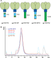
Various gp140-stem-His6 constructs. (A) Schematic diagrams demonstrating the organization of various gp140 constructs. gp140-FG, the C-terminal end of C97 gp140 is connected to a T4 fibritin foldon through an IEGRGSGG linker, followed by a GSG linker, GCN4, another GSG linker, and a His6 tag; gp140-GF, the C-terminal end of 92UG gp140 is connected to GCN4 through a GSG linker, followed by an IEGRGSG linker, a foldon, a glycine, and a His6 tag; gp140-Fbtc, the C-terminal end of 92UG gp140 is fused in phase to the C-terminal portion of a bacteriophage T4 fibritin unit (34), followed by a glycine and a His6 tag; gp140-Fbt, the C-terminal end of 92UG gp140 is fused in phase to the full-length fibritin unit, followed by a glycine and a His6 tag. The coloring scheme follows that in Fig. 1. (B) The constructs were expressed in 293T cells transiently transfected with the expression constructs. The supernatants were harvested at 5 days posttransfection and affinity purified by elution from Ni-NTA resin. The purified gp140 constructs were then subjected to gel filtration chromatography on a Superose 6 column (GE). All of these proteins eluted as a single sharp and symmetrical peak at about the correct elution volume. The variation in the peak is due to the different hydrodynamic radii of these proteins and to different loading volumes.
FIG 3
FIG 3
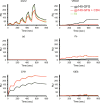
Antigenic properties of the C97ZA012 gp140-GFG trimer. The gp140-GFG protein was analyzed by a surface plasmon resonance assay for binding to the monoclonal antibodies 2G12 (specific for glycan), VRC01 (specific for the CD4 binding site; broadly neutralizing), b6 (specific for the CD4 binding site; nonneutralizing), 17b (CD4i), 3971 (specific for V3; nonneutralizing), and 10E8 (specific for the MPER; broadly neutralizing). gp140-GFG or the purified complex of gp140-GFG and CD4 was captured on the surface of a sensor chip coated with an anti-His6 antibody to avoid potential artifacts introduced by protein immobilization. Each antibody at various concentrations was passed over the gp140 surface individually without regeneration for single-cycle kinetic analysis. The recorded sensorgrams for gp140-GFG are shown in black, those for the gp140-GFG–CD4 complex in red, and the computational fit in green. Binding constants are summarized in Table 1. RU, response units.
FIG 4
FIG 4
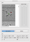
Graphical user interface of a program for picking a trimer from a rosette image. The rosette images are loaded in the upper left panel. The center of the rosette is selected and marked with a green dot; selected positions of individual trimers in the rosette are marked with red dots. The coordinate information from the image is displayed in the lower panel.
FIG 5
FIG 5
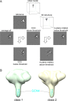
Structure determination for the C97ZA012 gp140 trimer by cryoEM. (A) Strategy to eliminate particles from the reconstruction in the early alignment cycles by using the SPIDER procedure. (First row) An initial model made of a long and thin cylinder connected to a short and thick cylinder was used to perform the first round of 3D reconstruction. The resulting structure served as the reference for the next cycle. (Second row) A representative projection is shown next to one of the refined 3D maps. (Third row) Particles were aligned to their best-matching projections. The “average all” panel represents the average for all particles that matched the projection shown in the second row. The “CCC above threshold” panel shows the average for the particles that have a cross-correlation coefficient (CCC) above the threshold value. For the particles that did not meet the threshold, the average image is shown as “CCC below threshold,” in the fourth row. The particles that met the CCC threshold were sorted by the angle-matching criterion. The average for particles below the angular threshold is represented in the “in-plane rotation below threshold” panel, which was included in the final 3D structure reconstruction. (Fourth row) The average for the rejected particles is shown for each criterion. (B) Side views of the two reconstructions corresponding to subclasses 1 and 2, with trimeric GCN4 (in cyan) fit into the tail-like density.
FIG 6
FIG 6
Further subclassification of three retained 3D classes. The particles that belonged to classes 1, 2, and 3 were extracted into separate stacks. Each class was subjected to further classification into three subclasses. The three structures (red, blue, and green) obtained after 60 refinement cycles were overlaid to evaluate the structural homogeneity of each class.
FIG 7
FIG 7
FSC curves for the reconstructions of classes 1 and 2.
FIG 8
FIG 8
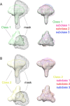
Focused classification of gp140 structures. Focused classification using a mask (shown in black mesh) covering one protomer of the trimer was performed for both class 1 (A; green) and class 2 (B; yellow). Three subclasses for each class are shown, in magenta, red, and blue. Differences within the mask are evident among subclasses, while density outside the mask remains the same.
FIG 9
FIG 9
Impact of the initial model on 3D reconstruction. Two different initial models were tested for 3D reconstruction. Model 1, a long and thin cylinder connected to a short and thick cylinder led to structure 1; model 2, a single cylinder gave rise to structure 2. The two structures are almost identical, as shown by the overlay.
FIG 10
FIG 10
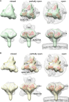
Conformation of the uncleaved C97ZA012 gp140 trimer. (A) CryoEM map of class 1 C97ZA012 gp140 trimer (light green) superimposed on the map of the cleaved, wild-type JR-FL EnvΔCT trimer in complex with PGT151 (structure EMD-3308 filtered to 21-Å resolution [shown as mesh[; the density for PGT151 was removed for clarity) (left), the map of the BG505 SOSIP.664 trimer in complex with soluble 2D CD4, 17b Fab, and 8ANC195 variant G32K5 Fab (structure EMD-3096 [mesh]) (middle), and the map of the BaL Env trimer on the virion surface in complex with soluble 2D CD4 (structure EMD-5455 [mesh]) (right). For the maps representing partially open and open conformations induced by ligand binding, the locations of three gp120s are shown by models, in red, of the gp120 core (PDB entries 5A8H and 3DNO, respectively). Locations of CD4 and antibodies are also indicated. (B) As in panel A, except that the map in light yellow is the cryoEM map of a class 2 C97ZA012 gp140 trimer.
FIG 11
FIG 11
Main difference between the maps for class 2 and the open conformation. The cryoEM map of a class 2 C97ZA012 gp140 trimer (yellow) is superimposed on the map of the BaL Env trimer on the virion surface in complex with soluble 2D CD4 (structure EMD-5455 [black mesh]). The main difference between the two maps, next to the trimer apex, is indicated.
FIG 12
FIG 12
Fit of the JR-FL EnvΔCT trimer into cryoEM maps of C97ZA012 gp140. The model of the JR-FL EnvΔCT trimer (PDB entry 5FUU), shown as a ribbon diagram in red, was fit by manual adjustment in UCSF Chimera into the density maps of the 3D reconstructions of class 1 (light green) and class 2 (light yellow). The location of V1V2, which presents the trimer-specific, broadly neutralizing epitopes, is indicated.

Similar articles

Cited by

References

    1. Harrison SC. 2005. Mechanism of membrane fusion by viral envelope proteins. Adv Virus Res 64:231–259. doi:10.1016/S0065-3527(05)64007-9. - DOI - PMC - PubMed
    1. Chan DC, Fass D, Berger JM, Kim PS. 1997. Core structure of gp41 from the HIV envelope glycoprotein. Cell 89:263–273. doi:10.1016/S0092-8674(00)80205-6. - DOI - PubMed
    1. Weissenhorn W, Dessen A, Harrison SC, Skehel JJ, Wiley DC. 1997. Atomic structure of the ectodomain from HIV-1 gp41. Nature 387:426–430. doi:10.1038/387426a0. - DOI - PubMed
    1. Kovacs JM, Nkolola JP, Peng H, Cheung A, Perry J, Miller CA, Seaman MS, Barouch DH, Chen B. 2012. HIV-1 envelope trimer elicits more potent neutralizing antibody responses than monomeric gp120. Proc Natl Acad Sci U S A 109:12111–12116. doi:10.1073/pnas.1204533109. - DOI - PMC - PubMed
    1. Chen J, Kovacs JM, Peng H, Rits-Volloch S, Lu J, Park D, Zablowsky E, Seaman MS, Chen B. 2015. HIV-1 envelope. Effect of the cytoplasmic domain on antigenic characteristics of HIV-1 envelope glycoprotein. Science 349:191–195. doi:10.1126/science.aaa9804. - DOI - PMC - PubMed

Publication types

MeSH terms

Substances

LinkOut - more resources