. 2017 Apr 28;91(10):e02335-16.
doi: 10.1128/JVI.02335-16. Print 2017 May 15.

The ND10 Component Promyelocytic Leukemia Protein Acts as an E3 Ligase for SUMOylation of the Major Immediate Early Protein IE1 of Human Cytomegalovirus

Affiliations

The ND10 Component Promyelocytic Leukemia Protein Acts as an E3 Ligase for SUMOylation of the Major Immediate Early Protein IE1 of Human Cytomegalovirus

Nina Reuter et al. J Virol. .

Abstract

Previous studies identified the nuclear domain 10 (ND10) components promyelocytic leukemia protein (PML), hDaxx, and Sp100 as factors of an intrinsic immune response against human cytomegalovirus (HCMV). This antiviral function of ND10, however, is antagonized by viral effector proteins like IE1p72, which induces dispersal of ND10. Furthermore, we have shown that both major immediate early proteins of HCMV, IE1p72 and IE2p86, transiently colocalize with ND10 subnuclear structures and undergo modification by the covalent attachment of SUMO. Since recent reports indicate that PML acts as a SUMO E3 ligase, we asked whether the SUMOylation of IE1p72 and IE2p86 is regulated by PML. To address this, PML-depleted fibroblasts, as well as cells overexpressing individual PML isoforms, were infected with HCMV. Western blot experiments revealed a clear correlation between the degree of IE1p72 SUMO conjugation and the abundance of PML. On the other hand, the SUMOylation of IE2p86 was not affected by PML. By performing in vitro SUMOylation assays, we were able to provide direct evidence that IE1p72 is a substrate for PML-mediated SUMOylation. Interestingly, disruption of the RING finger domain of PML, which is proposed to confer SUMO E3 ligase activity, abolished PML-induced SUMOylation of IE1p72. In contrast, IE1p72 was still efficiently SUMO modified by a SUMOylation-defective PML mutant, indicating that intact ND10 bodies are not necessary for this effect. Thus, this is the first report that the E3 ligase PML is capable of stimulating the SUMOylation of a viral protein which is supposed to serve as a cellular mechanism to compromise specific functions of IE1p72.IMPORTANCE The major immediate early proteins of human cytomegalovirus, termed IE1p72 and IE2p86, have previously been shown to undergo posttranslational modification by covalent coupling to SUMO moieties at specific lysine residues. However, the enzymatic activities that are responsible for this modification have not been identified. Here, we demonstrate that the PML protein, which mediates an intrinsic immune response against HCMV, specifically serves as an E3 ligase for SUMO modification of IE1p72. Since SUMO modification of IE1p72 has previously been shown to interfere with STAT factor binding, thus compromising the interferon-antagonistic function of this viral effector protein, our finding highlights an additional mechanism through which PML is able to restrict viral infections.

Keywords: human cytomegalovirus; immediate early protein IE1; nuclear domain 10; promyelocytic leukemia protein PML; sumoylation.


Figures

FIG 1
FIG 1
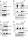
Direct correlation between the extent of IE1 SUMOylation and PML abundance. PML-kd cells (siPML2) and control HFFs (vector), along with HFFs overexpressing PML isoform I (HFF/PMLI), were either mock infected or infected with HCMV AD169 at an MOI of 0.1. At the indicated times postinfection (24 to 96 h postinfection [hpi]), the cell lysates were harvested and analyzed by Western blotting for PML, IE1, and IE2 protein levels. Beta-actin was included as an internal loading control. The ratio of IE1 SUMOylation was quantitated and is shown below the blot (% IE1-SUMO).
FIG 2
FIG 2
PML stimulates SUMO conjugation of IE1 in vivo. (A to C) Western blot analyses of cell extracts of HEK293T cells transfected with expression constructs for PML VI in combination with IE1 (A and B) or IE2 (C). For all Western blot experiments, lysates from nontransfected HEK293T cells (mock) served as a negative control, and detection of beta-actin was included as an internal loading control. (B and C) Coexpression of IE1, together with decreasing amounts of PML. The quantity of transfected PML-encoding DNA ranged from 1 μg to 10 ng, as indicated. The ratio of protein SUMOylation was quantitated and is shown below panels B and C (% IE1-SUMO and % IE2-SUMO, respectively).
FIG 3
FIG 3
HEK293T cells exhibit altered ND10 protein abundance in comparison to HFFs. (A to C) Equal amounts of protein lysates (prepared from 600.000 cells) of HEK293T cells and HFFs were compared by immunoblotting for PML (A), Sp100 (B), and hDaxx (C) protein expression levels. Beta-actin served as an internal loading control.
FIG 4
FIG 4
PML promotes SUMOylation of IE1 in vitro. (A to D) Bacterially purified IE1 was incubated with E1 (SAE1/SAE2), Ubc9, ATP, and either SUMO-1 (A and D) or SUMO-3 (B and C) in the presence or absence of GST-PML or GST-PML-SUMOmut (both isoform VI), as described in Materials and Methods. In panel D, increasing concentrations of PML-SUMOmut were used. The reaction mixtures were analyzed by Western blotting using anti-IE1 and anti-PML antibodies. (E) Coomassie blue-stained gel showing the purified proteins (15-fold amounts) that were used for the blots in panel D. Lanes: 1, purified IE1; 2 to 5, increasing amounts of PML-SUMOmut as used in lanes 3 to 6 of panel D.
FIG 5
FIG 5
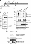
PML isoform-specific SUMOylation of IE1. (A) Schematic representation of exon organization and structural domains of the nuclear-localized PML isoforms I to VI, which all share the N-terminal region containing the RBCC motif (exons 1 to 6). The different C termini are generated by alternative use of 3′ exons. R, RING domain; B1 and B2, zinc binding boxes; CC, coiled-coil domain; S, SUMO; SIM, SUMO interaction motif; NLS, nuclear localization signal. (B) Cotransfection of HEK293T cells with plasmids encoding IE1 and the various nuclear-localized PML isoforms (FLAG-PML I to VI), followed by Western blotting detection of IE1, beta-actin, and PML I to VI. The last were detected by their FLAG moieties. (C) Relative IE1 SUMOylation from panel B (normalized for the expression level of the respective PML isoform). IE1 SUMOylation by PML isoform VI was set to 100%.
FIG 6
FIG 6
Disturbance of the PML-IE1 interaction interface abrogates IE1 SUMOylation. (A and C) Coimmunoprecipitation analysis of PML-kd/293T cells cotransfected with plasmids encoding Myc-tagged PML isoform VI (M-PML), together with either wt IE1, the PML interaction-defective IE1 mutant L174P, or the SUMOylation-deficient IE1 mutant K450R, as indicated. Two days posttransfection, the cells were lysed, and immunoprecipitation was performed using an anti-IE1 antibody. After electrophoresis, coprecipitated PML (M-PML) was visualized using an anti-Myc antibody (bottom). Immunoglobulin heavy chain (IgG HC) served as an internal control for the presence of the precipitating antibody. (B and D) Western blot analysis of extracts from HEK293T cells transfected with PML isoform VI alone (myc tagged) or in combination with either wt IE1 or the IE1 mutants L174 and K450R. Lysates from mock-transfected HEK293T cells served as specificity controls and detection of beta-actin expression as an internal loading control. (B) A FLAG-SUMO-1 expression construct was included in the cotransfection reaction, and SUMOylated PML was detected using an anti-FLAG antibody (top).
FIG 7
FIG 7
An intact RING finger domain of PML is required to stimulate IE1 SUMO conjugation. (A) Schematic representation of RINGmut based on PML isoform VI, where the key cysteine amino acids at positions 57 and 60 were mutagenized to similar nonpolar serine residues. (B) Coimmunoprecipitation analysis of PML-kd/293T cells cotransfected with plasmids encoding Myc-tagged PML RINGmut VI (M-RING) and wt IE1 or the PML interaction-negative mutant L174P. Two days posttransfection, the cells were lysed and immunoprecipitation was performed using an anti-IE1 antibody. After electrophoresis, coprecipitated PML RINGmut (M-RING) was visualized using an anti-Myc antibody (bottom). Immunoglobulin heavy chain (IgG HC) served as an internal control for the presence of the precipitating antibody. (C) Extracts from HEK293T cells transfected with IE1 and FLAG–SUMO-1 in combination with either wt PML or RINGmut (both isoform VI; myc tagged) were analyzed by Western blotting. Lysates from nontransfected HEK293T cells (mock) served as a specificity control. (D) Bacterially purified IE1 was incubated together with an in vitro SUMOylation kit containing E1 (SAE1/SAE2), Ubc9, ATP, and SUMO-1 in the presence or absence of PML GST-SUMOmut, GST-RINGmut (both isoform VI), or GST alone, as described in Materials and Methods. The reaction mixtures were analyzed by Western blotting using anti-IE1 and anti-PML antibodies.
FIG 8
FIG 8
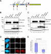
The PML RING finger mutant fails to orchestrate ND10. (A) Indirect immunofluorescence analysis of siPML2-transduced HFFs (siPML2), as well as nontransduced control HFFs (HFF), transfected with plasmid DNA coding for Myc-tagged PML RINGmut (isoform VI). Two days posttransfection, the cells were fixed and stained for the ND10 marker Sp100 or hDaxx, along with PML RINGmut. The last was visualized by applying an anti-Myc antibody. Cell nuclei were identified by detection of DAPI signals (scale bars = 10 μm). (B) Numbers of cells with either a diffuse or a dot-like subnuclear distribution of PML RINGmut. Twenty cells were evaluated for either transfected control HFFs or transfected siPML2 cells.
FIG 9
FIG 9
PML-promoted SUMOylation of IE1 in the absence of intact ND10 bodies. (A) Schematic representation of the SUMOylation-deficient PML mutant (SUMOmut) based on PML isoform VI, where the lysine residues of the covalent SUMO attachment sites at positions 65, 160, and 490 were exchanged for arginines. (B and C) Cotransfection of normal HEK293T cells (B) or siPML2-transduced HEK293T cells (C) with expression plasmids coding for IE1 in combination with either wt PML, RINGmut, or SUMOmut. Lysates of transfected and nontransfected cells (mock) were analyzed by Western blotting for PML, IE1, and beta-actin protein levels. (D) Immunostaining of normal HFFs or PML-kd HFFs (siPML2) transfected with a Myc-tagged version of PML SUMOmut. Two days posttransfection, the cells were fixed for detection of the ND10 factors Sp100 and hDaxx. PML SUMOmut was identified by its Myc tag moiety. Cell nuclei were visualized by DAPI staining (scale bars = 10 μm). (E) Direct quantification of the PML dots formed by PML SUMOmut upon transfection of vector- or siPML2-transduced HFFs with a SUMOmut expression construct. The graph shows mean values for 50 analyzed cells; standard deviations are indicated.

Similar articles

Cited by

References

    1. Hannoun Z, Maarifi G, Chelbi-Alix MK. 2016. The implication of SUMO in intrinsic and innate immunity. Cytokine Growth Factor Rev 29:3–16. doi:10.1016/j.cytogfr.2016.04.003. - DOI - PubMed
    1. Gareau JR, Lima CD. 2010. The SUMO pathway: emerging mechanisms that shape specificity, conjugation and recognition. Nat Rev Mol Cell Biol 11:861–871. doi:10.1038/nrm3011. - DOI - PMC - PubMed
    1. Wilkinson KA, Henley JM. 2010. Mechanisms, regulation and consequences of protein SUMOylation. Biochem J 428:133–145. doi:10.1042/BJ20100158. - DOI - PMC - PubMed
    1. Flotho A, Melchior F. 2013. Sumoylation: a regulatory protein modification in health and disease. Annu Rev Biochem 82:357–385. doi:10.1146/annurev-biochem-061909-093311. - DOI - PubMed
    1. Liang YC, Lee CC, Yao YL, Lai CC, Schmitz ML, Yang WM. 2016. SUMO5, a novel poly-SUMO isoform, regulates PML nuclear bodies. Sci Rep 6:26509. doi:10.1038/srep26509. - DOI - PMC - PubMed

MeSH terms

LinkOut - more resources