. 2017 Apr 28;292(17):7274-7282.
doi: 10.1074/jbc.M116.772434. Epub 2017 Mar 1.

Regulation of DENND3, the exchange factor for the small GTPase Rab12 through an intramolecular interaction

Affiliations

Regulation of DENND3, the exchange factor for the small GTPase Rab12 through an intramolecular interaction

Jie Xu et al. J Biol Chem. .

Abstract

The Rab family of small GTPases functions in multiple aspects of cellular membrane trafficking. Proteins bearing a differentially expressed in normal and neoplastic cells (DENN) domain have emerged as the largest family of Rab-activating guanine nucleotide exchange factors (GEFs). Rab12 functions in the initiation of starvation-induced autophagy, and our previous work revealed that its activator, DENN domain-containing protein 3 (DENND3), is phosphorylated and activated upon starvation. However, how the GEF activity of DENND3 toward Rab12 is regulated at the molecular level is still not understood. Here, we combine size-exclusion chromatography, Förster resonance energy transfer, pulldown, and in vitro GEF assays to demonstrate that regulation of GEF activity is achieved through an intramolecular interaction that is controlled by a key residue in DENND3, tyrosine 940. Our study sheds light on the regulation of Rab12 activation and lays the groundwork for characterizing the regulation of other DENN domain-containing proteins.

Keywords: DENN domain; DENND3; Rab12; autophagy; guanine nucleotide exchange factor (GEF); intramolecular interaction; membrane trafficking; oligomerization; phosphorylation; phosphotyrosine.


Conflict of interest statement

The authors declare that they have no conflicts of interest with the contents of this article

Figures
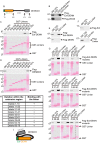
Figure 1.
Figure 1.
Characterization of an intramolecular interaction within DENND3. A, domain diagram of DENND3. Note the DENN domain between amino acids 80 and 520 and the N-terminal extension between amino acids 1 and 79. B, HEK-293T cells were transfected with FLAG-DENN domain and the N-terminal extension of DENND3 or FLAG-WD40 repeats, and lysates were incubated with GST, GST-partial linker (amino acids 538–611), or GST-linker (538–973) coupled to glutathione-Sepharose beads. Proteins specifically bound to the beads were processed for Western blotting with anti-FLAG antibody. An aliquot of the lysate (starting material; SM) equal to 10% of that added to the beads was analyzed in parallel. C and D, HEK-293T cells were transfected with FLAG-DENN domain with its N-terminal extension (C) or FLAG full-length DENND3 (D). Lysates were incubated with GST or GST-linker constructs coupled to glutathione-Sepharose beads and processed as described in B. E, HEK-293T cells were transfected with FLAG-DENN domain with its N-terminal extension (top panel) or FLAG-DENN domain alone (bottom panel). Lysates were incubated with GST or GST-linker coupled to glutathione-Sepharose beads and processed as described in B. F, HEK-293T cells were transfected with FLAG N-terminal extension, and lysates were incubated with GST or GST-linker coupled to glutathione-Sepharose beads (top panel), or HEK-293T cells were transfected with FLAG-linker, and lysates were incubated with GST or GST N-terminal extension coupled to glutathione-Sepharose beads (bottom panel) and processed as described in B. G, HEK-293T cells were transfected with FLAG-Ext-DENN with a series of deletions within the N-terminal extension. Lysates were incubated with GST or GST-linker coupled to glutathione-Sepharose beads and processed as described in B. H, summary table for the deletion constructs used in G. I, intramolecular interaction occurs between the C-terminal region of the linker and the Ext-DENN.
Figure 2.
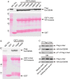
Figure 2.
Tyr-940 is a key residue involved in the intramolecular interaction of DENND3. A, cells were transfected with FLAG-Ext-DENN. Lysates were incubated with GST, GST-linker, or GST-linker with phosphomimetic or phosphorylation-defective mutants at conserved serine or threonine residues within amino acids 936–973 of DENND3, coupled to glutathione-Sepharose beads. Proteins specifically bound to the beads were processed for Western blotting with anti-FLAG antibody. An aliquot of the lysate (starting material; SM) equal to 10% of that added to the beads was analyzed in parallel. B, cells were transfected with FLAG-Ext-DENN. Lysates were incubated with GST, GST-linker, or GST-linker with phosphomimetic Y940D or phosphorylation-defective Y940F mutants, coupled to glutathione-Sepharose beads, and processed as described in A. C, cells co-transfected with FLAG-linker and HA-Ext-DENN domain were left unstarved, or were deprived of cell culture serum for 1.5 h, or were re-fed with serum for 0.5 h following the deprivation. Lysates were incubated with protein G beads coupled to anti-FLAG antibody (IP-FLAG). Proteins bound specifically to the beads were processed for Western blotting with anti-FLAG, anti-HA, or anti-phosphotyrosine antibodies. An aliquot of the cell lysate (starting material, SM) was analyzed in parallel.
Figure 3.
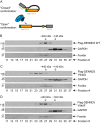
Figure 3.
Phosphorylation of Tyr-940 in the linker alters the conformation of DENND3. A, model depicting the hypothesis of the closed and open conformations. B–D, cells were transfected with FLAG-DENND3 wild-type (B), Y940D (C), or Y940F (D) mutants. Lysates were subjected to size-exclusion chromatography. The collected fractions were analyzed by Western blotting with the indicated antibodies.
Figure 4.
Figure 4.
Characterization of DENND3 oligomerization. A, lysates from HEK-293T cells co-transfected with FLAG-DENND3 and HA-DENND3 were incubated with protein G beads alone or protein G beads coupled to anti-FLAG antibody (IP-FLAG). Proteins specifically bound to the beads were processed for Western blotting with anti-FLAG and anti-HA antibodies. An aliquot of the cell lysate (starting material, SM) equal to 10% of that added to the beads was analyzed in parallel. B, lysates from HEK-293T cells co-transfected with FLAG-Ext-DENN and HA-DENND3 were incubated with protein G beads alone or protein G beads coupled to anti-FLAG antibody (IP-FLAG). Proteins bound specifically to the beads were processed as described in A. C, lysates from HEK-293T cells co-transfected with FLAG-linker and HA-DENND3 were incubated with protein G beads alone or protein G beads coupled to anti-FLAG antibody (IP-FLAG). Proteins bound specifically to the beads were processed as described in A. D, lysates from HEK-293T cells co-transfected with FLAG-WD40 and HA-DENND3 were incubated with protein G beads alone or protein G beads coupled to anti-FLAG antibody (IP-FLAG). Proteins bound specifically to the beads were processed as described in A. E, lysates from HEK-293T cells co-transfected with FLAG-Ext-DENN and GST-Ext-DENN were incubated with protein G beads alone or protein G beads coupled to anti-FLAG antibody (IP-FLAG). Proteins bound specifically to the beads were processed as described in A. F, HEK-293T cells were transfected with FLAG-Ext-DENN. Lysates were subjected to size-exclusion chromatography. The collected fractions were analyzed by Western blotting with the indicated antibodies.
Figure 5.
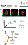
Figure 5.
With Tyr-940 phosphorylation DENND3 assumes an open conformation with enhanced GEF activity. A, diagram showing the principle underlying the test of the hypothesis of open/closed conformations through FRET experiments. B, HeLa cells were transfected with a FRET construct of DENND3 wild-type, Y940D, or Y940F mutant before FRET imaging. The color-coded images indicate the measured FRET signal. Warm colors represent high FRET signal; cool colors represent low FRET signal. The color scale bar represents the efficiency of the FRET signal. The mean values of FRET efficiency for wild type, Y940D, and Y940F are 0.1397, 0.1109, and 0.1460, respectively. Scale bar, 5 μm. C, FRET efficiency measurements from images as in B were quantified from n > 180 cells for each group over three repeats. The data are presented with box and whisker graphs. The box extends from the 25th to 75th percentiles. The line inside the box is plotted at the median. The whiskers are from the 10th to the 90th percentile. Statistical analysis employed one-way analysis of variance followed by Tukey's post-test. ****, p < 0.0001, *, p < 0.05. D, FLAG-DENND3 wild-type, Y940D, or Y940F mutants were immunoprecipitated from HEK-293T cells. Immunoprecipitations from mock-transfected cells were used as mock for the GDP/GTP exchange assay. To measure GEF activity, GDP-loaded Rab12 was incubated with immunoprecipitated DENND3 wild type, mutant or mock in the presence of GTPγS and [35S]GTPγS. At the time points indicated, an aliquot of the reaction was analyzed for nucleotide exchange. The enzymatic activity based on the amount of Rab12 loaded with GTPγS over time is shown. The curves were fit with a nonlinear regression one-phase association. The points represent mean ± S.E., n = 3. Duplicates of the immunoprecipitated DENND3 proteins were processed by SDS-PAGE and stained with Coomassie Blue dye, showing that equal amounts of DENND3 wild type and mutants were used for the GDP/GTP exchange assay (right bottom panel). E, model summarizing under nutrient starvation ULK-mediated phosphorylation and Tyr-940 phosphorylation on DENND3 and their functional consequence.

Comment in

Similar articles

Cited by

References

    1. Stenmark H. (2009) Rab GTPases as coordinators of vesicle traffic. Nat. Rev. Mol. Cell Biol. 10, 513–525 - PubMed
    1. Cherfils J., and Zeghouf M. (2013) Regulation of small GTPases by GEFs, GAPs, and GDIs. Physiol. Rev. 93, 269–309 - PubMed
    1. Levine T. P., Daniels R. D., Gatta A. T., Wong L. H., and Hayes M. J. (2013) The product of C9orf72, a gene strongly implicated in neurodegeneration, is structurally related to DENN Rab-GEFs. Bioinformatics 29, 499–503 - PMC - PubMed
    1. Marat A. L., Dokainish H., and McPherson P. S. (2011) DENN domain proteins: regulators of Rab GTPases. J. Biol. Chem. 286, 13791–13800 - PMC - PubMed
    1. Zhang D., Iyer L. M., He F., and Aravind L. (2012) Discovery of novel DENN proteins: implications for the evolution of eukaryotic intracellular membrane structures and human disease. Front. Genet. 3, 283. - PMC - PubMed

Publication types

MeSH terms

Grants and funding