. 2017 Feb 28;18(9):2175-2188.
doi: 10.1016/j.celrep.2017.02.003.

Vaccine Elicitation of High Mannose-Dependent Neutralizing Antibodies against the V3-Glycan Broadly Neutralizing Epitope in Nonhuman Primates

Affiliations

Vaccine Elicitation of High Mannose-Dependent Neutralizing Antibodies against the V3-Glycan Broadly Neutralizing Epitope in Nonhuman Primates

Kevin O Saunders et al. Cell Rep. .

Abstract

Induction of broadly neutralizing antibodies (bnAbs) that target HIV-1 envelope (Env) is a goal of HIV-1 vaccine development. A bnAb target is the Env third variable loop (V3)-glycan site. To determine whether immunization could induce antibodies to the V3-glycan bnAb binding site, we repetitively immunized macaques over a 4-year period with an Env expressing V3-high mannose glycans. Env immunizations elicited plasma antibodies that neutralized HIV-1 expressing only high-mannose glycans-a characteristic shared by early bnAb B cell lineage members. A rhesus recombinant monoclonal antibody from a vaccinated macaque bound to the V3-glycan site at the same amino acids as broadly neutralizing antibodies. A structure of the antibody bound to glycan revealed that the three variable heavy-chain complementarity-determining regions formed a cavity into which glycan could insert and neutralized multiple HIV-1 isolates with high-mannose glycans. Thus, HIV-1 Env vaccination induced mannose-dependent antibodies with characteristics of V3-glycan bnAb precursors.

Keywords: HIV; V3 glycan; glycan; long-term immunization; vaccination.


Figures

Figure 1
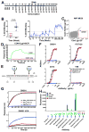
Figure 1. Repetitive vaccination elicits antibodies targeting the V3-glycan bnAb epitope
(A) CON-S repetitive vaccination regimen. The immunization modality is shown below the line and the week of vaccination is noted above the line. (B) Plasma blocking of 2G12 binding to Env. (Left) The percent blocking of 2G12 binding to B.JR-FL gp140C by macaque plasma after one DNA vaccination (week 2) or at the end of the vaccination regimen (week 204; Wilcoxon signed rank test between week 2 and 204, P<0.05 N= 10). (Right) The kinetics of 2G12 blocking antibodies in the plasma of vaccinated macaques. The mean ± SEM of triplicate measurements are shown (n= 10). Arrows on the x-axis indicate vaccination timepoints. (C) FACS plots of sorted single B cells from week 192 PBMCs from CON-S-immunized macaque M636 that bind ConC gp120 but not N332A glycan knockout mutant gp120. (D) Surface plasmon resonance binding to the study immunogen, CON-S gp140CFI, by DH501 and the DH501 unmutated common ancestor (UCA). (E) Diagram of the Man9GlcNAc2 glycosylated peptide that recapitulates the PGT128 epitope. N-linked glycans (green and blue) are shown at positions Asn301 (red N) and Asn332 (red N). The disulfide bond is shown by a straight horizontal line. (F) Direct ELISA binding to the V3 base peptide with Man9GlcNAc2 glycans at Asn301 and Asn332 (black, Man9 N332 N301 V3), at Asn301 only (red, Man9 N301 V3), or at neither site (blue, aglycone) by vaccine-induced macaque antibody DH501. (G) Biolayer interferometry binding of DH501 and its inferred UCA to the peptides shown in E. (H) Glycan luminex detection of antibody binding to 16 μM free glycan. The glycan-dependent bnAb PGT128 and peptide-binding antibody 19B were used as positive and negative controls, respectively. The complex glycan is Gal2Man3GlcNAc4. See also Supplemental Figure 1 and 2.
Figure 2
Figure 2. Genetic and plasma analyses determine DH501 was elicited after multiple protein boosts
(A) Autologous plasma blocking of DH501 binding to the CON-S immunogen. The plasma from sequential timepoints from macaque M636 from whom the DH501 glycan-reactive antibody was isolated was used to block DH501 binding to Env. Timepoints where next generation sequencing was performed in B are indicated by black arrows. The red arrow indicates where DH501 was isolated by single B cell sorting and RT-PCR. Values greater than 20% are considered positive. The mean ± SEM of triplicate measurements are shown (n= 10). Arrows on the x-axis indicate vaccination timepoints. (B) Comparison of the number of DH501 related sequences after DNA prime, rAd5 boost, and one protein boost, four protein boosts, or 12 protein boosts in macaque M636 from which DH501 was cloned. Sequencing was performed twice at each timepoint and DH501 clonal relatedness was determined using Clonanalyst (second column). Sequences were clustered to account for sequencing error. Number of sequence clusters represents the estimated minimum number of DH501 members at each timepoint (last column). (C) The maximum likelihood phylogenetic tree of the 73 total collapsed sequences (72 from NGS and DH501) that are clonally related to DH501. Time point at which the sequence was observed is indicated by the color code at the bottom. See also Supplemental Figure 3.
Figure 3
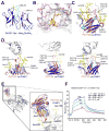
Figure 3. The molecular basis for high mannose and Env recognition by DH501
(A) The structure of DH501 Fv (blue) with the paratope oriented upward. CDR-H3 is highlighted in cyan, CDR-L1 in magenta, and bound constituents of the Man9GlcNAc2 compound in yellow. (B) Specific interactions between DH501 (magenta carbons behind a surface rendering) and bound mannose residues (yellow carbons). H-bonding interactions are shown with dashed lines and interacting residues on the DH501 heavy chain are labeled. (C) DH501 Fv (color scheme as in A) superimposed with PGT130 Fv in orange with its CDR-H3 in red and CDR-L1 in green. (D) DH501 Fv superimposed with the Fvs of V3-glycan broadly neutralizing antibodies in complex with gp120. Gp120 and glycan moieties are shown in gray and yellow respectively. DH501 is colored as in (A) and compared antibodies are shown in orange. Long CDRs with extended conformations in V3 glycan bnAbs are highlighted in red. N-linked Asn residues are numbered for identification of glycans. (E) DH501 Fv (blue) superimposed on PGT122 Fv (orange) bound to the BG505.6R.SOSIP.664 trimer (grey). DH501’s CDR-H3 (cyan) lacks the length of the PGT122s CDR-H3 (red) to deeply penetrate the glycan shield (yellow). (F) SPR binding of the DH501 Fab to a stabilized BG505 SOSIP.v4.1 trimer. Each trace represents a different concentration of Fab ranging from 50 to 200 μg/mL as indicated. The affinity measurements are displayed in the right corner of the graph. See also Supplemental Figure 4.
Figure 4
Figure 4. DH501 accommodates high mannose glycan by forming a negatively charged glycan-binding pocket
Paratope surfaces rendered by electrostatic surface potential along with gp120 and labeled glycans in green and yellow respectively. The electrostatic potential surface renders were calculated using CHARMM (Im et al., 1998; Jo et al., 2008a; Jo et al., 2008b) and expressed in units of kcal/(mol·e). The paratope is shown from a top view with the heavy chains oriented at the top and light chains at the bottom. CDRs with extended conformations protruding from the paratope surface are labeled in italics. V3-glycan bnAbs were characterized by one or more long CDRs and an amphipathic charge distribution. DH501 is distinguished by a pronounced pocket on its paratope bordered and formed almost entirely by the three heavy chain CDRs. The mannose residues of Man9GlcNAc2 that bind within the pocket are shown in yellow.
Figure 5
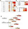
Figure 5. DH501 and early V3 glycan bnAb lineage antibodies require Man9GlcNAc2 glycosylation for neutralization of HIV-1
(A) Neutralization titers against a cross-clade panel of tier 1 and tier 2 viruses in the TZM-bl assay. Each virus was produced untreated or treated with kifunensine. PG9 (Man5GlcNAc2-reactive) and PGT128 (Man9GlcNAc2-reactive) were used as controls to demonstrate the modification of envelope glycosylation on virions. Representative titers are shown from up to 4 independent assays. (B) DH501 exhibits N301-dependent neutralization of kifunensine-treated B.JRFL. Neutralization of kifunensine-treated B.JR-FL (KIF-JR-FL) with intact wildtype N-linked glycosylation sites or with an alanine substitution at the indicated glycosylation site. SVA is a murine leukemia virus used as a negative control for neutralization. Values are the geometric mean for two independent experiments. (C) DH501 requires the GDIR motif for neutralization of kifunensine-treated B.JRFL. TZM-bl neutralization titers (IC50 μg/mL) for DH501 and PGT128 against wildtype and GDIR alanine mutant viruses produced in kifunensine-treated cells. (D) Neutralization of B.JR-FL and KIF-JR-FL pseudovirus by the broadly neutralizing N332 glycan-dependent DH270 lineage antibodies. Neutralization titer is shown as IC50 (μg/mL). The IC50 for B.JR-FL neutralization is shown in the first column followed by the KIF-JR-FL neutralization titer. Neutralization titers are color coded as >50 (white), 49-1 (light yellow), 0.9–0.1 (yellow), 0.09–0.023 (orange), <0.023 (red). Titers are the geometric mean of two independent experiments. See also Supplemental Figure 5.

Similar articles

Cited by

References

    1. Agrawal-Gamse C, Luallen RJ, Liu B, Fu H, Lee FH, Geng Y, Doms RW. Yeast-elicited cross-reactive antibodies to HIV Env glycans efficiently neutralize virions expressing exclusively high-mannose N-linked glycans. J Virol. 2011;85:470–480. - PMC - PubMed
    1. Alam SM, Aussedat B, Vohra Y, Meyerhoff RR, Cale EM, Walkowicz WE, Radakovich NA, Anasti K, Armand L, Parks R, et al. Mimicry of an HIV Broadly Neutralizing Antibody Epitope with a Synthetic Glycopeptide. Sci Transl Med. 2017 in press. - PMC - PubMed
    1. Astronomo RD, Kaltgrad E, Udit AK, Wang SK, Doores KJ, Huang CY, Pantophlet R, Paulson JC, Wong CH, Finn MG, Burton DR. Defining criteria for oligomannose immunogens for HIV using icosahedral virus capsid scaffolds. Chem Biol. 2010;17:357–370. - PMC - PubMed
    1. Astronomo RD, Lee HK, Scanlan CN, Pantophlet R, Huang CY, Wilson IA, Blixt O, Dwek RA, Wong CH, Burton DR. A glycoconjugate antigen based on the recognition motif of a broadly neutralizing human immunodeficiency virus antibody, 2G12, is immunogenic but elicits antibodies unable to bind to the self glycans of gp120. J Virol. 2008;82:6359–6368. - PMC - PubMed
    1. Blattner C, Lee JH, Sliepen K, Derking R, Falkowska E, de la Pena AT, Cupo A, Julien JP, van Gils M, Lee PS, et al. Structural delineation of a quaternary, cleavage-dependent epitope at the gp41-gp120 interface on intact HIV-1 Env trimers. Immunity. 2014;40:669–680. - PMC - PubMed

Publication types

MeSH terms