. 2017 Sep;222(7):2993-3005.
doi: 10.1007/s00429-017-1380-8. Epub 2017 Feb 28.

Store-operated calcium entry is essential for glial calcium signalling in CNS white matter

Affiliations

Store-operated calcium entry is essential for glial calcium signalling in CNS white matter

M Papanikolaou et al. Brain Struct Funct. 2017 Sep.

Abstract

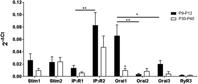
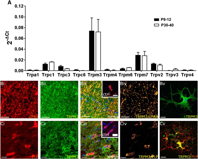
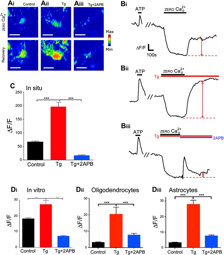
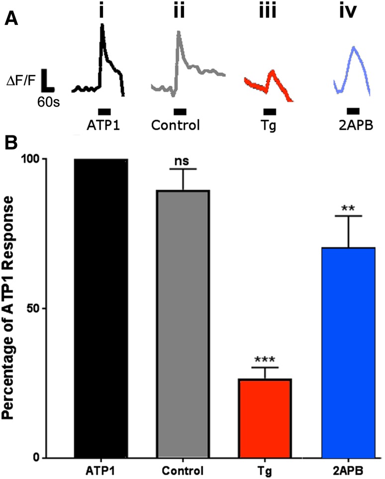
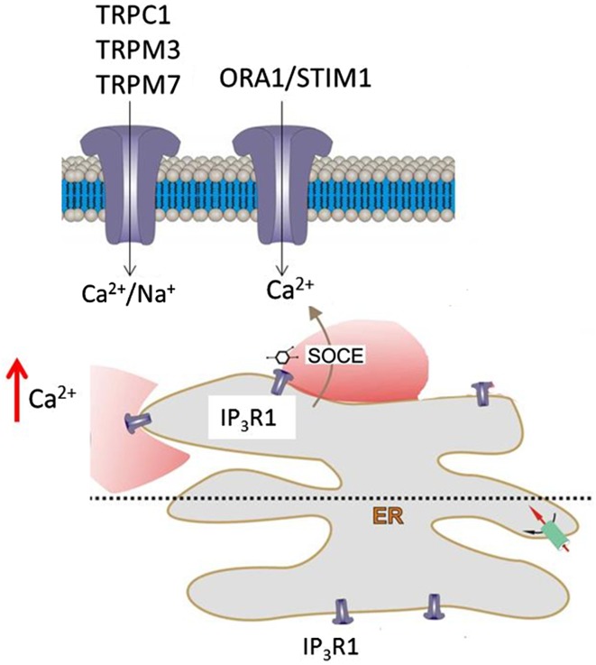
'Calcium signalling' is the ubiquitous response of glial cells to multiple extracellular stimuli. The primary mechanism of glial calcium signalling is by release of calcium from intracellular stores of the endoplasmic reticulum (ER). Replenishment of ER Ca2+ stores relies on store-operated calcium entry (SOCE). However, despite the importance of calcium signalling in glial cells, little is known about their mechanisms of SOCE. Here, we investigated SOCE in glia of the mouse optic nerve, a typical CNS white matter tract that comprises bundles of myelinated axons and the oligodendrocytes and astrocytes that support them. Using quantitative RT-PCR, we identified Orai1 channels, both Stim1 and Stim2, and the transient receptor potential M3 channel (TRPM3) as the primary channels for SOCE in the optic nerve, and their expression in both astrocytes and oligodendrocytes was demonstrated by immunolabelling of optic nerve sections and cultures. The functional importance of SOCE was demonstrated by fluo-4 calcium imaging on isolated intact optic nerves and optic nerve cultures. Removal of extracellular calcium ([Ca2+]o) resulted in a marked depletion of glial cytosolic calcium ([Ca2+]i), which recovered rapidly on restoration of [Ca2+]o via SOCE. 2-aminoethoxydiphenylborane (2APB) significantly decreased SOCE and severely attenuated ATP-mediated calcium signalling. The results provide evidence that Orai/Stim and TRPM3 are important components of the 'calcium toolkit' that underpins SOCE and the sustainability of calcium signalling in white matter glia.

Keywords: Astrocyte; CRAC; Calcium signalling; Glia; Oligodendrocyte; Store-operated calcium channel; TRP channel; White matter.


Conflict of interest statement

The authors declare that they have no conflicts of interest.

All applicable international, national, and/or institutional guidelines for the care and use of animals were followed. All procedures performed in studies involving animals were in accordance with the ethical standards of the institution at which the studies were conducted.

Figures

Fig. 1
Fig. 1
Expression of Orai/Stim transcripts in the mouse optic nerve. qRT-PCR was performed on lysates of acutely isolated optic nerves from WT postnatal mice (aged P9–P12) and adult mice (P30–P40). Data are from ten pooled optic nerves in each age group, run in triplicate, expressed as relative mRNA levels (2-ΔCt) compared to the housekeeping gene GAPDH (mean ± SEM, n = 3). Orai1 was the most highly expressed Orai isoform in the postnatal nerve (*p < 0.05, **p < 0.01, ANOVA and unpaired t tests) and was significantly downregulated with age (*p < 0.05, unpaired t test); there was no significant difference between the Orai isoforms in the adult. There was no significant difference between Stim1 and Stim2 expression in postnatal or adult nerves. IP3R2 was expressed at significantly greater levels than IP3R1 at both ages (**p < 0.01, unpaired t test); there was no significant difference between the age groups
Fig. 2
Fig. 2
Expression of Orai/Stim in optic nerve oligodendrocytes. Immunolabelling for Orai1 (a, green), Stim1 (b, green) and Stim2 (c, green) in PLP-DsRed mice to identify oligodendrocytes (ac, red), in optic nerve sections (ai iv, biiv, ciiv) and explant cultures (av, bv, cv). Confocal micrographs illustrate single channels (ai, aii, bi, bii, ci, cii) and merged cannels (aiii, biii, ciii). Expression of Orai1 and Stim1 is localized to oligodendroglial somata, whereas Stim2 immunostaining was primarily within the fascicles of myelinated axons, as demonstrated by the colocalization channels, illustrating voxels in which green and red channels are of equal intensity and appear yellow (aiv, biv, civ). Oligodendrocytes in explant cultures are immunopositive for Orai1 (av), Stim1 (bv) and Stim2 (cv). No immunostaining was observed in negative controls that were pre-incubated in blocking peptides for Orai1 (inset, aiii), Stim1 (inset, biii) and Stim2 (inset, ciii). Nuclei are stained with Hoechst blue. Scale bars a, b 10 µm, c 20 μm
Fig. 3
Fig. 3
Expression of Orai/Stim in optic nerve astrocytes. Double immunofluorescence labelling for Orai1 (a, green), Stim1 (b, green) and Stim2 (c, green), with GFAP to identify astrocytes (ac, red), in WT optic nerve sections (aiiv, bi–iv, ciiv) and explant cultures (av, bv, cv). Confocal micrographs illustrate single channels (ai, aii, bi, bii, ci, cii) and merged cannels (aiii, biii, ciii). Expression of Orai1 and Stim1 is localized to astrocyte processes, whereas astrocytes were immunonegative for Stim2, as demonstrated by the colocalization channels, illustrating voxels in which green and red channels are of equal intensity and appear yellow (aiv, biv, civ). Astrocytes in explant cultures are immunopositive for Orai1 (av) and Stim1 (bv), but are immunonegative for Stim2 (cv). No immunostaining was observed in negative controls that were pre-incubated in blocking peptides for Orai1 (inset, aiii), Stim1 (inset, biii) and Stim2 (inset, ciii). Nuclei are stained with Hoechst blue. Scale bars a, b 10 µm, c 20 μm
Fig. 4
Fig. 4
Expression of TRP channels in optic nerve glia. a qRT-PCR of acutely isolated optic nerves from WT mice aged P9–P12 and P30–P40; data are from 10 pooled optic nerves in each age group, run in triplicate, expressed as relative mRNA levels  (2-ΔCt) compared to the housekeeping gene GAPDH method (mean ± SEM, n = 3). TRPM3 was the most highly expressed TRP channel in the postnatal and adult nerve (***p < 0.001, ANOVA and unpaired t tests) and there was no developmental regulation of TRP channels, which had a rank order of expression in the adult of TRPM3 >>> TRPM7 (***p < 0.001, unpaired t test) > TRPC1 (*p < 0.05, unpaired t test) >> TRPV2 (**p < 0.01, unpaired t test); TRPC3, TRPM6 and TRPV3 were expressed at significantly lower levels, and TRPA1, TRPC6, TRPM4, and TRPV4 were barely detectable. Immunolabelling for TRPM3 (b, c, green) with GFAP to identify astrocytes in WT nerves (b, red) and in PLP-DsRed nerves to identify oligodendrocytes (c, red), in optic nerve sections (biiv, ciiv) and explant cultures (bv, cv). Confocal micrographs illustrate single channels (bi, bii, ci, cii) and merged cannels (biii, ciii). The colocalization channels illustrate voxels in which green and red channels are of equal intensity and appear yellow, showing expression of TRPM3 is localized to astroglial processes (bv) and oligodendroglial somata (cv). Immunostaining of explant cultures shows astrocytes are immunopositive for TRPM3 (bv) and that TRPM3 is localized to oligodendroglial somata and excluded from their distal processes (cv). No immunostaining was observed in negative controls that were pre-incubated in blocking peptides for TRPM3 (insets, biii, ciii). Nuclei are stained with Hoechst blue. Scale bars 20 μm
Fig. 5
Fig. 5
SOCE in optic nerve glia. Mouse optic nerves were isolated intact and loaded with Fluo-4 to analyse SOCE, using thapsigargin (10 µM for optic nerves, 2 μM for explant cultures) to block ER reuptake and 2APB (50 µM) to block SOCE channels. Confocal images of Fluo-4 fluorescence intensity illustrated in rainbow false colour (a, scale bars 10 μm) and representative traces of individual glia (b), illustrating that removal of extracellular [Ca2+]o results in a decline in cytosolic [Ca2+]i, which recovers rapidly on return to normal aCSF (ai, bi, red arrow), and this is markedly increased in the presence of thapsigargin (aii, bii, red arrow) and decreased in the presence of 2APB (aiii, biii, red arrow). Bar graphs showing the mean rise in cytosolic [Ca2+]i (indicated by red arrows in bi–iii) in aCSF control, thapsigargin and 2APB, in situ in the isolated intact optic nerve (c) and in vitro in explant cultures (d), illustrating results from all glia (di) and separately for oligodendrocytes (dii) and astrocytes (diii), identified by differential expression of PLP-DsRed; data are mean ± SEM change in fluorescence (ΔF/F), ***p < 0.01, unpaired t test with Welch’s correction
Fig. 6
Fig. 6
SOCE is essential for sustainability of ATP-mediated Ca2+ signalling in optic nerve glia. a Representative traces illustrating ATP-mediated rise in glial Ca2+ at the beginning of each experiment (ai, ATP1) and the second response tested at the end of the experiment, without application of any pharmacological agents (aii, control), compared to after application of thapsigargin to block replenishment of ER Ca2+ stores (aiii, Tg), or 2APB to block SOCE (aiv, 2APB). b Bar graph of mean (±SEM) responses to the second test pulse of ATP, expressed as a percentage of the first response to ATP at the beginning of the experiment (**p < 0.01, ***p < 0.001, unpaired t tests with Welch’s correction)
Fig. 7
Fig. 7
Mechanisms of SOCE in optic nerve glia. ATP-mediated calcium signalling in optic nerve glia is via P2Y G-protein-coupled receptors and the formation of IP3, which acts on IP3R1 on the ER to trigger release of Ca2+ into the cytosol. Subsequent replenishment of ER stores in astrocytes and oligodendrocytes is dependent on SOCE via TRPM3 and Orai1, which form the plasmalemmal Ca2+ channels, and mainly Stim1, which acts as the sensor of Ca2+ depletion, and uptake into the ER is via SERCA pumps. Oligodendrocytes also express Stim2, which may be localized to the myelin, whereas Orai1, Stim1 and TRPM3 are localized to oligodendroglial somata. Notably, calcium homeostasis in optic nerve glia depends on an apparent continuous Ca2+ influx from the extracellular milieu that is largely dependent on SOCE. Moreover, SOCE is essential for the sustainability of ATP-mediated Ca2+ signalling in optic nerve glia, which has a central role in white matter physiology and pathology

Similar articles

Cited by

References

    1. Abdullaev IF, Bisaillon JM, Potier M, Gonzalez JC, Motiani RK, Trebak M. Stim1 and Orai1 mediate CRAC currents and store-operated calcium entry important for endothelial cell proliferation. Circ Res. 2008;103:1289–1299. doi: 10.1161/01.RES.0000338496.95579.56. - DOI - PMC - PubMed
    1. Azim K, et al. Wnt/beta-catenin signaling determines dorsalization of the postnatal subventricular zone and neural stem cell specification into oligodendrocytes and glutamatergic neurons. Stem Cells. 2014;32:1301–1312. doi: 10.1002/stem.1639. - DOI - PubMed
    1. Bottenstein JE, Sato GH. Growth of a rat neuroblastoma cell line in serum-free supplemented medium. Proc Natl Acad Sci USA. 1979;76:514–517. doi: 10.1073/pnas.76.1.514. - DOI - PMC - PubMed
    1. Brasko C, Hawkins V, De La Rocha IC, Butt AM. Expression of Kir4.1 and Kir5.1 inwardly rectifying potassium channels in oligodendrocytes, the myelinating cells of the CNS. Brain Struct Funct. 2017;222:41–59. doi: 10.1007/s00429-016-1199-8. - DOI - PMC - PubMed
    1. Butenko O, et al. The increased activity of TRPV4 channel in the astrocytes of the adult rat hippocampus after cerebral hypoxia/ischemia. PloS One. 2012;7:e39959. doi: 10.1371/journal.pone.0039959. - DOI - PMC - PubMed

MeSH terms

LinkOut - more resources