. 2017 Jun;133(6):887-906.
doi: 10.1007/s00401-017-1687-9. Epub 2017 Feb 28.

Motor neuron intrinsic and extrinsic mechanisms contribute to the pathogenesis of FUS-associated amyotrophic lateral sclerosis

Affiliations

Motor neuron intrinsic and extrinsic mechanisms contribute to the pathogenesis of FUS-associated amyotrophic lateral sclerosis

Jelena Scekic-Zahirovic et al. Acta Neuropathol. 2017 Jun.

Abstract

Motor neuron-extrinsic mechanisms have been shown to participate in the pathogenesis of ALS-SOD1, one familial form of amyotrophic lateral sclerosis (ALS). It remains unclear whether such mechanisms contribute to other familial forms, such as TDP-43 and FUS-associated ALS. Here, we characterize a single-copy mouse model of ALS-FUS that conditionally expresses a disease-relevant truncating FUS mutant from the endogenous murine Fus gene. We show that these mice, but not mice heterozygous for a Fus null allele, develop similar pathology as ALS-FUS patients and a mild motor neuron phenotype. Most importantly, CRE-mediated rescue of the Fus mutation within motor neurons prevented degeneration of motor neuron cell bodies, but only delayed appearance of motor symptoms. Indeed, we observed downregulation of multiple myelin-related genes, and increased numbers of oligodendrocytes in the spinal cord supporting their contribution to behavioral deficits. In all, we show that mutant FUS triggers toxic events in both motor neurons and neighboring cells to elicit motor neuron disease.

Keywords: Amyotrophic lateral sclerosis; Fronto-temporal dementia; Mouse models; Non-cell autonomous mechanisms; RNA-binding proteins.


Figures
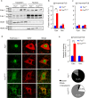
Fig. 1
Fig. 1
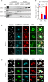
FUS localization in Fus ΔNLS/+ mice. a Immunoblot analysis of FUS protein subcellular localization in spinal cord of 2 Fus +/+ and 2 Fus ΔNLS/+ 4-month-old mice using two different antibodies targeting either the N-terminal part (N-ter. 1) of FUS or the C-terminal (C-ter. 1) NLS. Molecular weight markers are shown on the left. SOD1 and HDAC1 are used as loading controls for cytoplasmic and nuclear protein extracts fractions, respectively. b, c Quantification of FUS protein levels in cytoplasmic and nuclear fractions from immunoblots for Fus +/+ (blue bars) and Fus ΔNLS/+ (red bars). N = 6. *p < 0.05, ***p < 0.01 by Student’s unpaired t test. d Double immunostaining for the motoneuronal marker ChAT and FUS (N-terminal part) in the spinal cord ventral horn at 22 months of age. Note the cytoplasmic redistribution of truncated FUS in Fus ΔNLS/+ mice. Scale bar 7.5 µm. e Quantification of FUS (N-terminal part) staining intensity in different cellular compartments of motor neuron. N = 70 Fus +/+, N = 68 Fus ΔNLS/+. ***p < 0.01 by Student’s unpaired t test. f Distribution of FUS cytoplasmic/nuclear localization in motor neurons. N = 70 Fus +/+, N = 68 Fus ΔNLS/+
Fig. 2
Fig. 2
Subcellular redistribution of asymmetrically arginine dimethylated (ADMA) FUS. a Representative immunoblots on cytoplasmic and nuclear fractions of protein extracts from spinal cord of Fus +/+ and Fus ΔNLS/+ mice, using an antibody recognizing asymmetrically arginine dimethylated FUS (ADMA-FUS). HDAC1 is used as a loading control for nuclear fractions and SOD1 for cytoplasmic fractions. Molecular weight markers are shown on the left. b Quantification of ADMA-FUS protein levels in cytoplasmic and nuclear fractions from immunoblots for Fus +/+ (blue bars) and Fus ΔNLS/+ (red bars). N = 6. *p < 0.05, ***p < 0.01 by Student’s unpaired t test. c Triple immunostaining for the motoneuronal marker ChAT (green), for FUS (N-terminal part) (cyan) and for ADMA-FUS (red) in the spinal cord ventral horn. Scale bar 10 µm. d Triple immunostaining with antibodies to the N terminus of FUS (green), ChAT (red) and pan-Ubiquitin (cyan), showing diffuse cytoplasmic and nuclear punctate aggregates within motor neurons with relocated FUS. Scale bar 10 μm
Fig. 3
Fig. 3
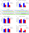
Fus ΔNLS/+ mice display a mild motor deficit. Age-dependent changes in the mean hanging time (s) (a) and holding impulse (N s) (b) in the four-limb wire inverted grid test in Fus +/+ and Fus ΔNLS/+ mice. N = 7 for 10 months; N = 5 for 22 months. *p < 0.05, ***p < 0.01 as compared to Fus +/+ by Student’s unpaired t test. c Representative gait patterns of Fus +/+and Fus ΔNLS/+ mice at 10 months (left panels) and 22 months (right panels) of age. The panels show the digitized prints with colorful phase lags representing the stance phase duration of each individual paw in a single-step cycle. dg Gait changes and variability in Fus ΔNLS/+mice. Stride length (d, distance between successive placements of the same paw in cm); swing speed (e, distance traveled by one paw per second), body speed (f, distance traveled by the animal per second, in cm/s), and body speed variation (g, regularity of body speed in %), are shown. N = 3 for 10 months; N = 5 for 22 months. All graphs show the overall sample means and standard errors at various ages (10 months; 22 months) for Fus +/+(blue bars) and Fus ΔNLS/+ (red bars) mice. *p < 0.05, ***p < 0.01 as compared to Fus+/+ by Student’s unpaired t test
Fig. 4
Fig. 4
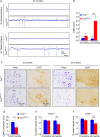
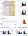
Muscle denervation and progressive degeneration of spinal motor neurons in Fus ΔNLS/+ mice. a Representative electromyograms of Fus ΔNLS/+ mice in gastrocnemius and tibialis anterior muscles. Note the presence of typical spontaneous denervation activities (fibrillation potentials) in Fus ΔNLS/+ mice. Scale bars 50 ms and 50 µV. b Graph showing EMG scores for Fus +/+ (blue bars) and Fus ΔNLS/+ (red bars) mice. Note that a significant difference was only detected for 22-month-old animals. ***p < 0.01 as compared to Fus +/+; N = 7 for 10 months; N = 9 for 22 months; Student’s unpaired t test. c Representative images of Nissl and ChAT staining of spinal cord ventral horn of 10-month-old (left panels) and 22-month-old (right panels) Fus +/+ and Fus ΔNLS/+ animals. In the 22-month-old Fus ΔNLS/+ mice degenerative changes (shrinking, chromatolysis) and loss of motor neurons occur. Scale bars 35 μm. d Compound muscle action potential (CMAP) amplitude. *p < 0.05 as compared to Fus +/+; N = 10 Fus +/+, N = 9 Fus ΔNLS/+. Student’s unpaired t test. Bar graphs showing means and standard errors of Nissl+ (e) and ChAT+ (f) motor neuron number in the ventral horn of the spinal cord at 10 months and 22 months for Fus +/+ (blue bars) and Fus ΔNLS/+ (red bars) mice. ***p < 0.01 as compared to Fus +/+; N = 3 for 10 months; N = 6 for 22 months; Student’s unpaired t test
Fig. 5
Fig. 5
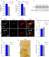
Lack of motor neuron disease in Fus +/ mice. a Expression levels of Fus mRNA in spinal cord. Fus mRNA levels were significantly reduced in Fus +/ mice as revealed by quantitative real-time PCR analysis. N = 7 Fus +/+, N = 9 Fus +/. *p < 0.05 by Student’s unpaired t test. b Quantification of FUS protein levels from immunoblots showed a lower amount of FUS in spinal cord of Fus +/ mice. N = 7 Fus +/+, N = 8 Fus +/. *p < 0.05 by Student’s unpaired t test. c Representative immunoblot for FUS on protein extracts from spinal cord of 100-week-old mice. TUBULIN was used as loading control. d Immunostaining for the neuronal marker NeuN and FUS on the spinal cord ventral horn of 100-week-old Fus +/+ and Fus +/ mice. Note preserved nuclear localization of FUS in Fus +/ mice. e Mean hanging time in the four-limb wire inverted grid test of Fus +/+ and Fus +/ mice. N = 4 Fus +/+, N = 3 Fus +/. p = not significant (NS) by Student’s unpaired t test. f Bar graphs showing means and standard errors for compound muscle action potential (CMAP) amplitude. No difference was observed between groups. N = 6 Fus +/+, N = 5 Fus +/. p = NS by Student’s unpaired t test. g Representative images of ChAT immunostaining on spinal cord ventral horn. Scale bar 35 μm. h Quantification of the number of motor neurons per spinal cord ventral horn in Fus +/+ and Fus +/ mice. The number of ChAT+ motor neurons was not altered in Fus +/ mice. N = 6 Fus +/+, N = 5 Fus +/. p = NS by Student’s unpaired t test
Fig. 6
Fig. 6
Motor neuron-selective reversal of the Fus ΔNLS allele to wild type delays but does not prevent Fus ΔNLS/+ motor phenotypes. a Representative images of spinal cord ventral horn of Fus +/+/ChAT-CRE, Fus ΔNLS/+/− and Fus ΔNLS/+/ChAT-CRE mice at 22 months stained with Nissl (left panels) or anti-choline acetyl transferase (ChAT, right panels). b, c Quantification of motor neurons per spinal cord ventral horn. The number of Nissl+ (b) and ChAT+ (c) motor neurons is rescued in Fus ΔNLS/+/ChAT-CRE mice while significantly reduced in Fus ΔNLS/+/− mice. N = 5 per genotype; ***p < 0.01 versus Fus +/+/ChAT-CRE, ### p < 0.01 versus Fus ΔNLS/+/ChAT-CRE; one-way ANOVA followed by Tukey post hoc test. In all graphs genotypes are represented as Fus +/+/ChAT-CRE (blue bars), Fus ΔNLS/+/− (red bars) and Fus ΔNLS/+/ChAT-CRE (green bars). d EMG recording traces in gastrocnemius muscle of 22-month-old animals. Note the absence of typical spontaneous denervation activities in Fus ΔNLS/+/ChAT-CRE versus Fus ΔNLS/+/− mice. Scale bars 50 ms and 50 µV. e EMG score showing significantly decreased spontaneous activity in Fus ΔNLS/+/ChAT-CRE as compared to Fus ΔNLS/+/− in 22-month-old animals. N = 5 Fus +/+/ChAT-CRE, N = 7 Fus ΔNLS/+/− and N = 6 Fus ΔNLS/+/ChAT-CRE. ***p < 0.01 versus Fus +/+/ChAT-CRE; # p < 0.05 versus Fus ΔNLS/+/ChAT-CRE; one-way ANOVA followed by Tukey post hoc test. Inverted grid test mean hanging time (f) and holding impulse (g). N = 7–8 for 10 months; N = 5–7 for 22 months. *p < 0.05, ***p < 0.01 versus Fus +/+/ChAT-CRE; # p < 0.05 versus Fus ΔNLS/+/ChAT-CRE; one-way ANOVA followed by Tukey post hoc test
Fig. 7
Fig. 7
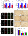
Axonal and myelin abnormalities in Fus ΔNLS/+ mice. a, b Normalized expression of myelin-related genes (Myocilin, NcMap, Pmp22, Pmp2, Cldn19, Prx, Dhh) in Fus ΔNLS/+ compared to their control littermates based on FPKM from RNAseq (a, n = 4) or RT quantitative PCR (b, n = 6–7). Error bars represent SEM. *p < 0.05, ***p < 0.01; Student’s t test. c Double immunolabeling for the oligodendrocyte marker carbonic anhydrase II (CAII) (green) and FUS (N-terminal part) (red) in the spinal cord. Scale bar 5 µm. d Representative images of spinal cord ventral horn of Fus +/+/ChAT-CRE, Fus ΔNLS/+ and Fus ΔNLS/+/ChAT-CRE mice at 22 months stained with anti-carbonic anhydrase II showing increased numbers of oligodendrocytes. Scale bars: left column 70 µm; right column 15 µm. e Oligodendrocyte density in whole spinal cord ventral quadrant, and separately in white and grey matter. Note increased numbers in white matter in 22-month-old Fus ΔNLS/+/−, not rescued in Fus ΔNLS/+/ChAT-CRE mice. N = 4 animals per genotype. ***p < 0.01 versus Fus +/+/ChAT-CRE; one-way ANOVA followed by Tukey post hoc test

Similar articles

Cited by

References

    1. Adlkofer K, Frei R, Neuberg DH, Zielasek J, Toyka KV, Suter U. Heterozygous peripheral myelin protein 22-deficient mice are affected by a progressive demyelinating to maculous neuropathy. J Neurosci. 1997;17:4662–4671. - PMC - PubMed
    1. Angeby-Moller K, Berge OG, Hamers FP. Using the CatWalk method to assess weight-bearing and pain behaviour in walking rats with ankle joint monoarthritis induced by carrageenan: effects of morphine and rofecoxib. J Neurosci Methods. 2008;174:1–9. doi: 10.1016/j.jneumeth.2008.06.017. - DOI - PubMed
    1. Baumer D, Hilton D, Paine SM, Turner MR, Lowe J, Talbot K, Ansorge O. Juvenile ALS with basophilic inclusions is a FUS proteinopathy with FUS mutations. Neurology. 2016;75:611–618. doi: 10.1212/WNL.0b013e3181ed9cde. - DOI - PMC - PubMed
    1. Belly A, Moreau-Gachelin F, Sadoul R, Goldberg Y. Delocalization of the multifunctional RNA splicing factor TLS/FUS in hippocampal neurones: exclusion from the nucleus and accumulation in dendritic granules and spine heads. Neurosci Lett. 2005;379:152–157. doi: 10.1016/j.neulet.2004.12.071. - DOI - PubMed
    1. Bergemalm D, Jonsson PA, Graffmo KS, Andersen PM, Brannstrom T, Rehnmark A, Marklund SL. Overloading of stable and exclusion of unstable human superoxide dismutase-1 variants in mitochondria of murine amyotrophic lateral sclerosis models. J Neurosci. 2006;26:4147–4154. doi: 10.1523/JNEUROSCI.5461-05.2006. - DOI - PMC - PubMed

Publication types

MeSH terms