Review
. 2017 May;18(5):285-298.
doi: 10.1038/nrm.2017.7. Epub 2017 Feb 22.

Biomolecular condensates: organizers of cellular biochemistry

Affiliations
Review

Biomolecular condensates: organizers of cellular biochemistry

Salman F Banani et al. Nat Rev Mol Cell Biol. 2017 May.

Abstract

Biomolecular condensates are micron-scale compartments in eukaryotic cells that lack surrounding membranes but function to concentrate proteins and nucleic acids. These condensates are involved in diverse processes, including RNA metabolism, ribosome biogenesis, the DNA damage response and signal transduction. Recent studies have shown that liquid-liquid phase separation driven by multivalent macromolecular interactions is an important organizing principle for biomolecular condensates. With this physical framework, it is now possible to explain how the assembly, composition, physical properties and biochemical and cellular functions of these important structures are regulated.


Conflict of interest statement

The authors declare no competing interests

Figures

Figure 1.
Figure 1.. Biomolecular Condensates in eukaryotic cells
A) Schematic of the numerous Condensates in the nucleus, cytoplasm and membranes of eukaryotic cells. Some compartments only occur in specific cell types, but are shown here for completeness. For example, Balbiani bodies and germ granules are specific to germ cells (green hues), and RNA transport granules and synaptic densities are seen in neuronal cell types (pink hues). See Supplementary information S6 (Table) for more information on individual Condensates. B) Caenorhabditis elegans germ granules, P granules, are perinuclear Condensates that behave like liquids. A montage of live time-lapse imaging of P granules under shear force (arrows, left top). P granules deform, drip, and fuse with one another around a nucleus (circular structure in the middle outlined in white) (Figure adapted with permissions from Brangwynne et al. 2009). See also Supplementary Information S1–S4 (Movies). Timepoints: 0s, 21s, 32s, 36s, 46s.
Figure 2.
Figure 2.. Different modes of multivalent interactions in synthetic and natural systems undergoing liquid-liquid phase separation
A) (Left) Nephrin contains three phospho-Tyr (pTyr) motifs (small blue circles), which interact with the SH2 domain (dark blue) on Nck. Nck also, contains three SH3 domains (blue), which bind to the numerous proline-rich motifs (PRM) (pink) in neural Wiskott-Aldrich syndrome protein (N-WASP). (Right) Engineered multivalent model systems, consisting of multiple SH3 or SUMO domains (blue), paired with multivalent ligands which contain multiple proline-rich or SUMO-interaction- motifs, PRM or SIM respectively (pink). See for details. B) Edc3 dimerizes via its YJefN domain (green rectangles) and binds to the helical leucine-rich motifs (purple triangles) in Dcp2 via its LSm domain (blue). See for details. C) Nucleophosmin (NPM1) assembles into pentamers via its oligomerizing domain (green triangles) and binds to proteins that contain positively charged Arg-rich linear motifs (R-motifs) (blue rectangles) via its negatively charged acidic, tracts (pink rectangles). NPM1 can also bind to potentially multivalent nucleic acids via its nucleotide binding domain (not shown). See for details. D) RNA binding protein PTB interacts with UCUCU tracts in RNA (connected by AAAA linkers) via its RNA recognition motifs (blue squares). See for details. E) Association of intrinsically disorder regions (IDRs) via cation-pi interactions between aromatic and basic residues, as in DDX4. F) Patterned intermolecular electrostatic interactions between acidic and basic tracts, as in the interactions between the Nephrin intracellular domain (NICD) and positively charged partners, such as supercharged GFP (scGFP). G) Patterned electrostatic interactions between acidic and basic tracts in a single molecular species, as in P granule protein Laf1. H) Polypeptide backbone interactions between β-strands in the polypeptide, as in FUS and hnRNPA1/2,,,. I) Phase diagram as a function of the concentrations of modules present in polymerizing multivalent components that are essential for Condensate formation. Phase separation will be promoted by increasing cellular concentration of component A. J) Regulation of Condensate formation by increase in critical concentration through increasing the valency of A and/or B or the affinity between A and B. Effective valency may be increased by the presence of a third interacting component as shown in the inset. K) Regulation of Condensate formation by decrease in the intrinsic solubility of component A. As molecule A becomes less soluble, phase separation can occur at lower concentrations of A.
Figure 3.
Figure 3.. A Model for Compositional Control of Biomolecular Condensates
Multivalent molecules comprising the scaffold of the Condensate contain complementary modules (blue and yellow, for example small ubiquitin-related modifier (SUMO) domains and SUMO-interaction motifs, respectively)) which allow the assembly of the scaffold to form the phase separated structure (large circles). Client molecules in this example harbour interaction modules complementary to the scaffold components but at lower valency, and are recruited to the structure through binding to free cognate sites in the scaffold (owing to stoichiometric excess of one of the modules). A) Stoichiometric excess of the scaffold component containing blue modules yields free blue scaffold sites. Clients containing yellow modules can be recruited to the body by binding to the blue scaffold sites that are unoccupied by scaffold–scaffold interactions. B) Stoichiometric excess of the scaffold component containing yellow modules yields free yellow scaffold sites. Clients containing blue modules can be recruited to the body by binding to the yellow scaffold sites that are unoccupied by scaffold–scaffold interactions. C) Higher valency of the blue client promotes stronger recruitment of this client when the yellow scaffold module is in stoichiometric excess (but not when the blue scaffold is in excess (not shown)). Figure modified with permissions from Banani, S. F. et al. Cell (2016).
Figure 4.
Figure 4.. Changing material properties of Biomolecular Condensates
Condensates composed of intrinsically disordered regions (IDRs) have the propensity to mature, changing their properties from liquid-like to solid-like. Initially, the components in the condensed phase exhibit only transient interactions and lack appreciable order. Thus the molecules freely rearrange (and exchange with the surrounding solution) and the molecular dynamics can be described as that of a liquid. Over time, the liquid becomes more solid-like. Several potential mechanisms for this ‘hardening’ and the concomitant decrease in molecular dynamics have been proposed, as described in the text. Briefly, these could include nucleation and elongation of amyloid-like fibres, kinetic trapping into amorphous glasses (‘vitrification’) or entanglement of the disordered polypeptides. ATP-dependent machineries such as chaperones and disaggregases are expected to act against these processes (other mechanisms that do not depend on ATP may act similarly).
Figure 5.
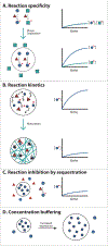
Figure 5.. Functional consequences of forming Biomolecular Condensates
A) Concentrating reactants inside Condensates can increase reaction kinetics and specificity. An enzyme with two alternative substrates is shown. Colocalizing the enzyme with one of its substrates within the condensed phase (black circle) accelerates rates of reaction with that substrate. Additionally, excluding the substrate of an alternative pathway can direct a specific reaction to occur inside Condensates. B) Changes in the physical properties of cellular bodies can affect the kinetics of reactions. For example, increased viscosity of cellular bodies by fibre formation (or other mechanisms of maturation, see text), may slow diffusion of molecules, decreasing reaction kinetics. C) Sequestering molecules inside Condensates can prevent reactions involving partners present in the bulk phase. This could control substrate flux through various pathways. D) The concentration of essential Condensates components in the bulk phase is clamped at the phase separation threshold (defined by the solubility limit of the molecule). Thus the concentration of these components in the bulk phase can be maintained despite fluctuations in expression or degradation.

Similar articles

Cited by

References

    1. Mao YS, Zhang B & Spector DL Biogenesis and function of nuclear bodies. Trends in Genetics 27, 295–306 (2011). - PMC - PubMed
    1. Decker CJ & Parker R P-bodies and stress granules: possible roles in the control of translation and mRNA degradation. Cold Spring Harb Perspect Biol 4, a012286 (2012). - PMC - PubMed
    1. Wu H Higher-order assemblies in a new paradigm of signal transduction. Cell 153, 287–292 (2013). - PMC - PubMed
    1. Pederson T The nucleolus. Cold Spring Harb Perspect Biol 3, (2011). - PMC - PubMed
    1. Dundr M et al. In vivo kinetics of Cajal body components. J Cell Biol 164, 831–842 (2004). - PMC - PubMed

Publication types