

Basal Forebrain Cholinergic System and Orexin Neurons: Effects on Attention
- PMID: 28197081
- PMCID: PMC5281635
- DOI: 10.3389/fnbeh.2017.00010
Basal Forebrain Cholinergic System and Orexin Neurons: Effects on Attention
Abstract
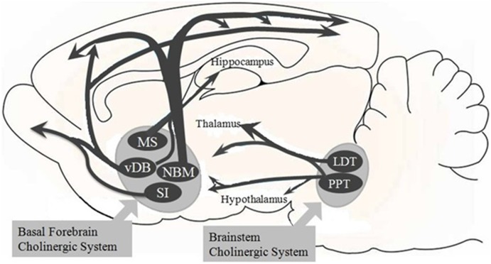
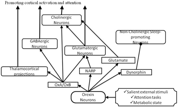
The basal forebrain (BF) cholinergic system has an important role in attentive functions. The cholinergic system can be activated by different inputs, and in particular, by orexin neurons, whose cell bodies are located within the postero-lateral hypothalamus. Recently the orexin-producing neurons have been proved to promote arousal and attention through their projections to the BF. The aim of this review article is to summarize the evidence showing that the orexin system contributes to attentional processing by an increase in cortical acetylcholine release and in cortical neurons activity.
Keywords: acetylcholine; attention; basal forebrain; lateral hypothalamus; orexin.
Figures
Similar articles
-
Orexin/hypocretin modulation of the basal forebrain cholinergic system: Role in attention.Brain Res. 2010 Feb 16;1314:112-23. doi: 10.1016/j.brainres.2009.08.046. Epub 2009 Aug 21. Brain Res. 2010. PMID: 19699722 Free PMC article. Review.
-
Orexin/hypocretin modulation of the basal forebrain cholinergic system: insights from in vivo microdialysis studies.Pharmacol Biochem Behav. 2008 Aug;90(2):156-62. doi: 10.1016/j.pbb.2008.01.008. Epub 2008 Jan 19. Pharmacol Biochem Behav. 2008. PMID: 18281084 Review.
-
Stimulation of cortical acetylcholine release by orexin A.Neuroscience. 2005;130(2):541-7. doi: 10.1016/j.neuroscience.2004.09.050. Neuroscience. 2005. PMID: 15664710
-
Orexin A-induced enhancement of attentional processing in rats: role of basal forebrain neurons.Psychopharmacology (Berl). 2016 Feb;233(4):639-47. doi: 10.1007/s00213-015-4139-z. Epub 2015 Nov 4. Psychopharmacology (Berl). 2016. PMID: 26534765 Free PMC article.
-
Cortical cholinergic inputs mediating arousal, attentional processing and dreaming: differential afferent regulation of the basal forebrain by telencephalic and brainstem afferents.Neuroscience. 2000;95(4):933-52. doi: 10.1016/s0306-4522(99)00487-x. Neuroscience. 2000. PMID: 10682701 Review.
Cited by
-
Hypocretin (Orexin) Replacement Therapies.Med Drug Discov. 2020 Dec;8:100070. doi: 10.1016/j.medidd.2020.100070. Epub 2020 Oct 17. Med Drug Discov. 2020. PMID: 38738170 Free PMC article.
-
Orexin Receptor Antagonists for the Prevention and Treatment of Alzheimer's Disease and Associated Sleep Disorders.Drugs. 2024 Nov;84(11):1365-1378. doi: 10.1007/s40265-024-02096-3. Epub 2024 Oct 4. Drugs. 2024. PMID: 39365407 Free PMC article. Review.
-
Towards a better understanding of anesthesia emergence mechanisms: Research and clinical implications.World J Methodol. 2018 Oct 12;8(2):9-16. doi: 10.5662/wjm.v8.i2.9. eCollection 2018 Oct 12. World J Methodol. 2018. PMID: 30345225 Free PMC article. Review.
-
The brain of the tree pangolin (Manis tricuspis). VIII. The subpallial telencephalon.J Comp Neurol. 2022 Oct;530(15):2611-2644. doi: 10.1002/cne.25353. Epub 2022 Jun 16. J Comp Neurol. 2022. PMID: 35708120 Free PMC article.
-
Reduced Insulin-Like Growth Factor-I Effects in the Basal Forebrain of Aging Mouse.Front Aging Neurosci. 2021 Sep 1;13:682388. doi: 10.3389/fnagi.2021.682388. eCollection 2021. Front Aging Neurosci. 2021. PMID: 34539376 Free PMC article.
References
-
- Alexandre C., Mochizuki T., Arrigoni E., Yamamoto M., Clark E., Scammell T. E. (2012). Orexin signaling in the basal forebrain promotes EEG activation and wakefulness. Sleep 35:A31.
-
- Babkoff H., Caspy T., Mikulincer M. (1991). Subjective sleepiness ratings: the effects of sleep deprivation, circadian rhythmicity and cognitive performance. Sleep 14, 534–539. - PubMed
Publication types
LinkOut - more resources
Full Text Sources
Other Literature Sources
Miscellaneous