. 2017 Feb 14;12(1):17.
doi: 10.1186/s13024-017-0158-z.

Defining the contribution of neuroinflammation to Parkinson's disease in humanized immune system mice

Affiliations

Defining the contribution of neuroinflammation to Parkinson's disease in humanized immune system mice

Gunjan Dhawan Manocha et al. Mol Neurodegener. .

Abstract

Background: Reactive microglia have been associated with the histological changes that occur in Parkinson's disease brains and mouse models of the disease. Multiple studies from autopsy brains have verified the presence of microgliosis in several brain regions including substantia nigra, striatum, hippocampus and various cortical areas. MPTP injections in rodents have also shown striato-nigral microgliosis correlating with the loss of dopaminergic neurons. However, consistent data with respect to cytokine and immune cell changes during Parkinson's disease have not been fully defined.

Results: In order to improve understanding of the role of neuroinflammation in Parkinson's disease, we employed the MPTP injection model using humanized CD34+ mice along with age-matched C57BL/6 mice. NSG mice engrafted with hu-CD34+ hematopoietic stem cells were injected with MPTP to quantify cytokine changes, neuron loss, gliosis, and behavioral dysfunction. The mice were also treated with or without the calcineurin/NFAT inhibitor, FK506, to determine whether modulating the immune response could attenuate disease. MPTP injections produced impairment of motor performance, increased microgliosis, elevated brain cytokine levels, and reduced tyrosine hydroxylase immunoreactivity in the substantia nigra and striatum of both humanized CD34+ mice and C57BL/6 mice with a strikingly different profile of human versus mouse cytokine elevations observed in each. Interestingly, FK506 injections significantly attenuated the MPTP-induced effects in the humanized CD34+ mice compared the C57BL/6 mice. In addition, analyses of human plasma from Parkinson's disease donors compared to age-matched, healthy controls demonstrated an increase in a number of pro-inflammatory cytokines in female patients similar to that observed in MPTP-injected female CD34+ mice.

Conclusions: This study demonstrates for the first time, induction of Parkinson's disease-like symptoms in female humanized CD34+ mice using MPTP. The profile of cytokine changes in the serum and brains of the humanized CD34+ mice following MPTP injection differed significantly from that occurring in the more commonly used C57BL/6 strain of mice. Moreover, several cytokine elevations observed in the MPTP injected humanized CD34+ mice were similarly increased in plasma of PD patients suggesting that these mice offer the more relevant model for the inflammatory aspects of human disease. Consistent with this, the effects of MPTP on loss of tyrosine hydroxylase immunoreactivity, loss of motor strength, and increase in proinflammatory cytokines were attenuated using an immunosuppressant drug, FK506, in the humanized CD34+ but not the C57BL/6 mice. Collectively, these findings suggest that MPTP injected, humanized CD34+ mice represent a more accurate model for assessing inflammatory changes in PD.

Keywords: FK506; Humanized mice; MPTP; Neuroinflammation; Parkinson’s disease.


Figures
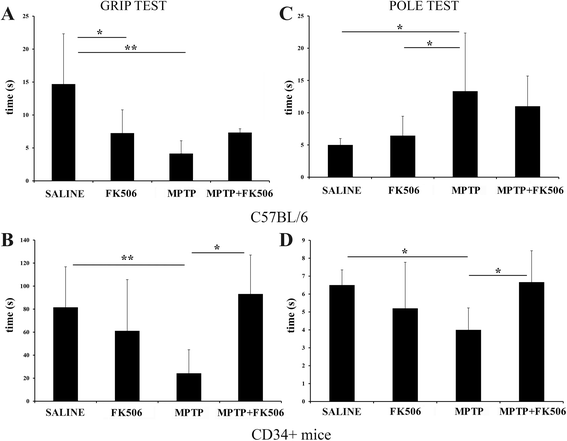
Fig. 1
Fig. 1
MPTP injected C57BL/6 and humanized CD34+ mice showed reduced motor strength based on the grip strength and pole tests. The hCD34+ mice and age-matched C57BL/6 female mice were intraperitoneal injected three times with saline vehicle or MPTP-HCL (18 mg/kg) at 2 h intervals followed by FK506 injections (10 mg/kg/day) for 5 days. Mice were tested for grip strength (a, b) and pole test (c, d) 7 days post MPTP injections. Results were averaged from 8 to 10 mice (CD34+) and 4-10 (C57BL/6) per group and plotted ± SD (*p < 0.05)
Fig. 2
Fig. 2
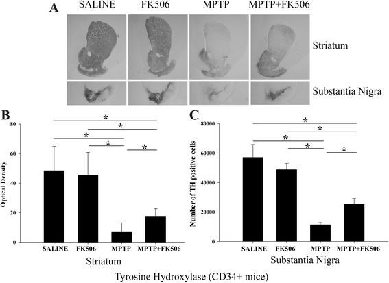
FK506 attenuated the decrease of TH immunoreactivity in the striatum and substantia nigra of MPTP injected humanized CD34+ mice. The hCD34+ mice female mice were intraperitoneal injected three times with saline vehicle or MPTP-HCL (18 mg/kg) at 2 h intervals followed by FK506 injections (10 mg/kg/day) for 5 days. Eight days post MPTP injections, brains were dissected out and right hemispheres fixed and immunostained using anti-TH antibody. a Representative images from striatum and substantia nigra are shown at 1× magnification. b Optical density of immunopositive staining from striatum and c number of TH-positive cells from substantia nigra were measured and averaged 4-5 sections per brain and 4–5 animals per group ± SD (*p < 0.05)
Fig. 3
Fig. 3
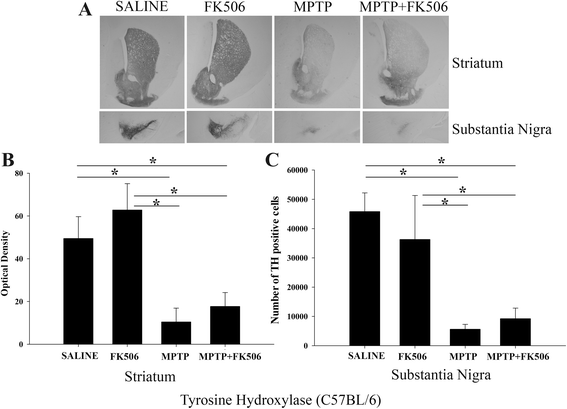
FK506 provided no protection of TH immunoreactivity in the striatum and substantia nigra of MPTP injected C57BL/6 mice. C57BL/6 female mice were intraperitoneal injected three times with saline vehicle or MPTP-HCL (18 mg/kg) at 2 h intervals followed by FK506 injections (10 mg/kg/day) for 5 days. Eight days post MPTP injections, brains were dissected out and right hemispheres fixed and immunostained using anti-TH antibody. a Representative images from striatum and substantia nigra are shown at 1× magnification. b Optical density of immunopositive staining from striatum and c number of TH-positive cells from substantia nigra were measured and averaged for 4–5 sections per brain from 4 to 5 animals per group ± SD (*p < 0.05)
Fig. 4
Fig. 4
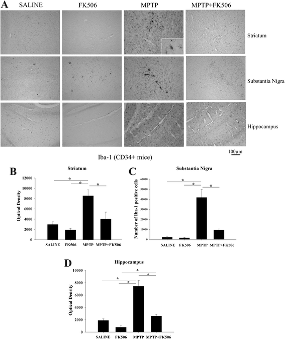
MPTP injections increased microgliosis in the striatum, substantia nigra, and hippocampus of humanized CD34+ mice. The hCD34+ mice were intraperitoneal injected three times with saline vehicle or MPTP-HCL (18 mg/kg) at 2 h intervals followed by FK506 injections (10 mg/kg/day) for 5 days. Eight days post MPTP injections, brains were dissected out and right hemispheres fixed and immunostained using anti-Iba-1 antibody (microglia marker). a Representative images from striatum, substantia nigra and hippocampus are shown at 10× magnification with 63× magnification insert. Optical density of immunopositive staining from b striatum and d hippocampus were measured from 3 to 4 optical fields at 10× magnification per section from 4 to 5 animals per group and c number of Iba-1 positive cells from the substantia nigra were counted and averaged ± SD (*p < 0.05)
Fig. 5
Fig. 5
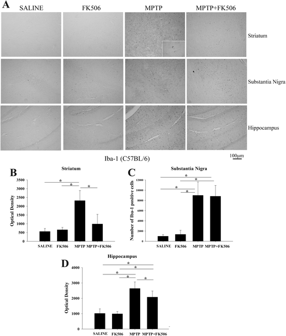
MPTP injections increased microgliosis in the striatum, substantia nigra, and hippocampus of C57BL/6 mice. C57BL/6 female mice were intraperitoneal injected three times with saline vehicle or MPTP-HCL (18 mg/kg) at 2 h intervals followed by FK506 injections (10 mg/kg/day) for 5 days. Eight days post MPTP injections, brains were dissected out and right hemispheres fixed and immunostained using anti-Iba-1 antibody (microglia marker). a Representative images from striatum, substantia nigra and hippocampus are shown at 10× magnification with 63× magnification insert. Optical density of immunopositive staining from b striatum and d hippocampus were measured for 3–4 optical fields at 10× magnification per section from 4 to 5 animals per group and c number of Iba-1 positive cells from substantia nigra were counted and averaged ± SD (*p < 0.05)
Fig. 6
Fig. 6
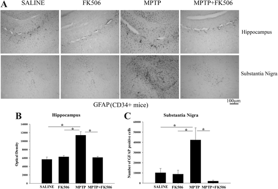
MPTP injections increased astrogliosis in the substantia nigra and hippocampus of humanized CD34+ mice. The hCD34+ mice were intraperitoneal injected three times with saline vehicle or MPTP-HCL (18 mg/kg) at 2 h intervals followed by FK506 injections (10 mg/kg/day) for 5 days. Eight days post MPTP injections, brains were dissected out and right hemispheres fixed and immunostained using anti-GFAP antibody (astrocyte marker). a Representative images from substantia nigra and hippocampus are shown at 10× magnification. Optical density of immunopositive staining from the b hippocampus was measured and averaged for 3–4 optical fields at 10× magnification per section from 4–5 animals per group and c number for GFAP positive cells from the substantia nigra were counted and averaged ± SD (*p < 0.05)
Fig. 7
Fig. 7
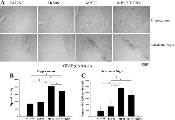
MPTP injections increased astrogliosis in the hippocampus of C57BL/6 mice. C57BL/6 female mice were intraperitoneal injected three times with saline vehicle or MPTP-HCL (18 mg/kg) at 2 h intervals followed by FK506 injections (10 mg/kg/day) for 5 days. Eight days post MPTP injections, brains were dissected out and right hemispheres fixed and immunostained using anti-GFAP antibody (astrocyte marker). a Representative images from substantia nigra and hippocampus are shown at 10× magnification. Optical densities of immunopositive staining from the b hippocampus was measured and averaged for 3–4 optical fields at 10× magnification per section from 4 to 5 animals per group and c number for GFAP positive cells from the substantia nigra were counted and averaged ± SD (*p < 0.05)
Fig. 8
Fig. 8
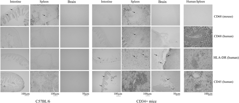
Human HLA-DR and human CD45 immunoreactivities were observed in the spleens, intestines, and occasionally in brain meninges of the humanized CD34+ mice. Intestines, spleens and brains (right hemispheres) were dissected out from 16 weeks old, female C57BL/6 and hCD34+ MPTP injected mice and fixed using 4% paraformaldehyde. Fixed tissue along with human spleen sections (positive controls) were immunostained using anti-CD68 (rodent specific), anti-CD68 (human specific), anti-HLA-DR (LN3, human specific), and anti-CD45 (human specific) antibodies. Intestine and spleen representative images at 10× magnification and brain representative images at 20× magnification are shown
Fig. 9
Fig. 9
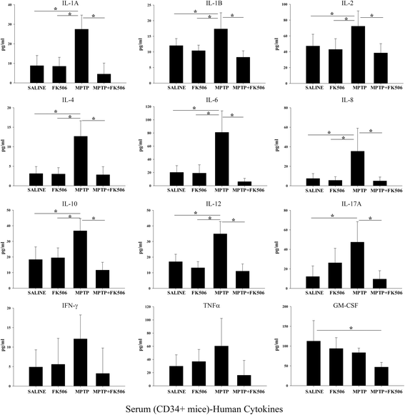
FK506 attenuated the MPTP-dependent increase of human inflammatory cytokine levels in humanized CD34+ mice serum. The hCD34+ mice were intraperitoneal injected three times with saline vehicle or MPTP-HCL (18 mg/kg) at 2 h intervals followed by FK506 injections (10 mg/kg/day) for 5 days. Eight days post MPTP injections, serum was collected and used for human specific multi-analyte cytokine ELISA arrays. Cytokine levels were determined from 6 animals per group ± SD (*p < 0.05)
Fig. 10
Fig. 10
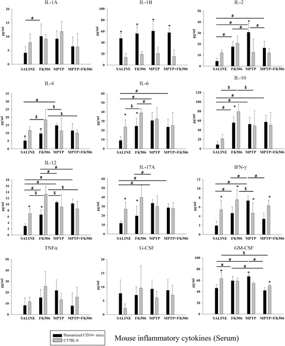
Serum of humanized CD34+ and C57BL/6 mice displayed differential changes in levels of mouse cytokines. Humanized CD34+ and C57BL/6 female mice were intraperitoneal injected three times with saline vehicle or MPTP-HCL (18 mg/kg) at 2 h intervals followed by FK506 injections (10 mg/kg/day) for 5 days. Eight days post MPTP injections, serum was collected and used for mouse specific multi-analyte cytokine ELISA arrays. Cytokine levels were determined from 4 to 6 animals per group ± SD (*p < 0.05 hCD34+ vs. C57BL/6; #p < 0.05 vs. hCD34+ mice, $p < 0.05 vs. C57BL/6)
Fig. 11
Fig. 11
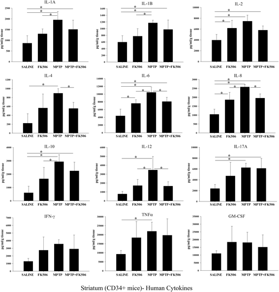
MPTP injections increased levels of human inflammatory cytokines in the striatum of humanized CD34+ mice. The hCD34+ mice were intraperitoneal injected three times with saline vehicle or MPTP-HCL (18 mg/kg) at 2 h intervals followed by FK506 injections (10 mg/kg/day) for 5 days. Eight days post MPTP injections, striatum were dissected out, lysed and used for human specific multi-analyte cytokine ELISA arrays. Cytokine levels were determined from 6 animals per group ± SD (*p < 0.05)
Fig. 12
Fig. 12
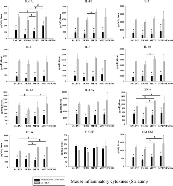
The striatums of humanized CD34+ and C57BL/6 mice displayed different murine cytokine levels after MPTP or FK506 injection. Humanized CD34+ and C57BL/6 female mice were intraperitoneal injected three times with saline vehicle or MPTP-HCL (18 mg/kg) at 2 h intervals followed by FK506 injections (10 mg/kg/day) for 5 days. Eight days post MPTP injections, striatum was dissected out, lysed and used for mouse specific multi-analyte cytokine ELISA arrays. Cytokine levels were determined from 4 to 6 animals per group ± SD (*p < 0.05 hCD34+ vs. C57BL/6; #p < 0.05 vs. hCD34+ mice, $p < 0.05 vs. C57BL/6 mice)
Fig. 13
Fig. 13
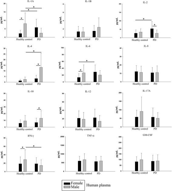
Human PD plasma had elevated levels of IL-1α, IL-2, IL-4 and IL-6 compared to healthy controls. Plasma from male and female Parkinson’s disease donors and age-matched healthy controls was used to perform cytokine ELISAs. Mean values from 14 to 34 patients per condition were averaged and graphed ± SD (*p < 0.05)

Similar articles

Cited by

References

    1. Castano A, Herrera AJ, Cano J, Machado A. Lipopolysaccharide intranigral injection induces inflammatory reaction and damage in nigrostriatal dopaminergic system. J Neurochem. 1998;70(4):1584–1592. doi: 10.1046/j.1471-4159.1998.70041584.x. - DOI - PubMed
    1. Herrera AJ, Castano A, Venero JL, Cano J, Machado A. The single intranigral injection of LPS as a new model for studying the selective effects of inflammatory reactions on dopaminergic system. Neurobiol Dis. 2000;7(4):429–447. doi: 10.1006/nbdi.2000.0289. - DOI - PubMed
    1. Marinova-Mutafchieva L, Sadeghian M, Broom L, Davis JB, Medhurst AD, Dexter DT. Relationship between microglial activation and dopaminergic neuronal loss in the substantia nigra: a time course study in a 6-hydroxydopamine model of Parkinson’s disease. J Neurochem. 2009;110(3):966–975. doi: 10.1111/j.1471-4159.2009.06189.x. - DOI - PubMed
    1. Wu DC, Jackson-Lewis V, Vila M, Tieu K, Teismann P, Vadseth C, Choi DK, Ischiropoulos H, Przedborski S. Blockade of microglial activation is neuroprotective in the 1-methyl-4-phenyl-1,2,3,6-tetrahydropyridine mouse model of Parkinson disease. J Neurosci. 2002;22(5):1763–1771. - PMC - PubMed
    1. Banati RB, Daniel SE, Blunt SB. Glial pathology but absence of apoptotic nigral neurons in long-standing Parkinson’s disease. Mov Disord. 1998;13(2):221–227. doi: 10.1002/mds.870130205. - DOI - PubMed

Publication types