. 2017 Jan 15;31(2):209-222.
doi: 10.1101/gad.288555.116. Epub 2017 Feb 6.

miRNAs cooperate in apoptosis regulation during C. elegans development

Affiliations

miRNAs cooperate in apoptosis regulation during C. elegans development

Ryan Sherrard et al. Genes Dev. .

Abstract

Programmed cell death occurs in a highly reproducible manner during Caenorhabditis elegans development. We demonstrate that, during embryogenesis, miR-35 and miR-58 bantam family microRNAs (miRNAs) cooperate to prevent the precocious death of mothers of cells programmed to die by repressing the gene egl-1, which encodes a proapoptotic BH3-only protein. In addition, we present evidence that repression of egl-1 is dependent on binding sites for miR-35 and miR-58 family miRNAs within the egl-1 3' untranslated region (UTR), which affect both mRNA copy number and translation. Furthermore, using single-molecule RNA fluorescent in situ hybridization (smRNA FISH), we show that egl-1 is transcribed in the mother of a cell programmed to die and that miR-35 and miR-58 family miRNAs prevent this mother from dying by keeping the copy number of egl-1 mRNA below a critical threshold. Finally, miR-35 and miR-58 family miRNAs can also dampen the transcriptional boost of egl-1 that occurs specifically in a daughter cell that is programmed to die. We propose that miRNAs compensate for lineage-specific differences in egl-1 transcriptional activation, thus ensuring that EGL-1 activity reaches the threshold necessary to trigger death only in daughter cells that are programmed to die.

Keywords: BH3-only; C. elegans; development; embryo; miRNA; programmed cell death.


Figures

Figure 1.
Figure 1.
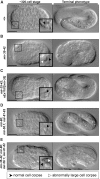
Embryos lacking miR-35 family miRNAs exhibit a large cell corpse phenotype. (A–E) Differential interface contrast (DIC) images of embryos of the genotypes +/+ (A), mir-35-41(nDf50) mir-42(nDf49) (B), mir-35-41(nDf50) mir-42(nDf49); nEx1187 [mir-35 (genomic)+sur-5::gfp] (C), mir-80(nDf53); mir-58.1(n4640); mir-81-82(nDf54) (D), and mir-35-41(nDf50) mir-42(nDf49); mir-80(nDf53); mir-58.1(n4640); mir-81-82(nDf54) (E). For each panel, a developing embryo (∼320-cell stage) is depicted at the left, with insets showing representative cell corpses. Normal cell corpses (black arrowheads) are present in all genetic backgrounds, but abnormally large cell corpses (white arrowheads) are also present (see B,E). The terminal phenotype of each embryo is shown at the right; those in A, C, and D survived to hatching, whereas those in B and E arrested. Bars, 10 µm.
Figure 2.
Figure 2.
Inappropriate cell death in mir-35 family mutants is enhanced upon loss of the mir-58 family and is dependent on the central apoptotic pathway. (A) The number of first-wave AB cell deaths was scored per embryo of each genotype. n = 5 or 10, as indicated. The number of abnormally large corpses per embryo was also scored until ventral enclosure. Values are averages ± SEM when applicable. (****) P ≤ 0.0001 via Student's t-test. (B,C) The number of large cell corpses forming over time in mir-35-41(nDf50) mir-42(nDf49) mutants (B) and mir-35-41(nDf50) mir-42(nDf49); mir-80(nDf53); mir-58.1(n4640); mir-81-82(nDf54) mutants (C) until ventral enclosure. Data are based on the time of initial detection for each of the 46 and 107 large cells corpses in B and C, respectively. Each circle represents the detection of a single large cell corpse. Detection times were grouped into 15-min bins (i.e., intervals). n = 10 embryos for each genotype.
Figure 3.
Figure 3.
The 3′ UTR of egl-1 is a target of miR-35 and miR-58 family miRNAs in vivo. (A) The 3′ UTR of egl-1 is illustrated in blue, with a reported C. elegans Argonaut ALG-1-binding site from a recent study (Grosswendt et al. 2014) indicated above. Sequences are given for predicted miR-35 family-binding sites and miR-58 family-binding sites, and conserved bases within each site are highlighted in gray across four Caenorhabditis species. The mutated sequences corresponding to egl-1mut mir-35 (red) and egl-1mut mir-58 (purple) are shown (for complete mutated sequences, see the Materials and Methods). (B) A schematic representation of the 3′ UTR reporters constructed for this study. (C) Analysis of single-copy 3′ UTR reporter expression during embryogenesis. The genetic background and transgene under investigation are indicated at the left of each image sequence, which shows representative embryos from three developmental stages. For each embryo, a DIC image (left) and GFP image (right) are shown. The following alleles were used: bcSi25 [Pmai-2gfp::h2b::mai-2 3′ UTR], bcSi26 [Pmai-2gfp::h2b::egl-1wt 3′ UTR], bcSi27 [Pmai-2gfp::h2b::egl-1mut mir-35 3′ UTR], bcSi46 [Pmai-2gfp::h2b::egl-1mut mir-35 3′ UTR], and bcSi47 [Pmai-2gfp::h2b::egl-1mut mir-35 mir-58 3′ UTR]. Asterisks indicate autofluorescence. Transgenic strains were homozygous for unc-119(ed3) and the cb-unc-119(+) selection marker. Bars, 10 µm.
Figure 4.
Figure 4.
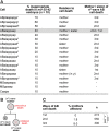
Abnormal cell death in mir-35 family mutants affects mothers and sisters of programmed cell death. (A) The identities of 46 larges corpses present across 10 mir-35-41(nDf50) mir-42(nDf49) embryos were determined as described in the Materials and Methods. Numerical superscripts 1–5 indicate pairs of bilaterally symmetric cells. (B) Lineage representation of “precocious death” affecting mother cells and “collateral death” affecting sister cells. (C) The average percentage of precociously dying mothers was determined per embryo for the three waves of AB cell death according to data presented in A. (*) P < 0.05; (****) P < 0.0001 via Fisher’s exact test.
Figure 5.
Figure 5.
egl-1 mRNA copy number crosses a threshold to trigger the precocious death of ABalappaap in mir-35 mir-58 double-family mutants. (A) smRNA FISH analysis in the ABalappaap lineage. The lineage is illustrated at the right, with the approximate time of each analysis (pre- and post-division) indicated. Representative images of the ABalappaap cell and its daughters are shown for each time point in four genetic backgrounds, as indicated. The egl-1 mRNA copy number corresponding to each image is given. Nuclei are labeled as belonging to the surviving daughter (anterior [a]) or the dying daughter (posterior [p]). Approximate cellular volumes are given at the right. Bars, 2 µm. (B) A fixed wild-type embryo showing the ABalappaap cell (arrowhead) as identified by smRNA FISH staining against mRNA transcribed from the Punc-3unc-3::gfp reporter. The anterior–posterior axis (A ↔ P) is indicated. Bar, 10 µm. (C) Quantification of egl-1 mRNA copy number in the ABalappaap cell and its two daughters (ABalappaapa and ABalappaapp [X]) for each of the indicated genotypes. An estimated threshold for triggering cell death in ABalappaap is represented by the red line. The mean value is given above each data set. Box and whiskers were plotted according to Tukey's test. (*) P < 0.05; (**) P < 0.01; (****) P < 0.0001 via Mann-Whitney test. The following alleles were used: mir-35-41(nDf50), mir-42 (nDf49), mir-80(nDf53), mir-58.1(n4640), and mir-81-82(nDf54). All strains analyzed carried the extrachromosomal array xdEx1091 [Punc-3unc-3::gfp+Psur-5rfp].
Figure 6.
Figure 6.
egl-1 mRNA levels are temporally dynamic in the MSpaap lineage and more abundant in mir family mutants. (A) smRNA FISH analysis in the MSpaap lineage. The lineage is illustrated at the right, with the approximate time of each analysis (predivision, dividing, and post-division) indicated. Representative images of the MSpaap cell and its daughters are shown for each time point in four genetic backgrounds, as indicated. Nuclei are labeled as belonging to the surviving daughter (anterior [a]) or the dying daughter (posterior [p]). The egl-1 mRNA copy number corresponding to each image is given. Approximate cellular volumes are given at the right. Bars, 2 µm. (B) A fixed wild-type embryo containing 167 nuclei showing the MSpaap cell (inset). The anterior–posterior axis (A ↔ P) is indicated Bar, 10 µm. (C) Quantification of egl-1 mRNA in the MSpaap cell and its two daughters (MSpaapa and MSpaapp [X]) for each of the indicated genotypes. The mean value is given above each data set. Box and whiskers were plotted according to Tukey's test. (*) P < 0.05; (**) P < 0.01; (****) P < 0.0001 via Mann-Whitney test. (D) Developmental time course showing egl-1 mRNA concentration in cells of the MSpaap lineage for the indicated genotypes. Division of the MSpaap cell is indicated by a vertical dashed line. Insets show a 2× zoom of the graph at the time of division. Graphs were generated from raw data as a centered moving average of order 5. Shaded areas represent SEM, when applicable. The following alleles were used: mir-35-41(nDf50), mir-42 (nDf49), mir-80(nDf53), mir-58.1(n4640), and mir-81-82(nDf54).
Figure 7.
Figure 7.
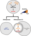
Genetic model of mir-35- and mir-58-dependent control of egl-1 BH3-only in the ABalappaap lineage. See the text for details.

Similar articles

Cited by

References

    1. Abbott AL, Alvarez-Saavedra E, Miska EA, Lau NC, Bartel DP, Horvitz HR, Ambros V. 2005. The let-7 microRNA family members mir-48, mir-84, and mir-241 function together to regulate developmental timing in Caenorhabditis elegans. Dev Cell 9: 403–414. - PMC - PubMed
    1. Alvarez-Saavedra E, Horvitz HR. 2010. Many families of C. elegans microRNAs are not essential for development or viability. Curr Biol 20: 367–373. - PMC - PubMed
    1. Brennecke J, Hipfner DR, Stark A, Russell RB, Cohen SM. 2003. bantam encodes a developmentally regulated microRNA that controls cell proliferation and regulates the proapoptotic gene hid in Drosophila. Cell 113: 25–36. - PubMed
    1. Brenner S. 1974. The genetics of Caenorhabditis elegans. Genetics 77: 71–94. - PMC - PubMed
    1. Chakraborty S, Lambie EJ, Bindu S, Mikeladze-Dvali T, Conradt B. 2015. Engulfment pathways promote programmed cell death by enhancing the unequal segregation of apoptotic potential. Nat Commun 6: 10126. - PMC - PubMed

Publication types

LinkOut - more resources