

Genetic advances in craniosynostosis
- PMID: 28160402
- PMCID: PMC5397362
- DOI: 10.1002/ajmg.a.38159
Genetic advances in craniosynostosis
Abstract
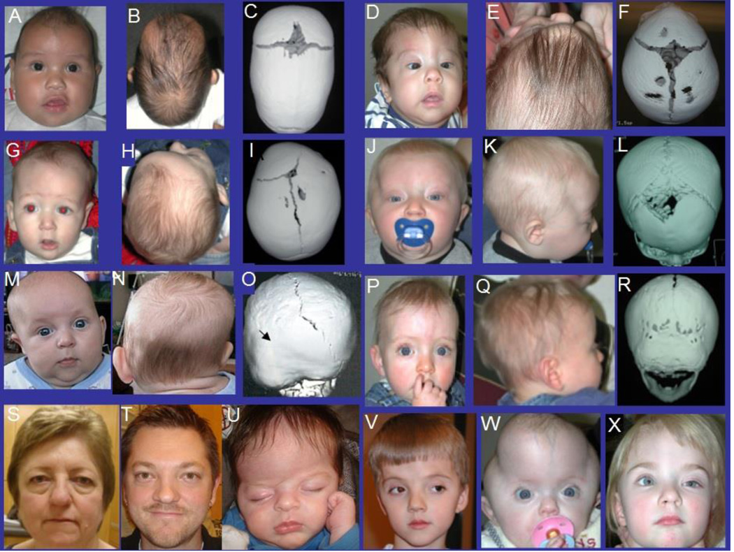
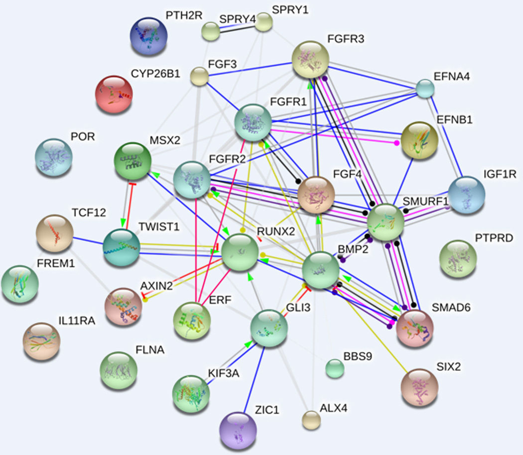
Craniosynostosis, the premature ossification of one or more skull sutures, is a clinically and genetically heterogeneous congenital anomaly affecting approximately one in 2,500 live births. In most cases, it occurs as an isolated congenital anomaly, that is, nonsyndromic craniosynostosis (NCS), the genetic, and environmental causes of which remain largely unknown. Recent data suggest that, at least some of the midline NCS cases may be explained by two loci inheritance. In approximately 25-30% of patients, craniosynostosis presents as a feature of a genetic syndrome due to chromosomal defects or mutations in genes within interconnected signaling pathways. The aim of this review is to provide a detailed and comprehensive update on the genetic and environmental factors associated with NCS, integrating the scientific findings achieved during the last decade. Focus on the neurodevelopmental, imaging, and treatment aspects of NCS is also provided.
Keywords: craniofacial; craniosynostosis; gene mutations; malformation; skull sutures.
© 2017 Wiley Periodicals, Inc.
Conflict of interest statement
The authors have no conflict of interest to declare.
Figures
Similar articles
-
Genetic analysis of non-syndromic craniosynostosis.Orthod Craniofac Res. 2007 Aug;10(3):129-37. doi: 10.1111/j.1601-6343.2007.00393.x. Orthod Craniofac Res. 2007. PMID: 17651129 Review.
-
Gain-of-function variants and overexpression of RUNX2 in patients with nonsyndromic midline craniosynostosis.Bone. 2020 Aug;137:115395. doi: 10.1016/j.bone.2020.115395. Epub 2020 Apr 30. Bone. 2020. PMID: 32360898 Free PMC article.
-
BBS9 gene in nonsyndromic craniosynostosis: Role of the primary cilium in the aberrant ossification of the suture osteogenic niche.Bone. 2018 Jul;112:58-70. doi: 10.1016/j.bone.2018.04.013. Epub 2018 Apr 17. Bone. 2018. PMID: 29674126 Free PMC article.
-
Signaling Mechanisms Underlying Genetic Pathophysiology of Craniosynostosis.Int J Biol Sci. 2019 Jan 1;15(2):298-311. doi: 10.7150/ijbs.29183. eCollection 2019. Int J Biol Sci. 2019. PMID: 30745822 Free PMC article. Review.
-
Recent Advances in Craniosynostosis.Pediatr Neurol. 2019 Oct;99:7-15. doi: 10.1016/j.pediatrneurol.2019.01.018. Epub 2019 Feb 2. Pediatr Neurol. 2019. PMID: 31421914 Review.
Cited by
-
Targeted Sequencing of Candidate Regions Associated with Sagittal and Metopic Nonsyndromic Craniosynostosis.Genes (Basel). 2022 May 3;13(5):816. doi: 10.3390/genes13050816. Genes (Basel). 2022. PMID: 35627201 Free PMC article.
-
Neuroimaging in Nonsyndromic Craniosynostosis: Key Concepts to Unlock Innovation.Diagnostics (Basel). 2024 Aug 23;14(17):1842. doi: 10.3390/diagnostics14171842. Diagnostics (Basel). 2024. PMID: 39272627 Free PMC article. Review.
-
Shaping modern human skull through epigenetic, transcriptional and post-transcriptional regulation of the RUNX2 master bone gene.Sci Rep. 2021 Oct 29;11(1):21316. doi: 10.1038/s41598-021-00511-3. Sci Rep. 2021. PMID: 34716352 Free PMC article.
-
A progressive and complex clinical course in two family members with ERF-related craniosynostosis: a case report.BMC Med Genet. 2020 May 5;21(1):90. doi: 10.1186/s12881-020-01015-z. BMC Med Genet. 2020. PMID: 32370745 Free PMC article.
-
Advanced parental age: Is it contributing to an increased incidence of non-syndromic craniosynostosis? A review of case-control studies.J Oral Biol Craniofac Res. 2021 Jan-Mar;11(1):78-83. doi: 10.1016/j.jobcr.2020.11.016. Epub 2020 Nov 28. J Oral Biol Craniofac Res. 2021. PMID: 33376670 Free PMC article. Review.
References
-
- Abbott MM, Rogers GF, Proctor MR, Busa K, Meara JG. Cost of treating sagittal synostosis in the first year of life. J Craniofac Surg. 2012;23(1):88–93. - PubMed
-
- Agrawal D, Steinbok P, Cochrane DD. Diagnosis of isolated sagittal synostosis: are radiographic studies necessary? Childs Nerv Syst. 2005;22(4):375–378. - PubMed
-
- Alderman BW, Lammer EJ, Joshua SC, Cordero JF, Ouimette DR, Wilson MJ, Ferguson SW. An epidemiologic study of craniosynostosis: risk indicators for the occurrence of craniosynostosis in Colorado. Am J Epidemiol. 1988;128(2):431–438. - PubMed
-
- Aldridge K, Kane AA, Marsh JL, Panchal J, Boyadjiev SA, Yan P, Govier D, Ahmad W, Richtsmeier JT. Brain morphology in nonsyndromic unicoronal craniosynostosis. Anat Rec A Discov Mol Cell Evol Biol. 2005;285(2):690–698. - PubMed
Publication types
MeSH terms
Grants and funding
LinkOut - more resources
Full Text Sources
Other Literature Sources
Medical
