. 2017 Feb 3:7:41989.
doi: 10.1038/srep41989.

The molecular basis of talin2's high affinity toward β1-integrin

Affiliations

The molecular basis of talin2's high affinity toward β1-integrin

Yaxia Yuan et al. Sci Rep. .

Abstract

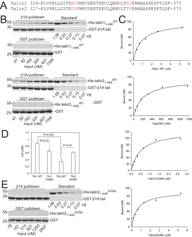
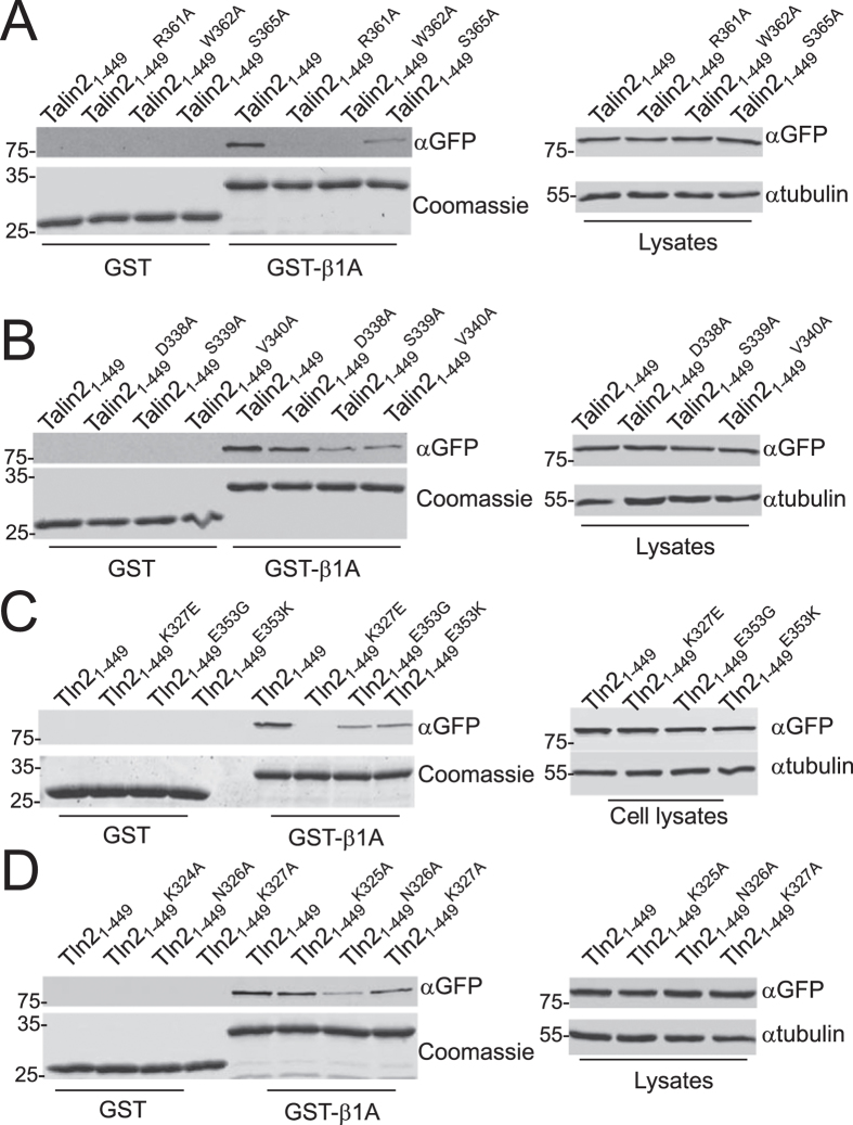
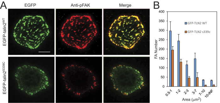
Talin interacts with β-integrin tails and actin to control integrin activation, thus regulating focal adhesion dynamics and cell migration. There are two talin genes, Tln1 and Tln2, which encode talin1 and talin2, and it is generally believed that talin2 functions redundantly with talin1. However, we show here that talin2 has a higher affinity to β1-integrin tails than talin1. Mutation of talin2 S339 to leucine, which can cause Fifth Finger Camptodactyly, a human genetic disease, completely disrupted its binding to β-integrin tails. Also, substitution of talin1 C336 with Ser enhanced the affinity of talin1, whereas substitution of talin2 S339 with Cys diminished that of talin2. Further computational modeling analysis shows that talin2 S339 formed a hydrogen bond with E353, which is critical for inducing key hydrogen bonds between talin2 N326 and β1-integrin R760, and between talin2 K327 and β1-integrin D759. Mutation at any of these residues significantly diminished the interaction of talin2 with β1- integrin tails. These hydrogen bonds were not observed in talin1/β1-integrin, but did exist in talin1C336S/β1-integrin complex. These results suggest that talin2 S339 forms a hydrogen bond with E353 to mediate its high affinity to β1-integrin.


Conflict of interest statement

The authors declare no competing financial interests.

Figures

Figure 1
Figure 1. Affinities of talin11–446, talin11–446C336S, talin21–449, and talin21–449S339C to β1-integrin tails.
The binding of purified His-tagged proteins to GST-β1-integrin tails was determined using GST pulldown assays. Bound protein was separated using SDS-PAGE, stained with Coomassie blue, and calibrated with standards run on the same gels. The binding curves and the Kd were analyzed with SigmaPlot. (A) Sequence alignment of the integrin-binding region of the F3 domains of talin1 and talin2. Unmatched residues are shown in Red. (B) GST-β1 tails-bound talin11–446 and talin21–449 were compared after SDS-PAGE and Coomassie blue staining. Cropped gel images are shown. The full-length gel images are included in Supplementary Fig. S3. (C) The specific binding curves of talin11–446, talin11–446C336S, talin21–449, and talin21–449S339C to β1-integrin tails. Please note that protein concentrations used in binding assays were different. (D) The dissociation constants of talin11–446, talin11–446C336S, talin21–449, and talin21–449S339C binding to β1A-integrin tails. Data are presented as mean ± SEM. P values from student’s t-test are shown. (E) GST-β1 tails-bound talin21–449S339L were detected by Coomassie blue staining. Cropped gel images are shown. The full-length gel images are included in Supplementary Fig. S3.
Figure 2
Figure 2. Tracked positional RMSD values for the wild type Talin1/Integrin, C336S mutant Talin1/Integrin and wild type Talin2/Integrin complex base on 40 ns MD simulation.
Figure 3
Figure 3. Superimposition of talin/integrin complexes.
(A) Sequence alignment of Mus musculus talin2 (mTalin2), Homo sapiens talin2 (hTalin2), and Homo sapiens talin1 (hTalin1) by PROMALS3D server. β strands of talin (β1-β7), α helix of β1-integrin as well as the name of the subunit are marked by black lines. β-sheet 1 and β-sheet 2 are colored in green and yellow respectively. The key interaction pair of Tln-S339/336 and Tln-E353/350 are colored in red, the key interaction pair of Tln-K324/327 and Int-D759 are colored in purple, and the Tln-N323/326 and Int-R760 interaction pair are colored in cyan. (B) (1) Typical talin2WT/integrin complex structure is derived from the last snapshot of the 40 ns MD trajectory by energy minimization. Talin2 and β1-integrin are represented as cyan and blue ribbons, respectively. Tln-S339, Tln-E353, Tln-K327, Tln-N326, Int-R760, and Int-D759 are showed in stick-ball model. (2) Superimposition of the talin2WT/integrin and talin1WT/integrin complexes. For talin2WT/integrin complex, talin2WT and β1-integrin are colored in cyan and blue, respectively. For talin1WT/integrin complex, talin1WT and β1-integrin are colored in golden and red, respectively. (3) Only β-sheet 1 of talin and β1-intergrin are shown. Although other parts of talin could be superimposed well, β-sheet 1 varies a lot in talin2WT and talin1WT. (4) Superimposition of the talin2WT/integrin and talin1C336S/integrin complex. Talin1C336S and β1-integrin are colored in green and purple, respectively. (5) Only β-sheet 1 of talin and β1-integrin are shown, and they both have similar conformation in talin2WT/integrin and talin1C336S/integrin complexes. (6) Superimposition of all three talin/integrin complexes. (7) Only β-sheet 1 of talin and β1-intergin are shown.
Figure 4
Figure 4. The hydrogen bonds between Tln-S339 and Tln-E353, Tln-K327 and Int-D759, and Tln-N326 and Int-R760 contribute to their affinity to β1-integrins.
(A) Tracked distance of the intra-molecular hydrogen bond formed by residue S336/339 and E350/353 of talin. Black, blue, and red tracks represent the distance (Unit in Å) between the Hɣ atom of S339/C336/S336 and the Oε atom of E353/350 in talin2WT/integrin, talin1WT/integrin, and talin1C336S/integrin complexes, respectively. (B) Tracked distance of the inter-molecular hydrogen bond formed between the positive-charged side chain of K324/327 in talin and the negative charged side chain of D759 in β1-integrin. Black, blue, and red tracks represent the distance between the Hζ atom of K327/324 of talin and the Oδ atom at D759 of β1-integrin in talin2WT/integrin, talin1WT/integrin, talin1C336S/integrin complexes, respectively. (C) Tracked distance of the inter-molecular hydrogen bond formed between the side chain of N323/326 in talin and side chain of R760 in β1-integrin. Black, blue, and red tracks represent the distance between the Oδ atom of N326/323 of talin and the Hη atom of R760 of β1-integrin in talin2WT/integrin, talin1WT/integrin, and talin1C336S/integrin complexes, respectively. (D) Hydrogen bond between S339 and E353 of talin with labeled distance in talin2WT/integrin complex. Talin is represent as cyan ribbons. S339 and E353 are shown in golden stick-ball style. (E) Side chains of C336 and E350 are far away from each other in talin1WT/integrin complex. (F) Hydrogen bond between S336 and E350 with labeled distance in talin1C336S/integrin complex. (G) Hydrogen bond between Tln-K327 and Int-D759, and Tln-N326 and Int-R760 with labeled distance in talin2WT/integrin complex. Talin and β1-integrin are presented as cyan ribbons and green ribbons respectively. (H) Side chains of Tln-K324, Tln-N323, Int-D759, and Int-R760 are far away from each other in talin1WT/integrin complex. (I) Hydrogen bond between Tln-K324 and Int-D759, and Tln-N323 and Int-R760 with labeled distance in talin1C336S/integrin complex.
Figure 5
Figure 5. The statiscal hydrogen bond profile among talin2WT/integrin, talin1WT/integrin and talin1C336S/integrin complexes.
(A) The difference of average inter-molecular hydrogen bond number between talin2WT/integrin and talin1WT/integrin. As the difference is dependent on the threshold of hydrogen bond definiation, the profile of various hydrogen bond distances and angle cutoffs is shown. Here the hydrogen bond distance is defined as the distance between hydrogen bond donor and acceptor, while the hydrogen bond angle is defined as the angle between donor, hydrogen, and acceptor. For a typical hydrogen bond distance cutoff of 3.0 Å and angle cutoff of 120°, talin2WT/integrin complex has about 1 more inter-molecular hydrogen bond than talin1WT/integrin complex. (B) The difference of the average inter-molecular hydrogen bond number between talin2WT/integrin and talin1C336S/integrin. It could be observed that these two complexes form an inter-molecular hydrogen bond in a similar level.
Figure 6
Figure 6. Experimental examination of the computational model.
(A) Binding of EGFP-talin21–449WT, -talin21–449R361A, -talin21–449W362A, and -talin21–449S365A to β1-integrin tails by GST pulldown assays. The EGFP fusion proteins were transiently expressed in CHO-K1 cells. (B) EGFP-talin21–449WT, -talin21–449D338A, -talin21–449S339A, and -talin21–449V340A were transiently transfected in to CHO-K1 cells. The binding of these proteins to β1-integrin tails was determined by GST pulldown assays. (C) Binding of EGFP-talin21–449WT, -talin21–449K327E, -talin21–449E353G, and -talin21–449E353K to β1-integrin tails by GST pulldown assays. (D) Binding of EGFP-talin21–449WT, -talin21–449K324A, -talin21–449N326A, and -talin21–449K327A to β1-integrin tails by GST pulldown assays. Cropped blot/gel images are shown. The full-length blot/gel images are included in Supplementary Fig. S4.
Figure 7
Figure 7. Substitution of talin2 S339 with Cys diminished focal adhesion formation.
Talin2-null U2 OS cells were transfected with EGFP-talin2WT and -talin2S339C, respectively, plated on fibronectin (5 μg/ml), fixed with paraformaldehyde, and stained with anti-phospho-FAK[pY397] antibody. Focal adhesions were examined with a TIRF microscopy. Scale bar, 20 μm. (A) The distribution of talin2, talin2S339C, and phospho-FAK in U2 OS cells. (B) Area distribution of phospho-FAK staining in U2 OS cells. Data are mean ± SEM of 3 experiments. In each group, FAs from 20 cells were analyzed and plotted.

Similar articles

Cited by

References

    1. Hynes R. O. Integrins: bidirectional, allosteric signaling machines. Cell 110, 673–687 (2002). - PubMed
    1. Truong H. & Danen E. H. J. Integrin switching modulates adhesion dynamics and cell migration. Cell Adh Migr 3, 179–181 (2009). - PMC - PubMed
    1. Scales T. M. E. & Parsons M. Spatial and temporal regulation of integrin signalling during cell migration. Curr Opin Cell Biol 23, 562–568 (2011). - PubMed
    1. Missan D. S. & DiPersio M. Integrin Control of Tumor Invasion. Crit Rev Eukaryot Gene Expr 22, 309–324 (2012). - PubMed
    1. Mercurio A. M. & Rabinovitz I. Towards a mechanistic understanding of tumor invasion—lessons from theα 6 β 4integrin. Semin Cancer Biol 11, 129–141 (2001). - PubMed

Publication types

LinkOut - more resources