

Fcγ receptor expression on splenic macrophages in adult immune thrombocytopenia
- PMID: 28142207
- PMCID: PMC5383444
- DOI: 10.1111/cei.12935
Fcγ receptor expression on splenic macrophages in adult immune thrombocytopenia
Abstract
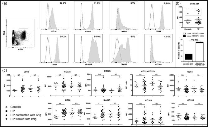
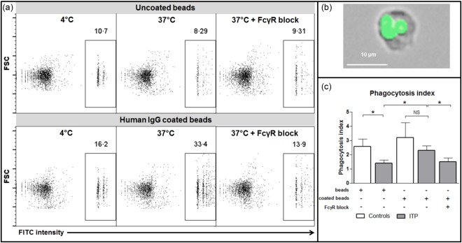
Splenic macrophages play a key role in immune thrombocytopenia (ITP) pathogenesis by clearing opsonized platelets. Fcγ receptors (FcγR) participate in this phenomenon, but their expression on splenic macrophages and their modulation by treatment have scarcely been studied in human ITP. We aimed to compare the phenotype and function of splenic macrophages between six controls and 24 ITP patients and between ITP patients according to the treatments they received prior to splenectomy. CD86, human leucocyte antigen D-related (HLA-DR) and FcγR expression were measured by flow cytometry on splenic macrophages. The major FcγR polymorphisms were determined and splenic macrophage function was assessed by a phagocytosis assay. The expression of the activation markers CD86 and HLA-DR was higher on splenic macrophages during ITP compared to controls. While the expression of FcγR was not different between ITP and controls, the phagocytic function of splenic macrophages was reduced in ITP patients treated with intravenous immunoglobulin (IVIg) within the 2 weeks prior to splenectomy. The FCGR3A (158V/F) polymorphism, known to increase the affinity of FcγRIII to IgG, was over-represented in ITP patients. Thus, these are the first results arguing for the fact that the therapeutic use of IVIg during human chronic ITP does not modulate FcγR expression on splenic macrophages but decreases their phagocytic capabilities.
Keywords: Fc receptors; autoimmunity; macrophages; spleen.
© 2017 British Society for Immunology.
Figures
Similar articles
-
RETRACTED: Hydroxychloroquine and azithromycin as a treatment of COVID-19: results of an open-label non-randomized clinical trial.Int J Antimicrob Agents. 2020 Jul;56(1):105949. doi: 10.1016/j.ijantimicag.2020.105949. Epub 2020 Mar 20. Int J Antimicrob Agents. 2020. Retraction in: Int J Antimicrob Agents. 2025 Jan;65(1):107416. doi: 10.1016/j.ijantimicag.2024.107416. PMID: 32205204 Free PMC article. Retracted. Clinical Trial.
-
Defining the optimum strategy for identifying adults and children with coeliac disease: systematic review and economic modelling.Health Technol Assess. 2022 Oct;26(44):1-310. doi: 10.3310/ZUCE8371. Health Technol Assess. 2022. PMID: 36321689 Free PMC article.
-
Depressing time: Waiting, melancholia, and the psychoanalytic practice of care.In: Kirtsoglou E, Simpson B, editors. The Time of Anthropology: Studies of Contemporary Chronopolitics. Abingdon: Routledge; 2020. Chapter 5. In: Kirtsoglou E, Simpson B, editors. The Time of Anthropology: Studies of Contemporary Chronopolitics. Abingdon: Routledge; 2020. Chapter 5. PMID: 36137063 Free Books & Documents. Review.
-
Comparing Two Approaches to Help Patients and Doctors Talk about Chronic Pain Treatments -- Voices in Pain Care [Internet].Washington (DC): Patient-Centered Outcomes Research Institute (PCORI); 2022 Apr. Washington (DC): Patient-Centered Outcomes Research Institute (PCORI); 2022 Apr. PMID: 39661719 Free Books & Documents. Review.
-
Immunomodulators and immunosuppressants for multiple sclerosis: a network meta-analysis.Cochrane Database Syst Rev. 2013 Jun 6;2013(6):CD008933. doi: 10.1002/14651858.CD008933.pub2. Cochrane Database Syst Rev. 2013. PMID: 23744561 Free PMC article. Review.
Cited by
-
Platelets in HIV: A Guardian of Host Defence or Transient Reservoir of the Virus?Front Immunol. 2021 Apr 23;12:649465. doi: 10.3389/fimmu.2021.649465. eCollection 2021. Front Immunol. 2021. PMID: 33968041 Free PMC article. Review.
-
Emerging Therapies in Immune Thrombocytopenia.J Clin Med. 2021 Mar 2;10(5):1004. doi: 10.3390/jcm10051004. J Clin Med. 2021. PMID: 33801294 Free PMC article. Review.
-
Cellular surface plasmon resonance-based detection of anti-HPA-1a antibody glycosylation in fetal and neonatal alloimmune thrombocytopenia.Front Immunol. 2023 Oct 5;14:1225603. doi: 10.3389/fimmu.2023.1225603. eCollection 2023. Front Immunol. 2023. PMID: 37868955 Free PMC article.
-
Serum from half of patients with immune thrombocytopenia trigger macrophage phagocytosis of platelets.Blood Adv. 2023 Jul 25;7(14):3561-3572. doi: 10.1182/bloodadvances.2022009423. Blood Adv. 2023. PMID: 37042934 Free PMC article.
-
Sialylated immunoglobulin G: a promising diagnostic and therapeutic strategy for autoimmune diseases.Theranostics. 2021 Mar 13;11(11):5430-5446. doi: 10.7150/thno.53961. eCollection 2021. Theranostics. 2021. PMID: 33859756 Free PMC article. Review.
References
-
- Nimmerjahn F, Ravetch JV. Antibody‐mediated modulation of immune responses. Immunol Rev 2010; 236:265–75. - PubMed
-
- Niederer HA, Clatworthy MR, Willcocks LC, Smith KG. FcgammaRIIB, FcgammaRIIIB, and systemic lupus erythematosus. Ann NY Acad Sci 2010; 1183:69–88. - PubMed
-
- Wenink MH, Santegoets KC, Roelofs MF et al The inhibitory Fc gamma IIb receptor dampens TLR4‐mediated immune responses and is selectively up‐regulated on dendritic cells from rheumatoid arthritis patients with quiescent disease. J Immunol 2009; 183:4509–20. - PubMed
-
- McGaha TL, Sorrentino B, Ravetch JV. Restoration of tolerance in lupus by targeted inhibitory receptor expression. Science 2005; 307:590–3. - PubMed
Publication types
MeSH terms
Substances
LinkOut - more resources
Full Text Sources
Other Literature Sources
Medical
Research Materials
