. 2017 Apr 7;45(6):3353-3368.
doi: 10.1093/nar/gkx043.

An in vitro fluorescence based study of initiation of RNA synthesis by influenza B polymerase

Affiliations

An in vitro fluorescence based study of initiation of RNA synthesis by influenza B polymerase

Stefan Reich et al. Nucleic Acids Res. .

Abstract

Influenza polymerase replicates, via a complementary RNA intermediate (cRNA), and transcribes the eight viral RNA (vRNA) genome segments. To initiate RNA synthesis it is bound to the conserved 5΄ and 3΄ extremities of the vRNA or cRNA (the 'promoter'). 5΄-3΄ base-pairing in the distal promoter region is essential to position the template RNA at the polymerase active site, as shown by a new crystal structure with the 3΄ end threading through the template entry tunnel. We develop fluorescence polarization assays to quantify initiation of cap-primed (transcription) or unprimed (replication) RNA synthesis by recombinant influenza B polymerase bound to the vRNA or cRNA promoter. The rate-limiting step is formation of a primed initiation complex with minimally ApG required to stabilize the 3΄ end of the template within the active-site. Polymerase bound to the vRNA promoter initiates RNA synthesis terminally, while the cRNA promoter directs internal initiation at a significantly lower rate. Progression to elongation requires breaking the promoter 5΄-3΄ base-pairing region and favourable compensation by the emerging template-product base-pairs. The RNA synthesis assay is adaptable to high-throughput screening for polymerase inhibitors. In a pilot study, we find that initiation at the cRNA promoter is unusually susceptible to inhibition by 2΄F-2΄dNTPs.


Figures

Figure 1.
Figure 1.
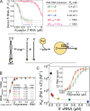
Interaction of influenza polymerase with the promoter. Influenza polymerase binds the promoter 5΄ and 3΄ ends separately and in competition with RNA–RNA intermolecular interactions. (A) Due to partial complementarity, the viral 5΄ and 3΄ RNA ends anneal to form a double-stranded ‘panhandle’ conformation as quantified by a simple FRET assay (v5΄, v3΄/c5΄, c3΄ correspond to the genomic/anti-genomic (complementary) 5΄ and 3΄ RNA ends, respectively; all RNAs are 18 nucleotides each if not indicated otherwise; numbering starts from the 5΄ end or 3΄ end of the 5΄ or 3΄ RNA respectively; see Material and Methods). (B) Using a fluorescence polarization based assay, the interaction of influenza polymerase and each promoter RNA was quantified and revealed each RNA separately to interact with the polymerase in a one-to-one stoichiometry and with high affinity (KD < 0.01 μM). (C) Strengthening intermolecular RNA duplex conformations by increasing the v5΄ RNA concentration impeded binding of polymerase and the v3΄ RNA with an IC50 = 0.3 nM correlating with the RNA duplex formation. Error bars indicate the standard deviation of the mean of at least duplicate experiments.
Figure 2.
Figure 2.
Schematic work flow of the high-throughput compatible RNA synthesis assay. Influenza B polymerase (yellow sphere) activated by 5΄ RNA (nt 1–14) and bound to the 3΄ template RNA (labelled by FAM-Ex-5 at its 5΄ end) is incubated with nucleoside triphosphates (NTPs) and optionally a primer or generally a molecule X whose effect on RNA synthesis is to be monitored. The reactions are quenched by 4 M NaCl final which perturbs polymerase–RNA interactions but permits RNA–RNA interactions. Fluorescence polarization (FP) is recorded after equilibration (development) and directly reads out the ratio of full-length product RNA over labelled template RNA (see Supplementary Figure S3).
Figure 3.
Figure 3.
Primed versus unprimed initiation of RNA synthesis by influenza B polymerase in vitro. (A) ApG, in a concentration-dependent manner (colored triangles), accelerates unprimed RNA synthesis (black triangles) to the level of capped RNA primed RNA synthesis reaction (black circles). (B) Affinities (KM) for the primer(s) in accelerating initiation of RNA synthesis show a preference of the capped RNA over non-capped RNA ending in AG-3΄ over minimally ApG. Error bars indicate the standard deviation of the mean of at least duplicate experiments (see Supplementary Figure S8). (C) Schematic depicting the initiation of RNA synthesis catalysed by influenza B polymerase at the vRNA promotor (v5΄ + v3΄). The 5΄-3΄ base-pairing region (involving v5΄ nt 11–13 and v3΄ nt 10–12) positions the 3΄ template RNA at the polymerase's active site. Formation of the first phosphodiester bond (yielding ApG) is rate-limiting for initiating RNA synthesis and bypassed by supplying an appropriate primer. ApG serves as a minimal primer and accelerates unprimed RNA synthesis by annealing to the U1C2 at the 3΄ template extremity. This ‘primed initiation complex’ (PRIC) conformation is further stabilized by favourable interactions of the polymerase with the primer's RNA-moiety and cap-structure.
Figure 4.
Figure 4.
Schematic model of influenza polymerase initiating RNA synthesis at the vRNA promoter. The 5΄-RNA is firmly anchored to the polymerase (beige sphere), the 3΄ template RNA positioned for terminal initiation at the active site by intermolecular interactions of the distal promoter RNAs (5΄-3΄ base-pairing region; highlighted, green base-pairs). Simultaneous binding of ATP at the post-translocation site and GTP at the active site (pre-translocation) followed by formation of the first phosphodiester bond is bypassed by an appropriate primer ending in AG-3΄. CTP, the next nucleotide to be incorporated and directed by G3 on the vRNA template is the first (and within the five ultimate nucleotides the only) distinction from the cRNA template. To allow for translocation of the template RNA and continued RNA synthesis (elongation), the 5΄-3΄ base-pairing region must dissipate which we propose is coupled to the newly emerging base-pairs between product and template RNA downstream the active site (pink). At the cRNA promoter, terminal fast initiation of RNA synthesis is prevented since with UTP directed to position A3, an A–U base pair is formed, instead of the G–C base-pair in the case of vRNA, which apparently does not compensate for breaking the cRNA promoter's 5΄-3΄ base-pairing region. At the expense of rate, the cRNA promoter re-directs RNA synthesis via internal initiation followed by re-align (see Figure 5).
Figure 5.
Figure 5.
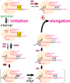
Schematic and hypothetical model of internal initiation of RNA synthesis at the cRNA promoter involving prime-and-re-align. Influenza B polymerase is depicted by a beige sphere with the location of the active site indicated by the arrow. The c5΄ end (pink) is firmly anchored to the polymerase, and helps position the c3΄ 1–18 template RNA (pink) for terminal or internal initiation at the active site. The depicted positioning of the c3΄ template RNA for internal initiation would be stabilized by a 5΄-3΄ base-pairing region involving (at least) c5΄ nucleotides G11, A12 and G13 and c3΄ nucleotides C12, U13, C14, in analogy to the vRNA promoter. The initial ATP would bind opposite of U4 at the active site in the pre-translocation conformation. Template RNA translocation by one step places the initial ATP into the post-translocation state, allowing binding of incoming GTP opposite to C5 at the active site and its incorporation to form pppApG. With the template RNA having translocated by 1 position, strain in the 5΄-3΄ base-pairing region would be generated. Further translocation of the template RNA and pppApG to the post-translocation conformation allows binding of incoming UTP opposite of A6 at the active site and its subsequent incorporation. However, this would build-up additional unfavourable strain in the 5΄-3΄ base-pairing region which would need to be relieved. However, breaking the strong 5΄-3΄ base-pairing region formed by the cRNA promoter (2x GC, 1x AU) is not the most favorable outcome at this stage since the newly formed base-pairs between the product RNA and the template RNA (2x AU, 1x GC) downstream of the active site do not compensate energetically. More favorably, the template RNA would translocate three steps backward while the product RNA stays in place. This alternative position of the template would then promote incorporation of the next nucleotide, through binding of ATP opposite of U4, as this restores three unstrained base-pairs in an alternative 5΄-3΄ base-pairing region. Translocation, binding of GTP opposite of C5 and formation of pppApGpUpApG would again build-up strain in the 5΄-3΄ base-pairing region. However, at this stage more newly formed base-pairs between the product RNA and the template RNA are available (compared to having initiated terminally) which would compensate for breaking the 5΄-3΄ base-pairing region and allow RNA synthesis to progress to elongation.
Figure 6.
Figure 6.
Terminal (vPol) versus internal (cPol) initiation of RNA synthesis. (A) The vRNA promoter (vPol; black; 3 independent rate-NTP-characteristics, circles, upward and downward triangles) or the cRNA promoter (cPol; pink; 2 independent rate-NTP-characteristics, upward and downward triangles) direct the initiation of RNA synthesis by influenza B polymerase with different enzymatic parameters (Table 1). The dependency of RNA synthesis (observed rate constants) on the substrate-concentration (NTP) was fitted according to a simple substrate-inhibition model proposed by HALDANE (19) (see Materials and Methods). The promoter RNA sequences responsible for the altered kcat, KM (NTPs) and KM (primer) are shown in the lower panel. Both the v5΄ and the c5΄ comprise a proximal hook structure (nt 1–10; blue colouring) followed by the distal 5΄-3΄ base-pairing region (nts 11–13/14; green colouring). The v3΄ and c3΄ template RNAs each direct the incorporation of NTPs based on complementarity. (B) Deletion of the ultimate U1 from the template RNA impairs initiation of RNA synthesis from the vRNA promoter but not cRNA promoter, indicating terminal and internal initiation mechanisms, respectively. Shown are five independent progress curves each for initiation at the vRNA and cRNA promoter (black and pink) and the corresponding U1 deletions, respectively (purple and orange). (C) vPol-like kinetics (black) were recovered by a cPol with either (i) A3 of c3΄ substituted by G3, as found in vRNA (c3΄A3G; green); or (ii) the distal cRNA promoter involving the 5΄-3΄ base-pairing region being swapped for the corresponding region of the vRNA promoter (5΄ nt 11–18, 3΄ nt 10–18; c3΄ vbp, purple); or (iii) the proximal cRNA promoter swapped for the corresponding region of the vRNA promoter (5΄ nt 1–10, 3΄ nt 1–9; v3΄ cbp, orange) (see Supplementary Figure S12). Only the c3΄ template RNA in combination with the cRNA promoter's 5΄-3΄ base-pairing region resulted in cPol-like slow progress curves indicating internal initiation (pink). The bi-phasic kinetics of RNA synthesis by some RNA combinations was attributed to strengthened RNA–RNA interactions which kinetically competed with polymerase for single-stranded template RNA (Figure 1, Supplementary Figure S13). In each case, five independent kinetics are shown.
Figure 7.
Figure 7.
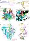
Structure of influenza B polymerase co-crystallized with the vRNA promoter and capped primer. (A) Schematic of RNAs used for crystallization of the putative transcription initiation state. Nucleotides 4–13 of the 13-mer capped RNA primer are in italics as they are not seen in the structure. (B) Overall view of the crystal structure with influenza B polymerase in ribbon representation colored according to domains as indicated. The RNA in space-filling representation with the capped primer (blue), 5΄ end (violet) and 3΄ end (yellow), as in (A). (C) Trajectory of the 3΄ end (template) as observed in the current structure (yellow) and in the previous FluB2 structure (orange) (8). (D) As (C) but in the context of the polymerase (surface and ribbon representation) showing the template passing through the entry tunnel into the active site cavity. (E) Overall view of the promoter bound to the PB1 subunit (cyan ribbons) showing the priming loop (purple) and active site motif C (red). The unobserved 3΄ terminal base (U1, red) is modeled. Based on superposition with the Norwalk virus polymerase primer-template/substrate (CTP) complex (PDB: 3BSO, (30)) the initial ApG of the product (orange), the incoming CTP (black) and the two catalytic divalent ions (green spheres) are modelled. (F) As (E) but only showing the RNA.

Similar articles

Cited by

References

    1. Desselberger U., Nakajima K., Alfino P., Pedersen F.S., Haseltine W.A., Hannoun C., Palese P.. Biochemical evidence that new influenza-virus strains in nature may arise by recombination (reassortment). Proc. Natl. Acad. Sci. U.S.A. 1978; 75:3341–3345. - PMC - PubMed
    1. Hay A.J., Gregory V., Douglas A.R., Lin Y.P.. The evolution of human influenza viruses. Philos. Trans. R. Soc. B. 2001; 356:1861–1870. - PMC - PubMed
    1. Young J.F., Palese P.. Evolution of human influenza—a viruses in nature—recombination contributes to genetic-variation of H1N1 strains. Proc. Natl. Acad. Sci. U.S.A. 1979; 76:6547–6551. - PMC - PubMed
    1. Belshe R.B. The need for quadrivalent vaccine against seasonal influenza. Vaccine. 2010; 28(Suppl. 4):D45–D53. - PubMed
    1. von Itzstein M. The war against influenza: discovery and development of sialidase inhibitors. Nat. Rev. Drug Discov. 2007; 6:967–974. - PubMed

Publication types

MeSH terms