. 2017 Jan 26;15(1):3.
doi: 10.1186/s12915-016-0345-3.

The golgin coiled-coil proteins capture different types of transport carriers via distinct N-terminal motifs

Affiliations

The golgin coiled-coil proteins capture different types of transport carriers via distinct N-terminal motifs

Mie Wong et al. BMC Biol. .

Abstract

Background: The internal organization of cells depends on mechanisms to ensure that transport carriers, such as vesicles, fuse only with the correct destination organelle. Several types of proteins have been proposed to confer specificity to this process, and we have recently shown that a set of coiled-coil proteins on the Golgi, called golgins, are able to capture specific classes of carriers when relocated to an ectopic location.

Results: Mapping of six different golgins reveals that, in each case, a short 20-50 residue region is necessary and sufficient to capture specific carriers. In all six of GMAP-210, golgin-84, TMF, golgin-97, golgin-245, and GCC88, this region is located at the extreme N-terminus of the protein. The vesicle-capturing regions of GMAP-210, golgin-84, and TMF capture intra-Golgi vesicles and share some sequence features, suggesting that they act in a related, if distinct, manner. In the case of GMAP-210, this shared feature is in addition to a previously characterized "amphipathic lipid-packing sensor" motif that can capture highly curved membranes, with the two motifs being apparently involved in capturing distinct types of vesicles. Of the three GRIP domain golgins that capture endosome-to-Golgi carriers, golgin-97 and golgin-245 share a closely related capture motif, whereas that in GCC88 is distinct, suggesting that it works by a different mechanism and raising the possibility that the three golgins capture different classes of endosome-derived carriers that share many cargos but have distinct features for recognition at the Golgi.

Conclusions: For six different golgins, the capture of carriers is mediated by a short region at the N-terminus of the protein. There appear to be at least four different types of motif, consistent with specific golgins capturing specific classes of carrier and implying the existence of distinct receptors present on each of these different carrier classes.

Keywords: Coiled-coil; Endosome-to-Golgi traffic; Golgi; Golgin; Intra-Golgi traffic; Vesicle tethering.


Figures
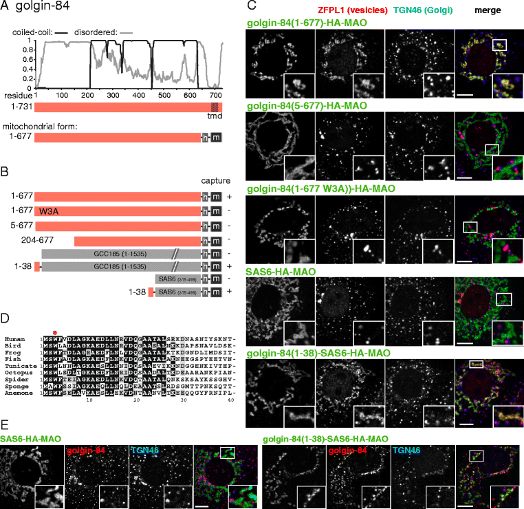
Fig. 1
Fig. 1
Mapping the part of golgin-84 that can capture vesicles. a Schematic diagram of human golgin-84 along with plots for the predicted degree of coiled-coil and disorder along its length. Also shown is the mitochondrial form in which the Golgi-targeting transmembrane domain (TMD) is replaced with a hemagglutinin (HA) tag (h) and the TMD of human monoamine oxidase A (m), a protein of the outer mitochondrial membrane. b Summary of the vesicle capture activity of the indicated variants of mitochondrial golgin-84. Capture at mitochondria was assayed by immunofluorescent staining of the Golgi integral membrane proteins ZFPL1, giantin and GalNAc-T2. Plus sign indicates that capture of all three markers was similar to the wild-type protein, minus sign indicates that no significant capture was observed. c Confocal micrographs of HeLa cells expressing the indicated golgin-84 variants and stained for the HA tag on the golgin-84 chimera as well as for ZFPL1 (in vesicles captured by golgin-84), and for TGN46 (a Golgi protein that is not captured). Cells were treated with nocodazole for 6 h prior to fixation to ensure that mitochondria were close to intra-Golgi transport vesicles. Key constructs from the set shown in (b) are included, with similar results obtained using the markers giantin and GalNAc-T2. Scale bars 10 μm. d Alignment of the N-terminus of human golgin-84 with that from the indicated species. Bird, G. gallus; frog, X. laevis; fish, T. rubripes; tunicate, C. savignyi; octopus, O. bimaculoides; spider, S. mimosarum; sponge, A. queenslandica; anemone, N. vectinis. Well conserved residues are shaded. e As c except that the cells were stained for golgin-84 using an antibody that binds outside of residues 1–38. This indicates that the N-terminus of golgin-84 is sufficient to capture vesicles containing golgin-84. Scale bars 10 μm
Fig. 2
Fig. 2
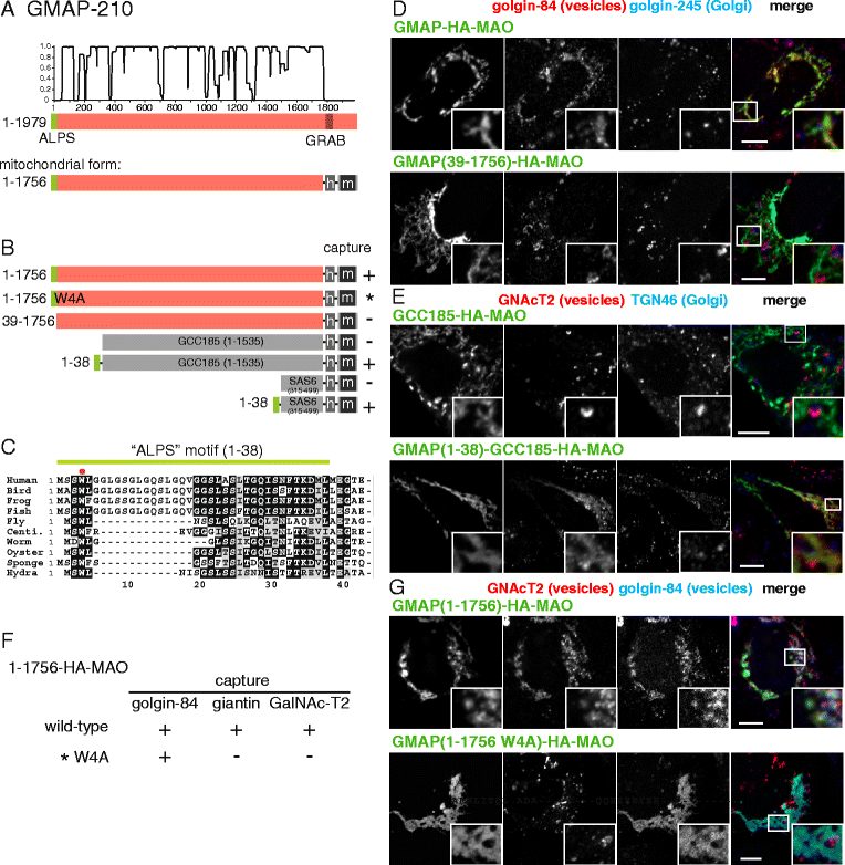
Mapping the vesicle capturing activities of GMAP-210. a Schematic diagram of human GMAP-210 with plots for the predicted degree of coiled-coil and disorder. In the mitochondrial form, the Golgi-targeting transmembrane domain (TMD) is replaced with a hemagglutinin (HA) tag (h) and the TMD of human monoamine oxidase A (m). b Summary of the vesicle capture activity of the indicated variants of mitochondrial GMAP-210. Capture at mitochondria was assayed by immunofluorescent staining of the Golgi integral membrane proteins golgin-84, giantin and GalNAc-T2. Plus sign indicates that capture of all three markers was similar to the wild-type protein, minus sign indicates that no significant capture was observed. c Alignment of the N-terminus of human GMAP-210 with that from the indicated species. Bird, G. gallus; fish D. rerio; frog, X. tropicalis; urchin, S. purpuratus; fly, D. melanogaster; centipede, S. maritima; worm, S. mansoni; oyster, C. gigas; sponge, A. queenslandica; hydra, H. vulgaris. Well conserved residues are shaded, and indicated as a green bar is the amphipathic lipid-packing sensor motif reported previously for the human protein [17], and by a red dot the conserved tryptophan mutated in this study. d, e Confocal micrographs of HeLa cells expressing the indicated GMAP-210 variants and stained for the HA tag on the chimera as well as for the indicated proteins in vesicles captured by GMAP-210, or for Golgi proteins that are not captured. Cells were treated with nocodazole for 6 h prior to fixation to ensure that mitochondria were close to intra-Golgi transport vesicles. Key constructs from the set shown in (b) are included, with similar results obtained using the marker giantin. Scale bars 10 μm. f, g As in (d and e), except comparing the complete GMAP-210 coiled-coil region with a variant in which Trp4 is mutated to alanine. This results in loss of tethering of some vesicle cargo but not golgin-84. Scale bars 10 μm
Fig. 3
Fig. 3
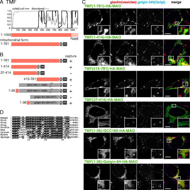
Mapping the vesicle capturing activity of TMF. a Schematic diagram of human TMF with plots for the predicted degree of coiled-coil and disorder. In the mitochondrial form, the Golgi-targeting transmembrane domain (TMD) is replaced with a hemagglutinin (HA) tag (h) and the TMD of human monoamine oxidase A (m). b Summary of the vesicle capture activity of the indicated variants of mitochondrial TMF. Capture at mitochondria was assayed by immunofluorescent staining of the Golgi integral membrane proteins golgin-84, giantin, and GalNAc-T2. Plus sign indicates that capture of all three markers was similar to the wild-type protein, minus sign indicates that no significant capture was observed. c Confocal micrographs of HeLa cells expressing the indicated TMF variants and stained for the HA tag on the golgin-84 chimera as well as for giantin that is in vesicles captured by TMF and for golgin-245, a protein that remains Golgi associated. Cells were treated with nocodazole for 6 h prior to fixation to ensure that mitochondria were close to intra-Golgi transport vesicles. Key constructs from the set shown in (b) are included, with similar results obtained using the markers golgin-84 and GalNAc-T2. Scale bars 10 μm. d Alignment of the N-terminus of human TMF with that from the indicated species. Bird, G. gallus; frog, X. tropicalis; fish D. rerio; urchin, S. purpuratus; bee, A. mellifera; oyster, C. gigas; hydra, H. vulgaris; sponge, A. queenslandica
Fig. 4
Fig. 4
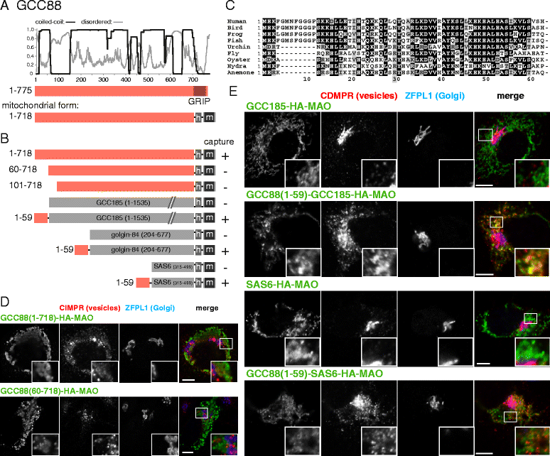
Mapping the part of GCC88 that can capture vesicles. a Schematic diagram of human GCC88 along with plots for the predicted degree of coiled-coil and disorder along its length. Also shown is the mitochondrial form as in Fig. 1a. b Summary of the vesicle capture activity of the indicated truncations and chimeras of mitochondrial GCC88. Capture at mitochondria was assayed by immunofluorescent staining of the integral membrane proteins CD-MPR, CI-MPR and Vti1a. A plus sign indicates that capture of all three markers was similar to the wild-type protein, a minus sign indicates that no significant capture was observed. c Alignment of the N-terminus of human GCC88 with that from the indicated species. Bird, G. gallus; frog, X. tropicalis; fish D. rerio; urchin, S. purpuratus; fly, D. melanogaster; oyster, C. gigas; hydra, H. vulgaris; anemone, N. vectinis. d, e Confocal micrographs of HeLa cells expressing the indicated GCC88 variants and stained for the hemagglutinin tag on the GCC88 chimera as well as for both CD-MPR (in vesicles captured by GCC88) and ZFPL1 (a cis-Golgi protein that is not captured). Key constructs from the set shown in (b) are included, with similar results also obtained using the vesicle markers CI-MPR and Vti1a. Scale bars 10 μm
Fig. 5
Fig. 5
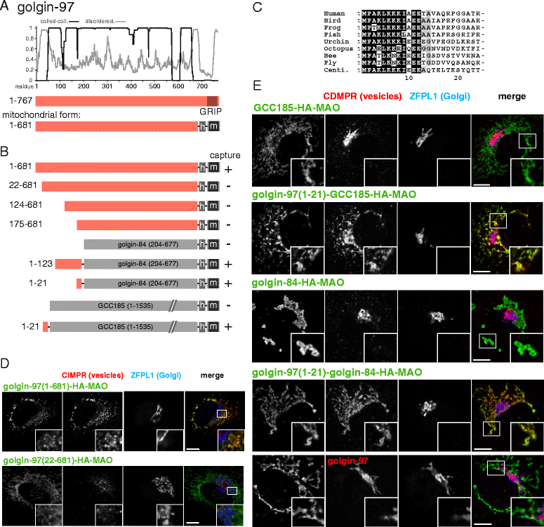
Mapping the part of golgin-97 that captures vesicles. a Schematic diagram of human golgin-97 along with plots for the predicted degree of coiled-coil and disorder along its length. Also shown is the mitochondrial form in which the GRIP domain has been replaced with a hemagglutinin (HA) tag and the human monoamine oxidase A transmembrane domain. b Summary of the vesicle capture activity of the indicated truncations and chimeras of golgin-97. Capture at mitochondria was assayed by immunofluorescent staining of the integral membrane proteins CD-MPR, CI-MPR and Vti1a. A plus sign indicates that capture of all three markers was similar to the wild-type protein, a minus sign indicates that no significant capture was observed. c Alignment of the N-terminus of human golgin-97 with that from the indicated species. Bird, G. gallus; frog, X. tropicalis; fish D. rerio; urchin, S. purpuratus; octopus, O. bimaculoides; bee, A. mellifera; fly, D. melanogaster; centipede, S. maritime. d, e Confocal micrographs of HeLa cells expressing the indicated golgin-97 variants and stained for the HA tag on the golgin-97 chimera as well as for CI-MPR or CD-MPR (in vesicles captured by golgin-97) along with ZFPL1 (a cis-Golgi protein that is not captured). Key constructs from the set shown in (b) are included, with similar results obtained using the vesicle markers CD-MPR, CI-MPR or Vti1a. Scale bars 10 μm
Fig. 6
Fig. 6
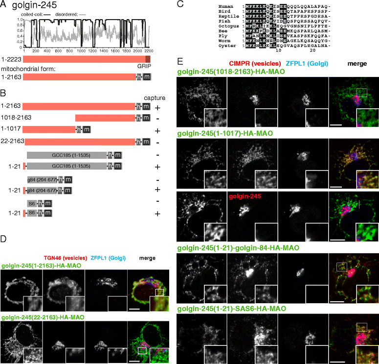
Mapping the part of golgin-245 that captures vesicles. a Schematic diagram of human golgin-245 along with plots for the predicted degree of coiled-coil and disorder along its length. Also shown is the mitochondrial form as in Fig. 1a. b Summary of the vesicle capture activity of the indicated truncations and chimeras of golgin-245. Capture at mitochondria was assayed by immunofluorescent staining of the integral membrane proteins CD-MPR, CI-MPR, TGN46 or Vti1a. A plus sign indicates that capture of all four markers was similar to the wild-type protein, a minus sign indicates that no significant capture was observed. c Alignment of the N-terminus of human golgin-245 with that from the indicated species. Bird, G. gallus; reptile, A. mississippiensis; fish D. rerio; octopus, O. bimaculoides; bee, A. mellifera; fly, D. melanogaster; worm, C. elegans; oyster, C. gigas. d, e Confocal micrographs of HeLa cells expressing the indicated golgin-245 variants and stained for the hemagglutinin tag on the golgin-245 chimera as well as for either TGN46 or CI-MPR (in vesicles captured by golgin-245) as well as for ZFPL1 (a cis-Golgi protein that is not captured). Key constructs from the set shown in (b) are included, with similar results obtained using the vesicle markers CI-MPR, TGN46, CD-MPR, and Vti1a. Scale bars 10 μm
Fig. 7
Fig. 7
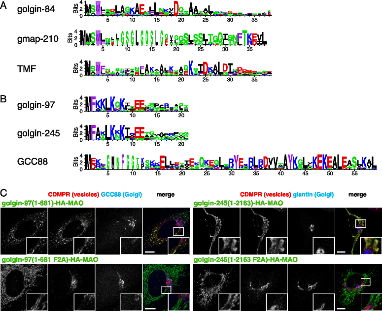
Patterns of conserved residues in the golgin vesicle capturing motifs. a Logo plots of the N-terminal regions of the three golgins that capture intra-Golgi transport vesicles. The height of the residue indicates how well it is conserved in orthologs from diverse metazoans, expressed as information content (bits). Residues are colored by their properties as follows: red, basic; blue, acidic; green, polar but uncharged; black, aliphatic; and purple, aromatic. b Logo plots of the N-terminal regions of the three GRIP domain golgins that capture endosome - to - Golgi transport vesicles. Residues as in (a). c Confocal micrographs of HeLa cells expressing the full - length mitochondrial forms of golgin-97 or golgin-245 or variants in which the conserved Phe2 residue is mutated to alanine, and stained for the hemagglutinin tag on the chimera. In both cases, this mutation results in loss of tethering of vesicles as indicated by CD-MPR. Scale bars 10 μm
Fig. 8
Fig. 8
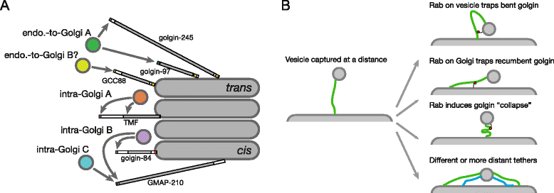
Summary of vesicle capture by golgins. a Summary of the vesicle capture activities of the six indicated golgins. Golgin-97 and golgin-245 seem likely to capture the same type of vesicle, whilst the region of GCC88 that has capture activity has a very different sequence and therefore either captures the same vesicle by a different mechanism, or a different type of vesicle with overlapping cargo. The remaining three golgins capture intra-Golgi vesicles, and it appears that those bound to TMF have a similar but not identical set of cargo to those captured by golgin-84. There appear to be two different types of vesicle captured by GMAP-210. The precise number of classes of intra-Golgi vesicle, however, remains unclear. In all cases, the vesicle is captured by an N-terminal motif, with TMF also having a capture activity in its coiled-coil region. b Some of the possible models for how a vesicle captured at the N-terminus of a golgin could move closer to the Golgi membrane so as to allow vesicle fusion. The Rab binding sites on the golgin could be used in various ways to hold the golgin N-terminus closer to the membrane, or to induce a conformational change in the golgin. Alternatively, the vesicle could be captured by a further, more distant, golgin, or by different, shorter golgins (blue), to hold the vesicle closer to the target membrane. Of course, all these models are speculative, and other models could also apply

Similar articles

Cited by

References

    1. Yu I-M, Hughson FM. Tethering factors as organizers of intracellular vesicular traffic. Annu Rev Cell Dev Biol. 2010;26:137–56. doi: 10.1146/annurev.cellbio.042308.113327. - DOI - PubMed
    1. Hong W, Lev S. Tethering the assembly of SNARE complexes. Trends Cell Biol. 2014;24:35–43. doi: 10.1016/j.tcb.2013.09.006. - DOI - PubMed
    1. Söllner T, Whiteheart SW, Brunner M, Erdjument-Bromage H, Geromanos S, Tempst P, et al. SNAP receptors implicated in vesicle targeting and fusion. Nature. 1993;362:318–24. doi: 10.1038/362318a0. - DOI - PubMed
    1. Stenmark H. Rab GTPases as coordinators of vesicle traffic. Nat Rev Mol Cell Biol. 2009;10:513–25. doi: 10.1038/nrm2728. - DOI - PubMed
    1. Barr FA. Review series: Rab GTPases and membrane identity: causal or inconsequential? J Cell Biol. 2013;202:191–9. doi: 10.1083/jcb.201306010. - DOI - PMC - PubMed

Publication types

LinkOut - more resources