. 2017 Jan 18:7:40756.
doi: 10.1038/srep40756.

A comprehensive platform for the analysis of ubiquitin-like protein modifications using in vivo biotinylation

Affiliations

A comprehensive platform for the analysis of ubiquitin-like protein modifications using in vivo biotinylation

Lucia Pirone et al. Sci Rep. .

Abstract

Post-translational modification by ubiquitin and ubiquitin-like proteins (UbLs) is fundamental for maintaining protein homeostasis. Efficient isolation of UbL conjugates is hampered by multiple factors, including cost and specificity of reagents, removal of UbLs by proteases, distinguishing UbL conjugates from interactors, and low quantities of modified substrates. Here we describe bioUbLs, a comprehensive set of tools for studying modifications in Drosophila and mammals, based on multicistronic expression and in vivo biotinylation using the E. coli biotin protein ligase BirA. While the bioUbLs allow rapid validation of UbL conjugation for exogenous or endogenous proteins, the single vector approach can facilitate biotinylation of most proteins of interest. Purification under denaturing conditions inactivates deconjugating enzymes and stringent washes remove UbL interactors and non-specific background. We demonstrate the utility of the method in Drosophila cells and transgenic flies, identifying an extensive set of putative SUMOylated proteins in both cases. For mammalian cells, we show conjugation and localization for many different UbLs, with the identification of novel potential substrates for UFM1. Ease of use and the flexibility to modify existing vectors will make the bioUbL system a powerful complement to existing strategies for studying this important mode of protein regulation.


Figures

Figure 1
Figure 1. Isolation and identification of bioSmt3 conjugates in Drosophila cultured cells.
(A) Schematic representation of the bioUbL strategy. See text for details. (B) Up: schematic representation of the bioUbL vectors for Drosophila cells. Left: the blot shows the enrichment in bioSmt3 conjugates in the elution panel using anti-biotin antibodies (lane 4; bracket). Arrowhead indicates free bioSmt3 in lane 4. Right: top twenty bioSmt3-modified proteins based on raw intensity. (C,D) bioSmt3 (pAc510x-bioSmt3-Lwr) visualized with fluorescently labeled streptavidin (D) localizes in a similar way than the endogenous Smt3 in S2R+ cells (C). Nuclei were marked with DAPI (blue). Green and red channels are shown independently in black and white (C’, D’). (E) GO analysis for biological process, cellular component and molecular function of the selected bioSmt3-conjugated protein set (n = 1054).
Figure 2
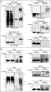
Figure 2. Validation of bioSmt3-modified proteins.
(A) Anti-Flag or anti-GFP Western blot of pulldowns performed in S2R+ cells transfected respectively with GFP-kuk, GFP-Ote, GFP-bocks or Flag-Cherry-tsr, together with pAc5-GAL4 and pAc510x-bioSmt3-Lwr (bioSmt3) (+) or Ac510x-FC-Lwr (−) as control. In the elution panels, arrowheads indicate the modified forms of each protein. Tubulin was used as a loading control. Molecular weight markers are shown to the left. (B) Western blot of pulldowns performed in S2R+ cells. Specific antibodies against endogenous proteins were used: Ultraspiracle (Usp), the Trithorax group protein Osa, Eukaryotic initiation factor 4E (eIF-4E), Failed axon connections (Fax) and Lamin (Lam). In elutions, arrowheads indicate the modified forms of the respective proteins. Molecular weight markers are shown to the left.
Figure 3
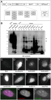
Figure 3. In vivo isolation and identification of bioSmt3 conjugates.
(A) Up, schematic representation of the bioSmt3 vector used for Drosophila transgenesis. Down, the blot shows the enrichment of bioSmt3 conjugates in the elution panel using anti-biotin antibodies (lane 4, bracket; strain used: hs-Gal4; UAS-bioSmt3). Asterisks indicate the three known endogenously biotinylated proteins in Drosophila. (B) BioSmt3 can substitute for endogenous Smt3. Left, larvae silenced for smt3 in the PG, arrested in development at the end of L3 larval stage. Right, the developmental arrest is rescued by the expression of bioSmt3 in the same genetic background. (CF). Confocal pictures of cells of the salivary glands of larvae expressing bioSmt3 (E,F) or GFP (C,D). bioSmt3 is detected by fluorescently-labeled streptavidin. Heat treatment (37 °C) increased the bioSmt3-positive bodies. (G) GO analysis for biological process, cellular component and molecular function of the selected 140 bioSmt3-conjugated proteins.
Figure 4
Figure 4. Isolation and localization of bioUbL-conjugates in mammalian cells.
(A) Schematic representation of the bioUbL plasmid collection. (B) BioUbL-conjugates revealed by anti-biotin Western blot. Pulldowns were performed in parallel using HEK 293FT cells expressing the different bioUbLs. Conjugates are indicated with a bracket. Asterisks indicate endogenously biotinylated proteins. Arrowheads indicate free bioUbLs. Molecular weight markers are shown to the left. (CJ) Cellular distribution of different bioUbLs. U2OS cells transfected with plasmids expressing the indicated bioUbLs (DJ) or BirA alone as a control (C). Conjugates are visualized using fluorescently-labeled streptavidin. (K) White nuclear bodies (white arrowheads indicate two examples) reflect the colocalization of bioSUMO1 (purple) with YFP-PML (green). Yellow arrowheads indicate the localization of bioSUMO1 in the nuclear membrane. (K’) and (K”) show independently the green and purple channels in black and white, respectively.
Figure 5
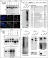
Figure 5. Isolation and identification of bioSUMO3-conjugates in mammalian cells.
(A) Western blot of pulldowns from HEK 293FT cells showing that the transcription factor SALL1 fused to YFP was SUMOylated in presence (+) of bioSUMO1 (lane 4) or bioSUMO3 (lane 5; bioSUMO1-BirA or bioSUMO3-BirA, respectively). Black arrowhead indicates the modified SALL1-YFP in the elution panel (lanes 4, 5), which is shifted in comparison to the non-modified SALL1-YFP in the input panel (white arrowhead, lanes 1–3). Molecular weight markers are shown to the left. (B,C) Partial colocalization between SALL1-YFP (green) and bioSUMO1 (purple) (bioSUMO1-BirA-UBC9) in U2OS cells (B) or with endogenous SUMO2/3 (C, purple). White arrowheads indicate colocalization. Nuclei were stained with DAPI (blue). (B’,C”) Green and purple channels are shown independently in black and white. (D) SUMOylation of PML by bioSUMO3 (bioSUMO3-BirA-GP) increases after ATO treatment. bioSUMO3-modified PML (black arrowheads) can be detected by anti-HA Western blot in the input (upper panel, lanes 1–4) and the elution (lower panel, lanes 1–4; NeutrAvidin pulldown), while the level of the non-modified form of PML is reduced (input panel, lanes 1–6; white arrowhead). Note that modification of PML by endogenous SUMO is also visible in the input panel after ATO treatment (lanes 5 and 6, grey arrowheads). Control indicates cells transfected with BirA-GP. Molecular weight markers are shown to the left. (E) Up: schematic representation of the bioSUMO3 vector for mammalian cells. Left: the Western blot shows the enrichment in bioSUMO3 conjugates in the elution panel using anti-biotin antibodies (lane 3, bracket). Arrowhead indicates free bioSUMO3. Right: bioSUMO3-modified proteins identified by nLC MS/MS on Orbitrap. (F) Validation of bioSUMO3-modified proteins. Specific antibodies against endogenous proteins were used: Ub, SIRT1, RANGAP1, PML and PARP. GAPDH is shown as a control in the input panel. In the elution panels, arrowheads indicate the modified forms of the respective proteins and the bracket indicates the ubiquitinated proteins (lanes 3). Molecular weight markers are shown to the left.
Figure 6
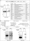
Figure 6. Isolation and identification of bioUFM1-conjugates in mammalian cells.
(A) Up: schematic representation of the bioUFM1 vector for mammalian cells. Below, left: Validation of HA-DDRGK. Right, top twenty bioUFM1-modified proteins. (B) GO analysis for biological process, cellular component and molecular function of the selected bioUFM1-conjugated protein set (n = 82). (C) Validation of bioUFM1-modified proteins CYB5R3 and PSMB5 fused to HA tag. In the elution panels (lanes 3 and 4), black arrowheads indicate the modified forms of the respective proteins. Residual non-specific interactions of non-modified forms are indicated by white arrowheads. Molecular weight markers are shown to the left.

Similar articles

Cited by

References

    1. Kleiger G. & Mayor T. Perilous journey: a tour of the ubiquitin-proteasome system. Trends in cell biology 24, 352–359, doi: 10.1016/j.tcb.2013.12.003 (2014). - DOI - PMC - PubMed
    1. Clague M. J. et al.. Deubiquitylases from genes to organism. Physiological reviews 93, 1289–1315, doi: 10.1152/physrev.00002.2013 (2013). - DOI - PubMed
    1. Komander D. & Rape M. The ubiquitin code. Annual review of biochemistry 81, 203–229, doi: 10.1146/annurev-biochem-060310-170328 (2012). - DOI - PubMed
    1. Hochstrasser M. Origin and function of ubiquitin-like proteins. Nature 458, 422–429, doi: 10.1038/nature07958 (2009). - DOI - PMC - PubMed
    1. van der Veen A. G. & Ploegh H. L. Ubiquitin-like proteins. Annual review of biochemistry 81, 323–357, doi: 10.1146/annurev-biochem-093010-153308 (2012). - DOI - PubMed

Publication types