Review
. 2017 Mar;18(3):175-186.
doi: 10.1038/nrm.2016.171. Epub 2017 Jan 18.

Telomeres in cancer: tumour suppression and genome instability

Affiliations
Review

Telomeres in cancer: tumour suppression and genome instability

John Maciejowski et al. Nat Rev Mol Cell Biol. 2017 Mar.

Erratum in

Abstract

The shortening of human telomeres has two opposing effects during cancer development. On the one hand, telomere shortening can exert a tumour-suppressive effect through the proliferation arrest induced by activating the kinases ATM and ATR at unprotected chromosome ends. On the other hand, loss of telomere protection can lead to telomere crisis, which is a state of extensive genome instability that can promote cancer progression. Recent data, reviewed here, provide new evidence for the telomere tumour suppressor pathway and has revealed that telomere crisis can induce numerous cancer-relevant changes, including chromothripsis, kataegis and tetraploidization.


Conflict of interest statement

Competing interests statement

The authors declare no competing interests.

Figures

Figure 1
Figure 1. Composition and structure of the human telomere system
Human telomeres comprise three components: telomeric DNA, the shelterin complex and the telomerase complex. Telomeric DNA consists of a long array of double-stranded TTAGGG repeats that culminates in a 50–300 nucleotide (nt) single-stranded 3′ overhang. This 3′ overhang invades double-stranded telomeric repeats to form a t-loop structure that is crucial for telomere function. Telomeric DNA protects chromosome ends through its association with the six-subunit shelterin complex. The length of telomeric repeats can be maintained by telomerase, which is composed of telomerase reverse transcriptase (TERT), telomerase RNA template component (TERC) and several accessory proteins (blue). TERT synthesizes telomeric DNA de novo using TERC as a template, whereas the accessory factors contribute to the biogenesis and nuclear trafficking of telomerase. DKC, dyskerin; NHP2, non-histone protein 2; NOP10, nucleolar protein 10; POT1, protection of telomeres 1; RAP1, repressor/activator protein 1; TCAB1, telomerase Cajal body protein 1; TIN2, TRF1-interacting nuclear factor 2; TRF, telomeric repeat-binding factor.
Figure 2
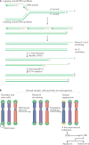
Figure 2. Telomere shortening as a barrier to tumorigenesis
a | The molecular basis of telomere shortening. Incomplete DNA synthesis at the end of the lagging strand (at the site of the terminal RNA primer) leaves a short 3′ overhang. Additional loss of telomeric DNA occurs through the processing of the leading-strand ends of telomeres to regenerate the 3′ overhang, which is necessary for t-loop formation and the structural integrity of the telomere. This process is carried out by the nuclease Apollo, which is bound to telomeric repeat-binding factor 2 (TRF2). Both the leading end and the lagging end of telomeres are further resected by exonuclease 1 (EXO1) to generate transient long overhangs. The CST (CTC1–STN1–TEN1) complex then binds to shelterin and mediates fill-in synthesis of the cytosine-rich strand (C-strand) at both ends. b | During development, telomerase is switched off through telomerase reverse transcriptase (TERT) silencing. As a result, telomeres experience the gradual attrition described in part a. After numerous population doublings, a few telomeres become too short (yellow) and lose their protective function. As a result, the kinases ATM and ATR are activated at the unprotected chromosome ends and this DNA damage response (DDR) signalling induces replicative arrest and senescence or apoptosis. This process limits the proliferative capacity of incipient cancer cells, thus functioning as a tumour suppressor pathway. Cells lacking p53 and RB function can avoid this replicative arrest.
Figure 3
Figure 3. Telomere crisis
Loss of the RB and p53 tumour suppressor pathways disables the ability of cells to respond with cell cycle arrest to ATR and ATM signalling. As the cells continue to divide, their telomeres continue to shorten. Once many telomeres become too short to function, the unprotected chromosome ends generate end-to-end fusions and dicentric chromosomes, leading to many forms of genome instability. Ultimately, telomerase reactivation provides a route out of telomere crisis by healing critically shortened telomeres and improving genomic stability, thereby increasing cell viability. The resulting tumour will have active telomerase and a heavily rearranged genome.
Figure 4
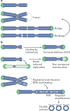
Figure 4. BFB cycles and chromosomal rearrangements during telomere crisis
a | Breakage–fusion–bridge (BFB) cycles can occur when telomere fusion generates a dicentric chromosome. During anaphase, the mitotic spindle pulls this dicentric chromosome towards opposite spindle poles, thereby generating the widely observed anaphase bridges. During cell division, the dicentric chromosome undergoes breakage and the broken ends fuse again, giving rise to another dicentric chromosome. b | BFB cycles can be interrupted by telomerase-mediated telomere healing. If this process occurs following breakage, it can result in the formation of a terminal chromosome deletion and loss of heterozygosity (LOH). Alternatively, broken chromosomes can be repaired by break-induced replication, yielding a non-reciprocal translocation. Repeated cycles of BFB that occur between sister chromatids can result in regional amplification and the generation of a homogeneously staining region (HSR) following chromosome staining. This HSR consists of multiple amplicons of inverted repeats. Excision of the amplified sequences out of the chromosome will generate circular double-minute chromosomes.
Figure 5
Figure 5. Chromothripsis and kataegis in telomere crisis
a | Dicentric chromosomes formed by telomere fusion rarely, if ever, break during mitosis and instead form chromatin bridges. b | Daughter nuclei connected by chromatin bridges undergo frequent nuclear envelope (NE) rupture in interphase (NERDI), resulting in the accumulation of 3′ repair exonuclease 1 (TREX1) on bridge DNA. TREX1-mediated resection of DNA leads to the formation of single-stranded DNA (ssDNA), which is bound by replication protein A (RPA), and bridge resolution. Bridge fragments are internalized into the nucleus where they remain associated with RPA for approximately 24 hours. c | Part of the dicentric chromosome that is present in the chromatin bridge undergoes extensive fragmentation followed by haphazard repair, which yields a chromothriptic chromosome in which many original chromosome fragments are lost and retained fragments are present in seemingly random order and orientation. Chromothriptic breakpoints are frequently associated with kataegis mutation clusters.
Figure 6
Figure 6. Tetraploidization during telomere crisis
Telomere crisis can lead to persistent DNA damage signalling when repair fails to join all the unprotected ends and dysfunctional telomeres persist. The persistent ATM and ATR signalling and activation of their downstream effector kinases checkpoint kinase 2 (CHK2) and CHK1, respectively, results in prolonged inhibition of cyclin-dependent kinase 1 (CDK1)–cyclin B (CYCB), thus blocking entry into mitosis. Eventually, cells bypass mitosis, enter a G1-like state and then undergo a second S phase. The resulting tetraploid cells have diplochromosomes in the first mitosis following endoreduplication. Subsequently, the cells undergo frequent chromosome losses, leading to the hyper-triploid cells that are frequently observed in cancer. The example karyotype shown is from Capan-2, a hyper-triploid pancreatic cancer cell line (http://www.pawefish.path.cam.ac.uk/PancCellLineDescriptions/Capan-2.html), courtesy of Vorapan Sirivatanauksorn and Paul Edwards.
None

Similar articles

Cited by

References

    1. Artandi SE, et al. Telomere dysfunction promotes non-reciprocal translocations and epithelial cancers in mice. Nature. 2000;406:641–645. Demonstrates that telomere attrition in p53-mutant mice promotes epithelial cancers through the formation of chromosome rearrangements. - PubMed
    1. Artandi SE, DePinho RA. Telomeres and telomerase in cancer. Carcinogenesis. 2010;31:9–18. - PMC - PubMed
    1. Maciejowski J, Li Y, Bosco N, Campbell PJ, de Lange T. Chromothripsis and kataegis induced by telomere crisis. Cell. 2015;163:1641–1654. Shows that dicentric chromosomes formed during telomere crisis persist through mitosis, are fragmented by TREX1 in G1 phase and give rise to chromothripsis and kataegis. - PMC - PubMed
    1. Hanahan D, Weinberg RA. Hallmarks of cancer: the next generation. Cell. 2011;144:646–674. - PubMed
    1. Yates LR, Campbell PJ. Evolution of the cancer genome. Nat. Rev. Genet. 2012;13:795–806. - PMC - PubMed

Publication types