. 2017 Jan 7;14(1):6.
doi: 10.1186/s12974-016-0782-5.

Enhanced neuroinflammation mediated by DNA methylation of the glucocorticoid receptor triggers cognitive dysfunction after sevoflurane anesthesia in adult rats subjected to maternal separation during the neonatal period

Affiliations

Enhanced neuroinflammation mediated by DNA methylation of the glucocorticoid receptor triggers cognitive dysfunction after sevoflurane anesthesia in adult rats subjected to maternal separation during the neonatal period

Yangzi Zhu et al. J Neuroinflammation. .

Abstract

Background: Mounting evidence indicates that children who experience abuse and neglect are prone to chronic diseases and premature mortality later in life. One mechanistic hypothesis for this phenomenon is that early life adversity alters the expression or functioning of the glucocorticoid receptor (GR) throughout the course of life and thereby increases sensitivity to inflammatory stimulation. An exaggerated pro-inflammatory response is generally considered to be a key cause of postoperative cognitive dysfunction (POCD). The aim of this study was to examine the effects of early life adversity on cognitive function and neuroinflammation after sevoflurane anesthesia in adult rats and to determine whether such effects are associated with the epigenetic regulation of GR.

Methods: Wistar rat pups were repeatedly subjected to infant maternal separation (early life stress) from postnatal days 2-21. In adulthood, their behavior and the signaling of hippocampal pro-inflammatory factors and nuclear factor-kappa B (NF-κB) after sevoflurane anesthesia were evaluated. We also examined the effects of maternal separation (MS) on the expression of GR and the DNA methylation status of the promoter region of exon 17 of GR and whether behavioral changes and neuroinflammation after anesthesia were reversible when the expression of GR was increased by altering DNA methylation.

Results: MS induced cognitive decline after sevoflurane inhalation in the Morris water maze and context fear conditioning tests and enhanced the release of cytokines and the activation of astrocyte intracellular NF-κB signaling induced by sevoflurane in the hippocampus of adult rats. Blocking NF-κB signaling by pyrrolidine dithiocarbamate (PDTC) inhibited the release of cytokines. MS also reduced the expression of GR and upregulated the methylation levels of the promoter region of GR exon 17, and such effects were reversed by treatment with the histone deacetylase inhibitor trichostatin A (TSA) in adult rats. Moreover, TSA treatment in adult MS rats inhibited the overactivation of astrocyte intracellular NF-κB signaling and the release of cytokines and alleviated cognitive dysfunction after sevoflurane anesthesia.

Conclusions: Early life stress induces cognitive dysfunction after sevoflurane anesthesia, perhaps due to the aberrant methylation of the GR gene promoter, which reduces the expression of the GR gene and facilitates exaggerated inflammatory responses.

Keywords: Cognitive dysfunction; DNA methylation; Glucocorticoid receptor; Neuroinflammation; Pro-inflammatory factors; Sevoflurane.


Figures
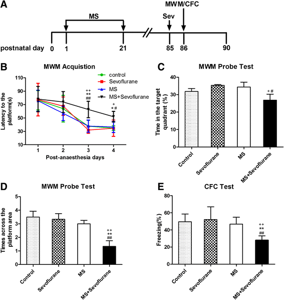
Fig. 1
Fig. 1
The effect of neonatal maternal separation on cognitive function after sevoflurane anesthesia in adult rats. a Schematic illustration of the experimental timeline. MS maternal separation, Sev anesthesia with 3% sevoflurane for 2 h, MWM Morris water maze, CFC context fear conditioning. b Anesthesia with 3% sevoflurane for 2 h in MS rats increased the escape latency in the acquisition phase of the MWM test on the third and fourth days after sevoflurane anesthesia. There was no statistically significant interaction of time and group between groups. Analysis was performed with repeated-measures two-way ANOVA. c The time in the target quadrant in the MS + sevoflurane group was less than those in the sevoflurane group and MS group by the MWM probe test. d The number of crossings of the platform area was lower in the MS + sevoflurane group than in the other three groups in the MWM probe test. e MS rats subjected to sevoflurane anesthesia exhibited reduced freezing to context when compared with normal rats that received sevoflurane anesthesia and MS rats without anesthesia. * P < 0.05, ** P < 0.01 versus control; + P < 0.05, ++ P < 0.01 versus sevoflurane; # P < 0.05, ## P < 0.01 versus MS. Error bars represent the means ± SD (n = 10). Statistical analyses were performed using a one-way ANOVA followed by Student–Newman–Keuls post hoc test
Fig. 2
Fig. 2
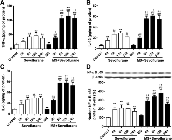
MS enhanced the release of cytokines and the expression of nuclear NF-κB p65 induced by sevoflurane in the hippocampus. a Sevoflurane anesthesia for 2 h increased the levels of TNF-α at 0, 6, 12, and 24 h after anesthesia in both control and MS rats. The levels of TNF-α in MS rats were higher than that in control rats at each time point after anesthesia. The changes in the levels of IL-1β (b), IL-6 (c), and nuclear NF-κB p65 (d) in the hippocampus were similar to those of TNF-α. * P < 0.05, ** P < 0.01 versus control; ## P < 0.01 versus MS; + P < 0.05, ++ P < 0.01 MS + sevoflurane versus sevoflurane (at the same time point). Error bars represent the means ± SD (n = 6). Statistical analyses were performed by one-way ANOVA followed by Student–Newman–Keuls post hoc test
Fig. 3
Fig. 3
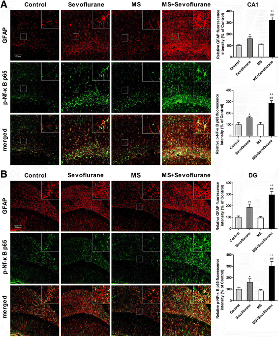
MS enhanced the activation of astrocytes and the astroglial NF-κB signaling pathway induced by 3% sevoflurane anesthesia for 2 h in the hippocampus. a Sevoflurane anesthesia increased the fluorescence intensity of glial fibrillary acidic protein (GFAP) and phosphorylated nuclear factor-kappa B (p-NF-κB p65) in the CA1 area of the hippocampus at 12 h after anesthesia in both the sevoflurane group and MS + sevoflurane group, and the fluorescence intensity of GFAP and p-NF-κB p65 in the MS + sevoflurane group was obviously higher than that in the sevoflurane group. Double immunofluorescence staining showed that p-NF-κB p65 (green) mainly co-localized (merged) with GFAP-positive reactive astrocytes (red) in the CA1 area of the hippocampus. b The changes in the fluorescence intensity of GFAP and p-NF-κB p65 in the DG area of the hippocampus were similar to those in the CA1 area of the hippocampus. * P < 0.05, ** P < 0.01 versus control; ## P < 0.01 versus MS; ++ P < 0.01 MS + sevoflurane versus sevoflurane. Error bars represent the means ± SD (n = 6). Statistical analyses were performed by one-way ANOVA followed by Student–Newman–Keuls post hoc test. CA1 cornu ammonis 1, DG dentate gyrus; bar = 100 μm
Fig. 4
Fig. 4
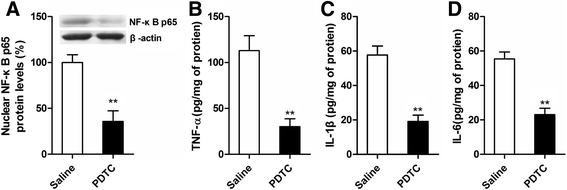
PDTC inhibits the enhancement effect of maternal separation on NF-κB activation and the release of pro-inflammatory cytokines induced by sevoflurane in MS rats. PDTC or vehicle (saline) was given to rats intraperitoneally 30 min before exposure to sevoflurane. a PDTC pretreatment decreased the nuclear NF-κB p65 protein levels in the hippocampus 12 h after sevoflurane anesthesia. b PDTC pretreatment decreased TNF-α levels in the hippocampus 12 h after sevoflurane anesthesia. c PDTC pretreatment decreased IL-1β levels in the hippocampus 12 h after sevoflurane anesthesia. d PDTC pretreatment decreased IL-6 levels in the hippocampus 12 h after sevoflurane anesthesia. ** P < 0.01 versus saline. Error bars represent the means ± SD (n = 6). Statistical analyses were performed with Student’s two-sample t test
Fig. 5
Fig. 5
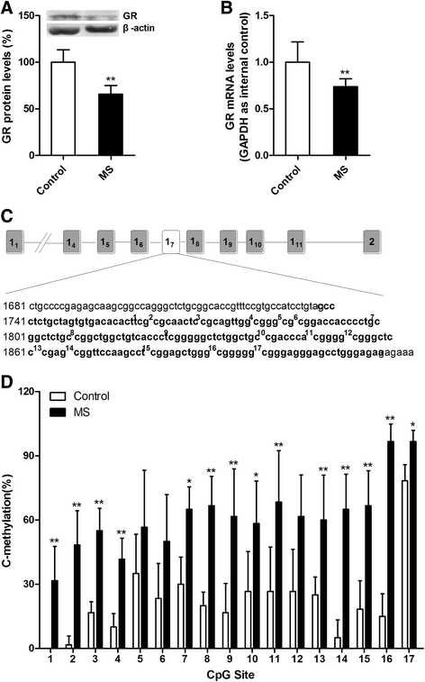
Maternal separation attenuated the expression of GR and increased the percentage of methylated clones within the exon 17 glucocorticoid receptor promoter region from the hippocampal tissues of adult rats. a GR protein levels in the hippocampal tissues of MS rats were lower compared with those of control rats. b qRT-PCR (GAPDH as an internal control) showed that MS decreased the mRNA levels of GR in the hippocampus compared with the control conditions. c The complete sequence map of exon 17 of the GR promoter region (bold), including the 17 CpG dinucleotides (superscript numbers). d The frequency of methylation observed at each CpG site in the exon 17 GR promoter region showed that MS rats have a higher percentage of methylated clones than control rats. * P < 0.05, ** P < 0.01 versus control. Error bars represent the means ± SD (n = 6). Statistical analyses were performed with Student’s two-sample t test
Fig. 6
Fig. 6
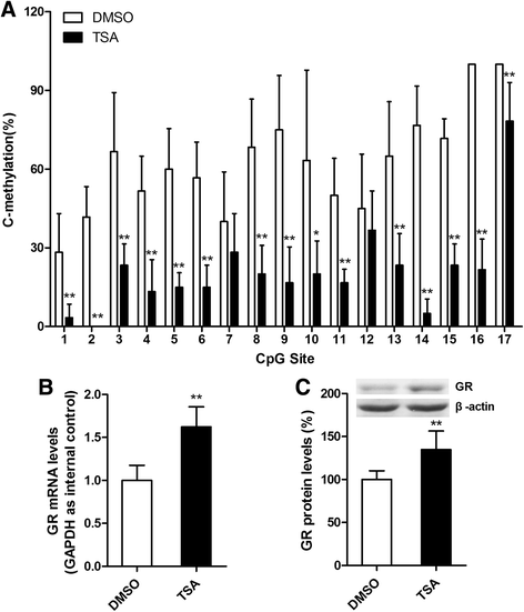
TSA decreased the percentage of methylated clones within the exon 17 GR promoter region and increased the expression of GR in the hippocampal tissues in adult MS rats. A volume of 2 μl of TSA (100 ng/ml in DMSO) was infused into the lateral ventricle of MS rats for seven consecutive days. a The frequency of methylation observed at each CpG site in the exon 17 GR promoter region showed that TSA decreased the percentage of methylated clones relative to the DMSO treatment. b qRT-PCR (GAPDH as internal control) showed that TSA increased the mRNA levels of GR in MS rats compared with the DMSO treatment. c TSA treatment increased the nuclear GR protein levels in the hippocampus of MS rats relative to the DMSO treatment. * P < 0.05, ** P < 0.01 versus DMSO. Error bars represent the means ± SD (n = 6). Statistical analyses were performed with Student’s two-sample t test
Fig. 7
Fig. 7
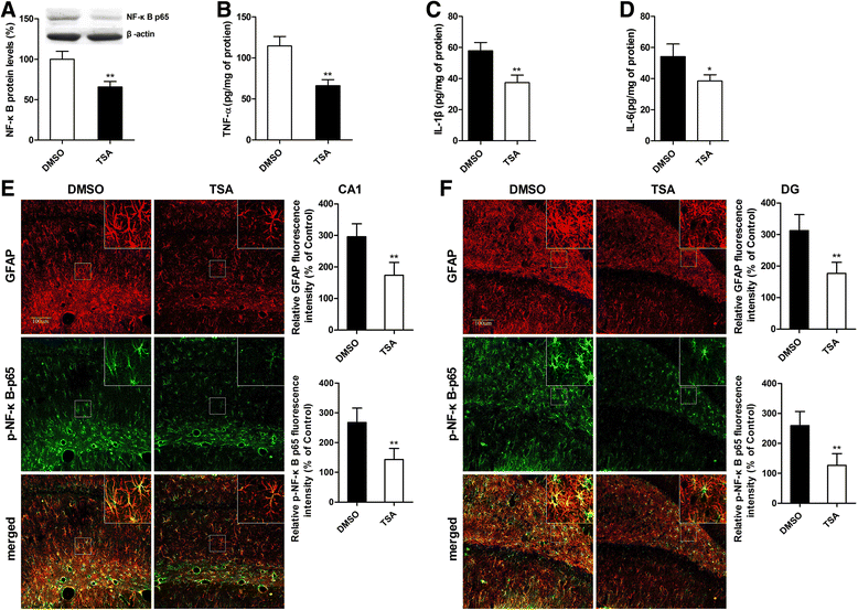
TSA reversed the effects of MS on the activation of NF-κB signaling and neuroinflammatory responses to sevoflurane in the hippocampal tissues in adult MS rats. a TSA pretreatment prevented the enhancement effect of MS on the levels of nuclear NF-κB p65 protein 12 h after anesthesia. b–d TSA pretreatment suppressed the increase in hippocampal TNF-α, IL-1β, and IL-6 levels 12 h after anesthesia in adult MS rats. e The fluorescence intensities of GFAP and p-NF-κB p65 in the TSA group rats were obviously lower than those in the DMSO group rats. Double immunofluorescence staining showed that p-NF-κB p65 (green) mainly co-localized (merged) with GFAP-positive reactive astrocytes (red) in the CA1 area of the hippocampus. f The changes in the fluorescence intensities of GFAP and p-NF-κB p65 in the DG area of the hippocampus were similar to those in the CA1 area of the hippocampus. * P < 0.05, ** P < 0.01 versus DMSO. Error bars represent the means ± SD (n = 6). Statistical analyses were performed with Student’s two-sample t test
Fig. 8
Fig. 8
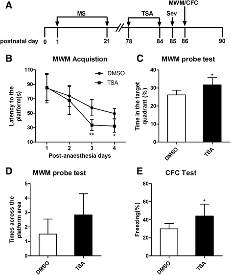
TSA attenuated sevoflurane-induced cognitive impairment in adult rats with neonatal maternal separation. a Schematic illustration of the experimental timeline. MS maternal separation, Sev anesthesia with 3% sevoflurane for 2 h, TSA trichostatin A, MWM Morris water maze, CFC context fear conditioning. b TSA group rats had a shorter escape latency than DMSO group rats on the third and fourth days after sevoflurane anesthesia. There was a statistically significant interaction of time and group between the different groups (* P < 0.05). Analysis was performed by repeated-measures two-way ANOVA. c The time in the target quadrant in the TSA group was more than that in the DMSO group by the MWM probe test. d There was no significant difference in the number of crossings of the platform area between the two groups of rats. e TSA group rats exhibited increased freezing to context relative to DMSO group rats. * P < 0.05, ** P < 0.01 versus DMSO. Error bars represent the means ± SD (n = 10). Statistical analyses were performed with Student’s two-sample t test

Similar articles

Cited by

References

    1. Steinmetz J, Christensen KB, Lund T, Lohse N, Rasmussen LS. Long-term consequences of postoperative cognitive dysfunction. Anesthesiology. 2009;110:548–55. doi: 10.1097/ALN.0b013e318195b569. - DOI - PubMed
    1. Monk TG, Weldon BC, Garvan CW, Dede DE, van der Aa MT, Heilman KM, et al. Predictors of cognitive dysfunction after major noncardiac surgery. Anesthesiology. 2008;108:18–30. doi: 10.1097/01.anes.0000296071.19434.1e. - DOI - PubMed
    1. Terrando N, Eriksson LI, Ryu JK, Yang T, Monaco C, Feldmann M, et al. Resolving postoperative neuroinflammation and cognitive decline. Ann Neurol. 2011;70:986–95. doi: 10.1002/ana.22664. - DOI - PMC - PubMed
    1. Peng L, Xu L, Ouyang W. Role of peripheral inflammatory markers in postoperative cognitive dysfunction (POCD): a meta-analysis. PLoS One. 2013;8:e79624. doi: 10.1371/journal.pone.0079624. - DOI - PMC - PubMed
    1. Vacas S, Degos V, Feng X, Maze M. The neuroinflammatory response of postoperative cognitive decline. Br Med Bull. 2013;106:161–78. doi: 10.1093/bmb/ldt006. - DOI - PMC - PubMed

MeSH terms