Human neural progenitors derived from integration-free iPSCs for SCI therapy
- PMID: 28073086
- PMCID: PMC5629634
- DOI: 10.1016/j.scr.2017.01.004
Human neural progenitors derived from integration-free iPSCs for SCI therapy
Abstract
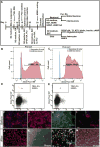
As a potentially unlimited autologous cell source, patient induced pluripotent stem cells (iPSCs) provide great capability for tissue regeneration, particularly in spinal cord injury (SCI). However, despite significant progress made in translation of iPSC-derived neural progenitor cells (NPCs) to clinical settings, a few hurdles remain. Among them, non-invasive approach to obtain source cells in a timely manner, safer integration-free delivery of reprogramming factors, and purification of NPCs before transplantation are top priorities to overcome. In this study, we developed a safe and cost-effective pipeline to generate clinically relevant NPCs. We first isolated cells from patients' urine and reprogrammed them into iPSCs by non-integrating Sendai viral vectors, and carried out experiments on neural differentiation. NPCs were purified by A2B5, an antibody specifically recognizing a glycoganglioside on the cell surface of neural lineage cells, via fluorescence activated cell sorting. Upon further in vitro induction, NPCs were able to give rise to neurons, oligodendrocytes and astrocytes. To test the functionality of the A2B5+ NPCs, we grafted them into the contused mouse thoracic spinal cord. Eight weeks after transplantation, the grafted cells survived, integrated into the injured spinal cord, and differentiated into neurons and glia. Our specific focus on cell source, reprogramming, differentiation and purification method purposely addresses timing and safety issues of transplantation to SCI models. It is our belief that this work takes one step closer on using human iPSC derivatives to SCI clinical settings.
Keywords: Neural repair; Neuroprotection; Spinal cord injury; iPSC.
Copyright © 2017 The Authors. Published by Elsevier B.V. All rights reserved.
Figures


Similar articles
-
Cell therapy for spinal cord injury by using human iPSC-derived region-specific neural progenitor cells.Mol Brain. 2020 Sep 3;13(1):120. doi: 10.1186/s13041-020-00662-w. Mol Brain. 2020. PMID: 32883317 Free PMC article.
-
Transplanted Human Induced Pluripotent Stem Cell-Derived Neural Progenitor Cells Do Not Promote Functional Recovery of Pharmacologically Immunosuppressed Mice With Contusion Spinal Cord Injury.Cell Transplant. 2015;24(9):1799-812. doi: 10.3727/096368914X684079. Epub 2014 Sep 8. Cell Transplant. 2015. PMID: 25203632
-
Effects of the Post-Spinal Cord Injury Microenvironment on the Differentiation Capacity of Human Neural Stem Cells Derived from Induced Pluripotent Stem Cells.Cell Transplant. 2016 Oct;25(10):1833-1852. doi: 10.3727/096368916X691312. Cell Transplant. 2016. PMID: 27075820
-
iPSC-derived neural precursor cells: potential for cell transplantation therapy in spinal cord injury.Cell Mol Life Sci. 2018 Mar;75(6):989-1000. doi: 10.1007/s00018-017-2676-9. Epub 2017 Oct 9. Cell Mol Life Sci. 2018. PMID: 28993834 Free PMC article. Review.
-
Applications of induced pluripotent stem cell technologies in spinal cord injury.J Neurochem. 2017 Jun;141(6):848-860. doi: 10.1111/jnc.13986. Epub 2017 Apr 5. J Neurochem. 2017. PMID: 28199003 Review.
Cited by
-
Establishment of stable iPS-derived human neural stem cell lines suitable for cell therapies.Cell Death Dis. 2018 Sep 17;9(10):937. doi: 10.1038/s41419-018-0990-2. Cell Death Dis. 2018. PMID: 30224709 Free PMC article.
-
Exploiting urine-derived induced pluripotent stem cells for advancing precision medicine in cell therapy, disease modeling, and drug testing.J Biomed Sci. 2024 May 9;31(1):47. doi: 10.1186/s12929-024-01035-4. J Biomed Sci. 2024. PMID: 38724973 Free PMC article. Review.
-
Urine-Derived Stem Cells: Applications in Regenerative and Predictive Medicine.Cells. 2020 Feb 28;9(3):573. doi: 10.3390/cells9030573. Cells. 2020. PMID: 32121221 Free PMC article. Review.
-
Design and Validation of a Process Based on Cationic Niosomes for Gene Delivery into Novel Urine-Derived Mesenchymal Stem Cells.Pharmaceutics. 2021 May 11;13(5):696. doi: 10.3390/pharmaceutics13050696. Pharmaceutics. 2021. PMID: 34064902 Free PMC article.
-
Stem cell therapy for chronic skin wounds in the era of personalized medicine: From bench to bedside.Genes Dis. 2019 Sep 17;6(4):342-358. doi: 10.1016/j.gendis.2019.09.008. eCollection 2019 Dec. Genes Dis. 2019. PMID: 31832514 Free PMC article. Review.
References
-
- Ban H, Nishishita N, Fusaki N, Tabata T, Saeki K, Shikamura M, Takada N, Inoue M, Hasegawa M, Kawamata S, Nishikawa S. Efficient generation of transgene-free human induced pluripotent stem cells (iPSCs) by temperature-sensitive Sendai virus vectors. Proc Natl Acad Sci U S A. 2011;108:14234–14239. - PMC - PubMed
Publication types
MeSH terms
Substances
Grants and funding
LinkOut - more resources
Full Text Sources
Other Literature Sources
Medical
Research Materials

