Review
. 2017 Jul 5;9(7):a028035.
doi: 10.1101/cshperspect.a028035.

Pathology of Neurodegenerative Diseases

Affiliations
Review

Pathology of Neurodegenerative Diseases

Brittany N Dugger et al. Cold Spring Harb Perspect Biol. .

Abstract

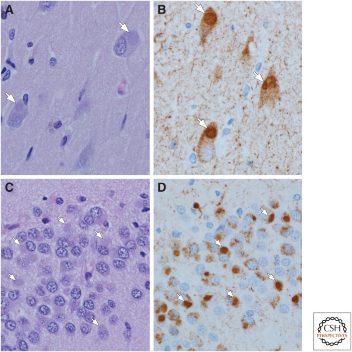
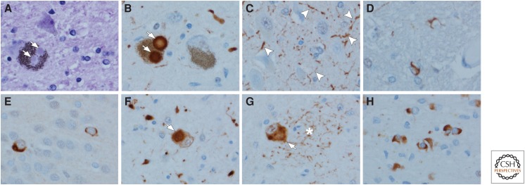
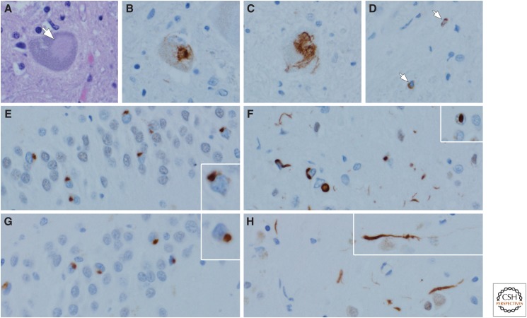
Neurodegenerative disorders are characterized by progressive loss of selectively vulnerable populations of neurons, which contrasts with select static neuronal loss because of metabolic or toxic disorders. Neurodegenerative diseases can be classified according to primary clinical features (e.g., dementia, parkinsonism, or motor neuron disease), anatomic distribution of neurodegeneration (e.g., frontotemporal degenerations, extrapyramidal disorders, or spinocerebellar degenerations), or principal molecular abnormality. The most common neurodegenerative disorders are amyloidoses, tauopathies, α-synucleinopathies, and TDP-43 proteinopathies. The protein abnormalities in these disorders have abnormal conformational properties. Growing experimental evidence suggests that abnormal protein conformers may spread from cell to cell along anatomically connected pathways, which may in part explain the specific anatomical patterns observed at autopsy. In this review, we detail the human pathology of select neurodegenerative disorders, focusing on their main protein aggregates.


Figures

Figure 1.
Figure 1.
Amyloidoses: Gerstmann–Sträussler–Scheinker disease (GSS) (A,B), Creutzfeldt–Jakob disease (CJD) (C,D), and Alzheimer’s disease (AD) (E,F). In GSS, dense-cored amyloid plaques can be detected with hemotoxylin and eosin (H&E) staining, arrow (A), but immunohistochemistry for human PrP (monoclonal antibody 3F4) reveals more clearly the multicentric nature of the deposits (B). One of the hallmark histologic features of CJD is the spongiform change in affected cortical and subcortical areas (C) with a perineuronal synaptic pattern of PrP deposition (arrows) in an adjacent section (D). In AD, amyloid deposits detected with immunohistochemistry (monoclonal antibody 6F/3D) are heterogeneous and include those with dense cores, especially in primary cortices (E), as well as poorly circumscribed and noncompact diffuse plaques in the cortex (asterisks) (F). In addition to parenchymal deposits, most cases of AD also have amyloid angiopathy (F).
Figure 2.
Figure 2.
Tau pathology in AD: Neurofibrillary tangles (NFTs) (A,B) and neuritic plaques (C,D). Flame-shaped intracellular NFTs (arrow) can be detected as bundles of basophilic filaments in pyramidal neurons of the hippocampus (A). Immunohistochemistry for phospho-tau shows not only NFTs (white arrow), but also pervasive neuropil threads and a pretangle (black arrow) that are not visible with routine histology. Neuritic plaques (asterisk) can also be detected with routine histology (C) because of central dense amyloid, clusters of swollen cell processes, activated microglia, and surrounding reactive astrocytes (arrow). Immunohistochemistry for phospho-tau shows clusters of irregular, swollen cell processes around a central unstained region (D) (i.e., amyloid core [asterisk]). Note also neuropil threads and several NFTs and pretangles (arrows). (Immunohistochemistry with CP13, whose epitope is near phosphoserine 202.)
Figure 3.
Figure 3.
Tau pathology in chronic traumatic encephalopathy (CTE) (A,B) and aging-related tau astrogliopathy (ARTAG) (C,D). The characteristic lesion in CTE is a focal cluster of neuronal and glial tau concentrated at the depths of a sulcus (s) (A), with preferential involvement of superficial cortical layers. Higher magnification shows neuronal (arrows) and astroglial tau preferentially around blood vessels (v) (B). In ARTAG, tau-positive astrocytes are concentrated at the pial surface in the transverse fissure at the base of the brain (C). At the pial surface, the astrocytes are thorn-shaped (C, left lower inset), whereas the astrocytes in gray matter have granular cytoplasmic processes (C, right lower inset). Tau-positive astrocytes are concentrated at the glia limitans around penetrating blood vessels (v) (D). (Immunohistochemistry with CP13, whose epitope is near phosphoserine 202.)
Figure 4.
Figure 4.
4R tauopathies: Progressive supranuclear palsy (PSP) (AD), corticobasal degeneration (CBD) (EH), and argyrophilic grain disease (AGD) (I–L). In PSP, the typical neuronal lesion is a globose NFT visible with phospho-tau immunohistochemistry (A) or with hemotoxylin and eosin (H&E) (B). Oligodendroglial coiled bodies (C) and tufted astrocytes (D) are typical glial lesions in PSP. In CBD, the typical neuronal lesion is a pretangle visible with tau immunohistochemistry (E). Cortical neurons with ballooning degeneration can be detected with H&E (F) and phospho-tau (G). The typical astrocytic lesion is the astrocytic plaque (H), characterized by tau accumulation in distal processes of astrocytes. In AGD, the defining lesions are round to spindle-shaped grains (I) that are most abundant in limbic areas. There are also pretangles. Ballooned neurons similar to those in CBD are also detected in limbic areas with H&E (J) and phospho-tau (K). Tau-positive astrocytes are frequent, and they have ramified granular tau in cell processes (L). (Immunohistochemistry with CP13, whose epitope is near phosphoserine 202.)
Figure 5.
Figure 5.
3R tauopathy: Pick’s disease (AD). Pick bodies are slightly basophilic, well-circumscribed cytoplasmic inclusions (arrows) that can be detected in pyramidal neurons of the hippocampus (A,B) and granule neurons of the dentate fascia (C,D) (arrows). Hemotoxylin and eosin (H&E) reveals cytoplasmic inclusions (A,C), whereas phospho-tau immunohistochemistry shows not only Pick bodies (arrows), but also diffuse cytoplasmic immunoreactivity and immunoreactivity in fine neuropil threads (B,D). (Immunohistochemistry with CP13, whose epitope is near phosphoserine 202.)
Figure 6.
Figure 6.
Synucleinopathies: Lewy body disease (AD) and multiple system atrophy (MSA) (E–H). Brainstem-type Lewy bodies are hyaline inclusions (arrows) that can be detected in neuromelanin-containing neurons of the substantia nigra upon hemotoxylin and eosin (H&E) (A) or with α-synuclein immunohistochemistry (arrows) (B). In addition to Lewy bodies, α-synuclein immunohistochemistry reveals Lewy neurites (arrowheads), for example, in the CA2/3 sector of the hippocampus (C). Sparse oligodendroglial inclusions can be detected in the basal ganglia and the ventral tegmental region of the midbrain in most cases (D). Neuronal cytoplasmic inclusions are detected in MSA with α-synuclein immunohistochemistry. They are infrequent in the dentate fascia of the hippocampus (E), and frequent in neurons of the pontine nuclei (arrow) (F), and the inferior olivary nucleus (arrow) (G). Neuronal inclusions are usually accompanied by dystrophic neurites (asterisk in G). The hallmark lesions of MSA are α-synuclein-immunoreactive glial cytoplasmic inclusions in affected white matter of the basal ganglia, brainstem, or cerebellum (H). (Immunohistochemistry with polyclonal α-synuclein antibody [NACP] specific to an epitope in the C-terminus corresponding to residues 98-15.)
Figure 7.
Figure 7.
Transactivation response DNA binding protein 43 (TDP-43) proteinopathies: amyotrophic lateral sclerosis (ALS) (A–D) and frontotemporal lobar degeneration with TDP-43 neuronal inclusions (FTLD-TDP) (F–H). In ALS, Lewy-like hyaline inclusions (arrow) can be detected in anterior horn cells of the spinal cord (A). Immunohistochemistry for TDP-43 reveals dense inclusions (B) and skein-like inclusions (C) in motor neurons of the brainstem and spinal cord. White matter motor tracts often have sparse TDP-43-immunoreactive oligodendroglial inclusions (arrows) (D). In FTLD-TDP Type A, neurons of the dentate fascia often have granular or ring-shaped neuronal cytoplasmic inclusions (E, inset), whereas the affected cortices have more pleomorphic neuronal cytoplasmic inclusions, short curved dystrophic neurites, and neuronal intranuclear inclusions (F, inset). In FTLD-TDP Type C, round, well-circumscribed (Pick-body-like) neuronal cytoplasmic inclusions are frequent in the dentate fascia of the hippocampus (G), whereas they are sparse in the affected cortices, where long, thick dystrophic neurites (H, inset) are the most characteristic feature. Type B (not shown) has predominantly neuronal cytoplasmic inclusions with minimal dystrophic neurites and no intranuclear inclusions. (TDP-43 immunohistochemistry with polyclonal MC2085 antibody to a neoepitope in caspase-cleaved TDP-43.)

Similar articles

Cited by

References

    1. Adler CH, Connor DJ, Hentz JG, Sabbagh MN, Caviness JN, Shill HA, Noble B, Beach TG. 2010. Incidental Lewy body disease: Clinical comparison to a control cohort. Mov Disord 25: 642–646. - PMC - PubMed
    1. Adler CH, Beach TG, Hentz JG, Shill HA, Caviness JN, Driver-Dunckley E, Sabbagh MN, Sue LI, Jacobson SA, Belden CM, et al. 2014. Low clinical diagnostic accuracy of early vs. advanced Parkinson disease: Clinicopathologic study. Neurology 83: 406–412. - PMC - PubMed
    1. Alafuzoff I, Ince PG, Arzberger T, Al-Sarraj S, Bell J, Bodi I, Bogdanovic N, Bugiani O, Ferrer I, Gelpi E, et al. 2009a. Staging/typing of Lewy body related α-synuclein pathology: A study of the BrainNet Europe Consortium. Acta Neuropathol 117: 635–652. - PubMed
    1. Alafuzoff I, Thal DR, Arzberger T, Bogdanovic N, Al-Sarraj S, Bodi I, Boluda S, Bugiani O, Duyckaerts C, Gelpi E, et al. 2009b. Assessment of β-amyloid deposits in human brain: A study of the BrainNet Europe Consortium. Acta Neuropathol 117: 309–320. - PMC - PubMed
    1. Alafuzoff I, Pikkarainen M, Neumann M, Arzberger T, Al-Sarraj S, Bodi I, Bogdanovic N, Bugiani O, Ferrer I, Gelpi E, et al. 2015. Neuropathological assessments of the pathology in frontotemporal lobar degeneration with TDP43-positive inclusions: An inter-laboratory study by the BrainNet Europe consortium. J Neural Transm (Vienna) 122: 957–972. - PubMed

MeSH terms

Substances