. 2017 Jan 17;114(3):E307-E316.
doi: 10.1073/pnas.1612730114. Epub 2017 Jan 4.

SNX-1 and RME-8 oppose the assembly of HGRS-1/ESCRT-0 degradative microdomains on endosomes

Affiliations

SNX-1 and RME-8 oppose the assembly of HGRS-1/ESCRT-0 degradative microdomains on endosomes

Anne Norris et al. Proc Natl Acad Sci U S A. .

Abstract

After endocytosis, transmembrane cargo reaches endosomes, where it encounters complexes dedicated to opposing functions: recycling and degradation. Microdomains containing endosomal sorting complexes required for transport (ESCRT)-0 component Hrs [hepatocyte growth factor-regulated tyrosine kinase substrate (HGRS-1) in Caenorhabditis elegans] mediate cargo degradation, concentrating ubiquitinated cargo and organizing the activities of ESCRT. At the same time, retromer associated sorting nexin one (SNX-1) and its binding partner, J-domain protein RME-8, sort cargo away from degradation, promoting cargo recycling to the Golgi. Thus, we hypothesized that there could be important regulatory interactions between retromer and ESCRT that balance degradative and recycling functions. Taking advantage of the naturally large endosomes of the C. elegans coelomocyte, we visualized complementary ESCRT-0 and RME-8/SNX-1 microdomains in vivo and assayed the ability of retromer and ESCRT microdomains to regulate one another. We found in snx-1(0) and rme-8(ts) mutants increased endosomal coverage and intensity of HGRS-1-labeled microdomains, as well as increased total levels of HGRS-1 bound to membranes. These effects are specific to SNX-1 and RME-8, as loss of other retromer components SNX-3 and vacuolar protein sorting-associated protein 35 (VPS-35) did not affect HGRS-1 microdomains. Additionally, knockdown of hgrs-1 had little to no effect on SNX-1 and RME-8 microdomains, suggesting directionality to the interaction. Separation of the functionally distinct ESCRT-0 and SNX-1/RME-8 microdomains was also compromised in the absence of RME-8 and SNX-1, a phenomenon we observed to be conserved, as depletion of Snx1 and Snx2 in HeLa cells also led to greater overlap of Rme-8 and Hrs on endosomes.

Keywords: Hrs; RME-8; SNX-1; clathrin; endosome.


Conflict of interest statement

The authors declare no conflict of interest.

Figures
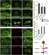
Fig. 1.
Fig. 1.
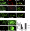
SNX-1 and RME-8 are important for retrograde recycling in the C. elegans coelomocyte. MIG-14::GFP expressed in coelomocytes was visualized in wild-type, snx-1(tm847), and rme-8(b1023) mutant backgrounds. MIG-14 labels endosomes, internal puncta, and the plasma membrane. Note uneven distribution around the periphery of the large circular endosomes in the Inset (AA′′). Fluorescence micrographs of hybrid-model retromer cargo composed of GFP, a portion of the human CD4-GFP extracellular domain, and the cytoplasmic and transmembrane domains of human CIMPR, expressed in wild-type, snx-1(tm847), and rme-8(b1023) mutant coelomocytes. hCIMPR-CD4-GFP labels endosomes, internal puncta, and, to a lesser extent, the coelomocyte plasma membrane. Note uneven distribution around the periphery of the large circular endosomes in the Inset (BB′′). C. elegans TGN38/46 homolog TGN-38::GFP was expressed in the coelomocytes of wild-type, snx-1(tm847), and rme-8(b1023) mutants. TGN-38::GFP labels endosomes unevenly, strongly labels internal puncta, and weakly labels the plasma membrane (CC′′). (Scale bar: 5 µm.) Quantification of average intensity, under the same image acquisition conditions, of each cargo in wild-type vs. mutant backgrounds (D and E). MIG-14, hCIMPR, and TGN-38 coexpressed with Golgi marker AMAN-2. Puncta (small arrows) colocalizes with Golgi marker AMAN-2::RFP (large arrowheads indicate Golgi adjacent to cargo-labeled endosomes) (FH′′). (I) Diagrammatic representation of three cargo constructs. All experiments were performed at 25 °C.
Fig. S1.
Fig. S1.
hCI-MPR labels Golgi in wild-type (A and A′′) and snx-1(tm847) (B and B′′) mutants. Large arrowheads indicate the endosomally localized hCI-MPR, which is absent in the snx-1(tm847) mutant. Small arrows indicate the hCI-MPR that colocalizes with Golgi marker AMAN-2, which is present in wild-type and snx-1(tm847) mutants. TGN-38 GFP panel from Fig. 1C is represented at lower scaling to show the morphological structures that trap TGN-38 in rme-8(b1023ts) and snx-1(tm847) mutants (CC′′). MIG-14-GFP levels are restored in snx-1(tm847) mutants by depletion of HGRS-1. Coelomocytes expressing MIG-14 GFP wild type (D and D′) or snx-1(tm847) (E and E′) after treatment with vector (D and E) or HGRS-1 RNAi (D′ and E′). (F) Quantification of MIG-14 GFP levels in DE′. (Scale bar: 5 µm.)
Fig. 2.
Fig. 2.
Retromer-associated proteins RME-8 and SNX-1 specifically label endosomal microdomains complementary to that of ESCRT-0 protein HGRS-1. Images are shown of coelomocytes within intact living animals expressing citrine::HGRS-1 and tagRFP::SNX-1 that have been allowed to take up Cy5-BSA from the body cavity for 10 min (AA′′). A line scan displaying pixel intensity around the periphery of one endosome (as visualized in A′′, Inset) shows complementary localization of HGRS-1 and SNX-1 on the Cy5-BSA filled endosome (A′′′). Citrine::RME-8 and tagRFP::HGRS-1 label distinct microdomains (BB′′). A line scan of the endosome limiting membrane (B′′, Inset) indicates complementary localization of HGRS-1 and RME-8 (B′′′). Endogenous VPS-35 tagged with GFP labels microdomains distinct from citrine::HGRS-1 (CC′′). Line scan of the endosome in C′′, Inset (C′′′). Citrine::HGRS-1 and CLIC-1::tagRFP colocalize in discrete microdomains on the endosome (DD′′). Line scan of the endosome in D′′, Inset (D′′′). (Scale bar: 5 µm.) Diagrammatic representation of the localization of the endosomal components to the right of the relevant line scans (E). (F) Quantification of colocalization of the endosomal components of AD.
Fig. 3.
Fig. 3.
HGRS-1 and SNX-1 microdomains are enriched in relevant cargo. (AA′′) Peaks in intensity of MIG-14::tagRFP (pseudocolored green) along the endosomal periphery coincide with GFP::SNX-1 (pseudocolored red). (A′′′) Two-color line scan around the endosomal periphery (A′′, Inset). (BB′′) MIG-14::GFP and tagRFP::HGRS-1 display complementary microdomain enrichment. (B′′′) Two-color line scan around the endosomal periphery (B′′, Inset). (CC′′) Cy3-BSA is enriched in citrine::HGRS-1–labeled microdomains after 5 min of uptake. (C′′′) Two-color line scan around the endosomal periphery (C′′, Inset). (Scale bar: 5 µm.) (D) A diagrammatic representation of the panels is displayed to the right. (E) Quantification of colocalization of relevant cargo and endosomal components of AC.
Fig. 4.
Fig. 4.
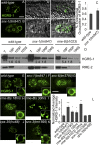
HGRS-1 endosomal occupancy is controlled by SNX-1, SNX-6, and RME-8. (AA′′ and BB′′) Fluorescence and DIC images of coelomocytes expressing citrine::HGRS-1 in wild-type and snx-1 mutant backgrounds. Quantification of average citrine::HGRS-1 endosomal occupancy in wild-type and snx-1 mutant strains (C). Membrane association of endogenous HGRS-1 was measured by Western blot after lysis and ultracentrifugation to separate membranes from cytosol in wild-type, snx-1(tm847), and rme-8(b1023) mutant strains. The fraction is indicated above the lane. Equivalent samples were loaded in each lane. The ratios of 100P vs. supernatant and 18P vs. supernatant are indicated below the two fractions. In rme-8 and snx-1 mutants, the 18P and 100P fractions contain proportionally more HGRS-1 than in wild type. RME-2 is a transmembrane yolk receptor that fractionates only with the membranous fractions (D). (EK) Citrine::HGRS-1 imaged in wild-type, snx-1(tm847), snx-6(tm3790), rme-8(b1023), snx-3(tm1595), and vps-35(hu68) mutant strains with the same exposure and scaling conditions at 25 °C. (L) Quantification of citrine::HGRS-1 intensity in wild-type and mutant backgrounds. (Scale bar: 5 µm.)
Fig. S2.
Fig. S2.
HGRS-1 intensity is not affected by SNX-1 or RME-8 overexpression, and total protein levels are unchanged in snx-1 and snx-3 mutants. Coelomocytes expressing citrine::HGRS-1 alone (A) or in the presence of SNX-1::tagRFP or tagRFP::RME-8 (B and C). (D) Levels of citrine::HGRS-1 from AC quantified. Measurement of levels of RME-8 in wild type vs. citrine- and tagRFP-tagged transgenes. Actin was used as a loading control (E). Animals expressing GFP:HGRS-1 in wild-type, snx-1, and snx-3 mutants were lysed, run on SDS/PAGE, and probed for GFP. Actin was used as a loading control (F). (G) Quantification of F. (Scale bar: 5 µm.)
Fig. S3.
Fig. S3.
HGRS-1 is not important for limiting RME-8 or SNX-1 endosomal domains. Strains expressing GFP::SNX-1 were treated with empty vector RNAi control (A) or hgrs-1 RNAi (A′). Strains expressing GFP::RME-8 were treated with empty vector RNAi control (B) or hgrs-1 RNAi (B′). Strains expressing GFP::HGRS-1 were treated with empty vector RNAi control (C) or hgrs-1 RNAi (C′). The fluorescence intensity of GFP::SNX-1, GFP::RME-8, and GFP::HGRS-1 was quantified in control vs. hgrs-1 RNAi (D). (Scale bar: 5 μm.)
Fig. 5.
Fig. 5.
RME-8 and SNX-1 control microdomain separation. Strains expressing citrine::HGRS-1 and tagRFP::RME-8 display complementary microdomains (AA′′′). (BB′′′) snx-1(tm847) mutants expressing citrine::HGRS-1 and tagRFP::RME-8 display an increase in overlap of the normally separate domains. (CC′′′) Coelomocytes expressing tagRFP::HGRS-1 (pseudocolored green) and GFP::RME-8(DNAJ-HPD-AAA) (pseudocolored red) display an increase in overlap of the normally separate domains, as well as an altered morphology of the endosomal components HGRS-1 and RME-8. (DD''') Citrine::HGRS-1 and tagRFP::SNX-1 in wild-type coelomocytes display complementary microdomain localization. (EE′′′) rme-8(b1023) mutant coelomocytes expressing citrine::HGRS-1 and tagRFP::SNX-1 display an increase in overlap of the normally separate domains 18 h after temperature shift. (FF′′′) At 30 h after temperature shift, endosomes are no longer recognizable, and citrine::HGRS-1 and tagRFP::SNX-1 display an increase in overlap of the normally separate domains. (GG′′′) Strains expressing citrine::HGRS-1 and tagRFP::SNX-1 in snx-3(tm1595) worms display wild-type complementary localization. (H) Overlap of endosomal microdomains is quantified. (I) A diagrammatic representation of endosomal microdomain localization of the panels to the left. (Scale bar: 5 µm.)
Fig. S4.
Fig. S4.
Clathrin accumulates in the absence of RME-8 in coelomocytes, and loss of clathrin impairs microdomain separation. Strains expressing citrine::HGRS-1 and tagRFP::SNX-1 display complementary microdomains (AA′′′). chc-1(b1025ts) mutants expressing citrine::HGRS-1 and tagRFP::SNX-1, shifted to the nonpermissive temperature overnight, display an increase in overlap of the normally separate domains (BB′′′). Quantification of the overlap of citrine::HGRS-1 and tagRFP::SNX-1 normalized to wild type in the chc-1(b1025ts) mutant background (C). Clathrin light-chain (Clic-1)::tagRFP imaged in wild-type and rme-8(b1023) mutant strains with the same exposure and scaling conditions shifted to 25 °C for the indicated time (DD′′). Quantification of CLIC-1::tagRFP in wild-type and mutant backgrounds (E). (Scale bar: 5 µm.)
Fig. 6.
Fig. 6.
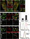
The role of SNX-1 in microdomain separation is conserved in HeLa cells. Confocal microscopy of endogenous Hrs and RME-8 display a shoulder to shoulder localization in the perinuclear region in control cells. Arrows indicate the juxtaposition of Hrs and RME-8 immunostaining (AA′′′). Under conditions of SNX1/2 siRNA, RME-8– and Hrs-labeled domains colocalize in enlarged structures in the perinuclear area. Arrows indicate the enlarged endosomes positive for colocalized RME-8 and Hrs (BB′′′). dSTORM (C′–H′) vs. epifluorescent (CH) images of endogenous Hrs and RME-8 transfected with control scrambled siRNA (CE and C′–E′) and SNX1/2 siRNA (FH and F′–H′). Arrows indicate RME-8 and Hrs that are close and/or colocalized. Nearest-neighbor analysis strength of interaction of Hrs and RME-8 is significantly increased upon SNX1/2 siRNA (I). Quantification of line scans shown in E′ (J) and H′ (K).
Fig. S5.
Fig. S5.
Western blot of HeLa cell lysates probed for Snx1 or Snx2 indicate efficient knockdown.

Similar articles

Cited by

References

    1. Mosesson Y, Mills GB, Yarden Y. Derailed endocytosis: An emerging feature of cancer. Nat Rev Cancer. 2008;8(11):835–850. - PubMed
    1. Seaman MN, Marcusson EG, Cereghino JL, Emr SD. Endosome to Golgi retrieval of the vacuolar protein sorting receptor, Vps10p, requires the function of the VPS29, VPS30, and VPS35 gene products. J Cell Biol. 1997;137(1):79–92. - PMC - PubMed
    1. Hierro A, et al. Functional architecture of the retromer cargo-recognition complex. Nature. 2007;449(7165):1063–1067. - PMC - PubMed
    1. Arighi CN, Hartnell LM, Aguilar RC, Haft CR, Bonifacino JS. Role of the mammalian retromer in sorting of the cation-independent mannose 6-phosphate receptor. J Cell Biol. 2004;165(1):123–133. - PMC - PubMed
    1. Swarbrick JD, et al. VPS29 is not an active metallo-phosphatase but is a rigid scaffold required for retromer interaction with accessory proteins. PLoS One. 2011;6(5):e20420. - PMC - PubMed

Publication types

MeSH terms

Substances