. 2017 Jan 4;37(1):58-69.
doi: 10.1523/JNEUROSCI.2305-16.2016.

Tau Isoforms Imbalance Impairs the Axonal Transport of the Amyloid Precursor Protein in Human Neurons

Affiliations

Tau Isoforms Imbalance Impairs the Axonal Transport of the Amyloid Precursor Protein in Human Neurons

Valentina Lacovich et al. J Neurosci. .

Abstract

Tau, as a microtubule (MT)-associated protein, participates in key neuronal functions such as the regulation of MT dynamics, axonal transport, and neurite outgrowth. Alternative splicing of exon 10 in the tau primary transcript gives rise to protein isoforms with three (3R) or four (4R) MT binding repeats. Although tau isoforms are balanced in the normal adult human brain, imbalances in 3R:4R ratio have been tightly associated with the pathogenesis of several neurodegenerative disorders, yet the underlying molecular mechanisms remain elusive. Several studies exploiting tau overexpression and/or mutations suggested that perturbations in tau metabolism impair axonal transport. Nevertheless, no physiological model has yet demonstrated the consequences of altering the endogenous relative content of tau isoforms over axonal transport regulation. Here, we addressed this issue using a trans-splicing strategy that allows modulating tau exon 10 inclusion/exclusion in differentiated human-derived neurons. Upon changes in 3R:4R tau relative content, neurons showed no morphological changes, but live imaging studies revealed that the dynamics of the amyloid precursor protein (APP) were significantly impaired. Single trajectory analyses of the moving vesicles showed that predominance of 3R tau favored the anterograde movement of APP vesicles, increasing anterograde run lengths and reducing retrograde runs and segmental velocities. Conversely, the imbalance toward the 4R isoform promoted a retrograde bias by a significant reduction of anterograde velocities. These findings suggest that changes in 3R:4R tau ratio has an impact on the regulation of axonal transport and specifically in APP dynamics, which might link tau isoform imbalances with APP abnormal metabolism in neurodegenerative processes.

Significance statement: The tau protein has a relevant role in the transport of cargos throughout neurons. Dysfunction in tau metabolism underlies several neurological disorders leading to dementia. In the adult human brain, two tau isoforms are found in equal amounts, whereas changes in such equilibrium have been associated with neurodegenerative diseases. We investigated the role of tau in human neurons in culture and found that perturbations in the endogenous balance of tau isoforms were sufficient to impair the transport of the Alzheimer's disease-related amyloid precursor protein (APP), although neuronal morphology was normal. Our results provide evidence of a direct relationship between tau isoform imbalance and defects in axonal transport, which induce an abnormal APP metabolism with important implications in neurodegeneration.

Keywords: APP; Alzheimer's; axonal transport; splicing; tau; tauopathies.


Figures

Figure 1.
Figure 1.
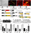
3R or 4R tau modulation by trans-splicing in highly polarized human neurons. A, B, Human neuronal cultures at five days after plating (DIV5) showing projections and proper polarization in bright-field (A) and after immunostaining with axonal and dendritic markers (tau and MAP2) (B). White arrow indicates the axon without MAP2 staining and yellow arrows show dendrites. C, Enriched human neuronal cultures at DIV18 showing highly polarized projections and dense connectivity. D, Neuron at DIV19 showing colocalization of APP–YFP (green) with LV-dsRED transduction (red). E, Map of LVs used for neuronal transduction at DIV11. LV-dsRED: control vector carrying the reporter cassette; LV-PTM3R: LV carrying a pre-trans-splicing molecule with exons 11–13; or LV-PTM4R: with exons 10–13. TSD: trans-splicing domain consisting of a binding domain complementary to the 3′ end of intron 9, a branch point and a 3′ splice acceptor site. F, Representative gel of RT-PCR products obtained with primers spanning tau exons 9–13 simultaneously amplifying the 3R and 4R isoforms. RT-PCR was performed with mRNA extracted from neurons transduced with each LV. G, Relative content of 3R and 4R tau isoforms determined by real time RT-PCR with specific primers for 3R (E10−) and 4R (E10+) tau mRNA. Delta Ct was calculated relative to human ApoB gene expression (mean ±SEM; n = 3) in three independent experiments. One-way ANOVA followed by Dunnett's comparison versus control neurons performed separately for each isoform; *p < 0.05 and **p < 0.001 versus 3R CTRL; #p < 0.001 vs 4R CTRL. H, I, Western blot analyses of human neuron homogenates. Shown is the detection of the 3R (H) and 4R (I) tau protein with isoform-specific antibodies normalized to actin quantified by optical density; **p < 0.001 (n = 3); one-way ANOVA followed by Dunnett's comparison with control (CTRL) neurons (transduced with LV-dsRED).
Figure 2.
Figure 2.
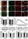
Normal neuronal polarization after 3R:4R tau isoform modulation. A, Confocal images of enriched neuronal cultures at DIV19 without transduction or transduced with LV-PTM3R or LV-PTM4R (from top to bottom). Immunofluorescence staining shows APP, tau, and Hoechst number for nuclei. B, Confocal high-magnification images from control and transduced neurons stained for APP and tau showing polarization and extension integrity. C, D, Sum of neuronal projection extension expressed in micrometers (H) and number of primary projection extensions from cell bodies (I) in neurons at DIV19 that were transduced at DIV11 with LV control, LV-PTM3R, and LV-PTM4R. E, Sholl analysis performed at DIV19 in transduced neurons showing the number of projection intersections versus distance from cell bodies (in micrometers). Data are shown as mean ± SEM; n = 10). F, G, Western blot of neuronal homogenates showing total tau and APP protein levels after each LV transduction conditions compared with NTC neurons. Tubulin (tub) was used as a loading control. Data in G are expressed as percentage of NTC (mean ± SEM; n = 3). H, Western blot using antibodies specific to tau phosphorylated at Ser 202 (CP13) or Ser 396/404 (PHF-1). Quantitative analyses are related to β-actin signal as a loading control (mean ± SEM; n = 3).
Figure 3.
Figure 3.
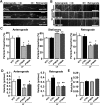
Impairment of axonal transport of APP vesicles in neurons under tau 3R:4R imbalance. A, Representative images from movies used to analyze the axonal transport of APP vesicles in neurons transduced with LV-control (dsRED), LV-PTM3R, and LV-PTM4R. Scale bar, 10 μm (see Movies 1–3). B, Kymographs of time versus distance from a 30 s movie generated at 8 frames/s in axons from neurons transfected with APP–YFP. Scale bar, 10 μm. C, Average proportion per kymograph of anterograde, stationary, and retrograde APP–YFP vesicles from NTCs or transduced with LV carrying dsRED, PTM3R, or PTM4R. D, Average velocity of anterograde and retrograde moving vesicles per kymograph. Kymographs analyzed: n = 59 NTCs, 78 dsRED, 57 PTM3R, and 58 PTM4R. One-way ANOVA followed by Dunnett's comparison versus NTCs, *p < 0.05, **p < 0.02, ***p < 0.01. E, Average number of APP–YFP vesicles per micrometer of axonal length in NTCs, dsRED, PTM3R, and PTM4R (n = 25 for each). One-way ANOVA was used for statistical analysis.
Figure 4.
Figure 4.
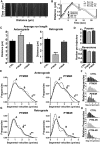
Kinesin and dynein transport properties are differentially impaired by tau isoform ratio modulation in human neurons. A, Representative kymographs of time versus distance showing moving and stationary APP vesicles and their trajectories using MATLAB (dotted lines). B, Representative recovered trajectory plotted as a function of distance versus time showing the properties extracted using custom-made MATLAB scripts. CE, Run lengths, segmental velocities, pauses, and reversions were determined from a total of 1283 trajectories (see Materials and Methods for details). C, Anterograde and retrograde average run lengths of vesicles moving in a net direction obtained from APP–YFP trajectories in neurons transduced with LV-dsRED (control), PTM3R, or PTM4R. n = ctrl: antero 1551, retro 1548; PTM3R: antero 572, retro 555; PTM4R: antero 465, retro 503. One-way ANOVA followed by Dunnett's comparison versus Ctrl *<0.05, ***<0.01. D, Average number of pauses and reversions per trajectory. Pauses n = ctrl: 2752; PTM3R: 981; PTM4R: 913. Reversion: n = ctrl: 895; PTM3R: 307; PTM4R: 262. EH, Distribution of segmental velocities of moving APP–YFP vesicles. A Gaussian mixture model with three modes (AC) was used to represent the segmental velocities distributions for anterograde (E) and retrograde (G) APP vesicle transport in control (full line) or transduced with PTM3R (light gray) or PTM4R (dark gray) LVs (dotted lines). The center and fraction of the different modes are displayed in Table 1. Relative frequencies of anterograde (F) and retrograde (H) segmental velocities used for model construction. Segmental velocities n = ctrl: antero 1671, retro 1344; PTM3R: antero 795, retro 549; PTM4R: antero 916, retro 752. Significant differences (**) obtained from comparison of nonoverlapping confidence intervals with the control group (LV-dsRED transduced neurons).
Figure 5.
Figure 5.
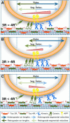
Hybrid competition–regulation model of tau-isoform-dependent APP axonal transport. We propose a model in which the APP vesicle is driven by two groups of motors, kinesin and dynein, along tau-decorated MTs. Motors behave in a tug-of-war/coordination scenario, displaying a differential tau isoform-dependent interaction with MTs. A, In the control condition, the two groups of motors are balanced to result in higher anterograde runs (dark arrows) and segmental velocities (light arrows) compared with the retrograde ones, giving an anterograde bias (red triangle) for the movement of APP vesicles. B, Shifts in the ratio toward 3R would not change the amount of tau bound to MT, but rather would reduce its binding strength, facilitating the recruitment of extra active motors. Extra kinesin load leads to an anterograde bias (red triangle) that favors the distal delivery of APP vesicles. C, Increased 4R tau level induces a higher binding strength of tau to MT that facilitates anterograde motor detachment and strongly impairs kinesin velocities (light arrows), whereas no significant changes in retrograde runs or velocities are observed. This results in a retrograde bias (red triangle) that should favor retrieval of APP vesicles to the cell body.

Comment in

Similar articles

Cited by

References

    1. Andreadis A. Tau gene alternative splicing: expression patterns, regulation and modulation of function in normal brain and neurodegenerative diseases. Biochim Biophys Acta. 2005;1739:91–103. doi: 10.1016/j.bbadis.2004.08.010. - DOI - PubMed
    1. Avale ME, Rodríguez-Martín T, Gallo JM. Trans-splicing correction of tau isoform imbalance in a mouse model of tau mis-splicing. Hum Mol Genet. 2013;22:2603–2611. doi: 10.1093/hmg/ddt108. - DOI - PMC - PubMed
    1. Bodea LG, Eckert A, Ittner LM, Piguet O, Götz J. Tau physiology and pathomechanisms in frontotemporal lobar degeneration. J Neurochem. 2016;138:71–94. doi: 10.1111/jnc.13600. - DOI - PMC - PubMed
    1. Bull ND, Guidi A, Goedert M, Martin KR, Spillantini MG. Reduced axonal transport and increased excitotoxic retinal ganglion cell degeneration in mice transgenic for human mutant p301s tau. PLoS One. 2012;7:e34724. doi: 10.1371/journal.pone.0034724. - DOI - PMC - PubMed
    1. Dawson HN, Cantillana V, Chen L, Vitek MP. The tau N279K exon 10 splicing mutation recapitulates frontotemporal dementia and parkinsonism linked to chromosome 17 tauopathy in a mouse model. J Neurosci. 2007;27:9155–9168. doi: 10.1523/JNEUROSCI.5492-06.2007. - DOI - PMC - PubMed

Publication types