

Histone deacetylases 1 and 2 cooperate in regulating BRCA1, CHK1, and RAD51 expression in acute myeloid leukemia cells
- PMID: 28030834
- PMCID: PMC5351634
- DOI: 10.18632/oncotarget.14062
Histone deacetylases 1 and 2 cooperate in regulating BRCA1, CHK1, and RAD51 expression in acute myeloid leukemia cells
Abstract
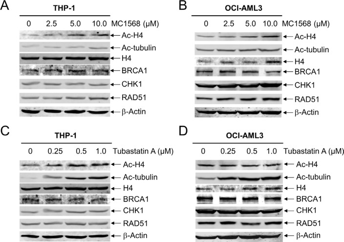
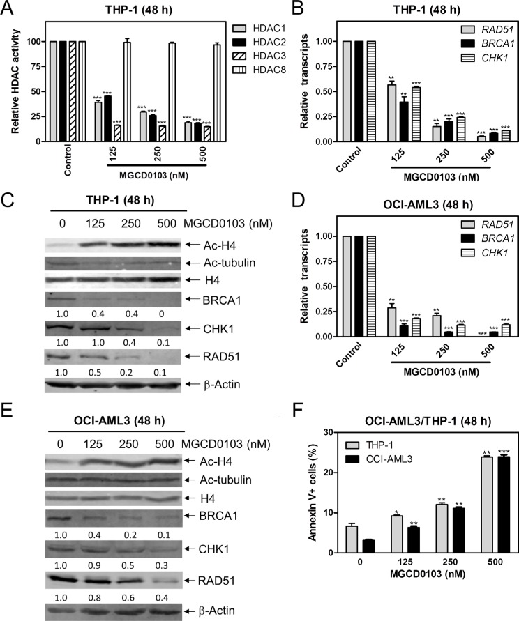
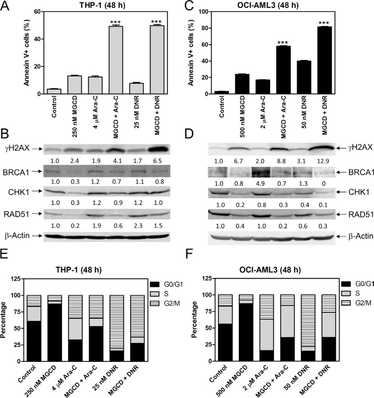
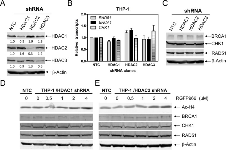
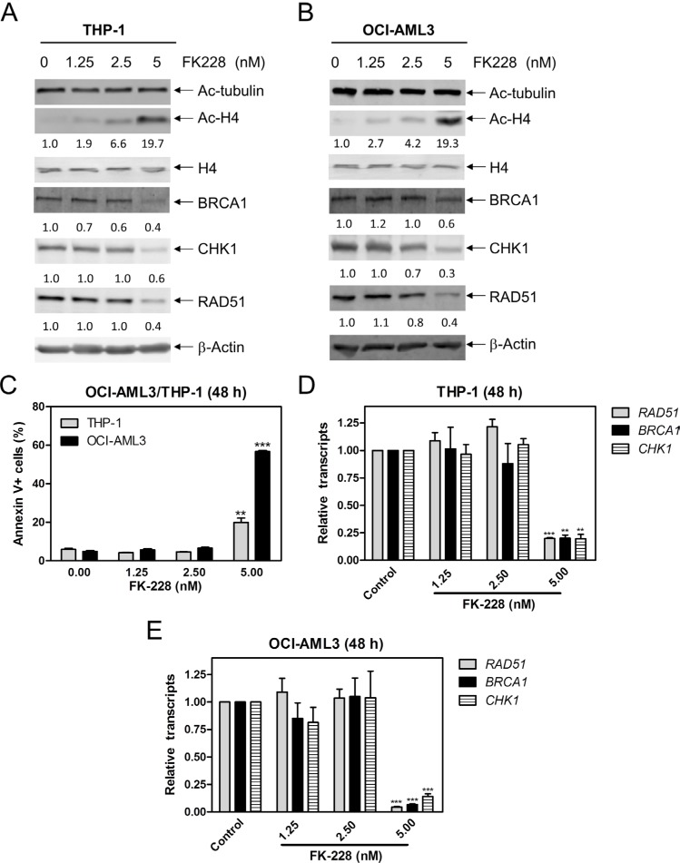
Resistance to chemotherapy and a high relapse rate highlight the importance of finding new therapeutic options for the treatment of acute myeloid leukemia (AML). Histone deacetylase (HDAC) inhibitors (HDACIs) are a promising class of drugs for the treatment of AML. HDACIs have limited single-agent clinical activities, but when combined with conventional or investigational drugs they have demonstrated favorable outcomes. Previous studies have shown that decreasing expression of important DNA damage repair proteins enhances standard chemotherapy drugs. In our recent studies, the pan-HDACI panobinostat has been shown to enhance conventional chemotherapy drugs cytarabine and daunorubicin in AML cells by decreasing the expression of BRCA1, CHK1, and RAD51. In this study, we utilized class- and isoform-specific HDACIs and shRNA knockdown of individual HDACs to determine which HDACs are responsible for decreased expression of BRCA1, CHK1, and RAD51 following pan-HDACI treatment in AML cells. We found that inhibition of both HDAC1 and HDAC2 was necessary to decrease the expression of BRCA1, CHK1, and RAD51, enhance cytarabine- or daunorubicin-induced DNA damage and apoptosis, and abrogate cytarabine- or daunorubicin-induced cell cycle checkpoint activation in AML cells. These findings may aid in the development of rationally designed drug combinations for the treatment of AML.
Keywords: BRCA1; CHK1; HDAC; RAD51; acute myeloid leukemia.
Conflict of interest statement
The authors declare no competing financial interests.
Figures

Similar articles
-
Panobinostat enhances cytarabine and daunorubicin sensitivities in AML cells through suppressing the expression of BRCA1, CHK1, and Rad51.PLoS One. 2013 Nov 11;8(11):e79106. doi: 10.1371/journal.pone.0079106. eCollection 2013. PLoS One. 2013. PMID: 24244429 Free PMC article. Clinical Trial.
-
Inhibition of histone deacetylases 1 and 6 enhances cytarabine-induced apoptosis in pediatric acute myeloid leukemia cells.PLoS One. 2011 Feb 16;6(2):e17138. doi: 10.1371/journal.pone.0017138. PLoS One. 2011. PMID: 21359182 Free PMC article.
-
HDAC Inhibition Induces MicroRNA-182, which Targets RAD51 and Impairs HR Repair to Sensitize Cells to Sapacitabine in Acute Myelogenous Leukemia.Clin Cancer Res. 2016 Jul 15;22(14):3537-49. doi: 10.1158/1078-0432.CCR-15-1063. Epub 2016 Feb 8. Clin Cancer Res. 2016. PMID: 26858310 Free PMC article.
-
Panobinostat for the treatment of acute myelogenous leukemia.Expert Opin Investig Drugs. 2016 Sep;25(9):1117-31. doi: 10.1080/13543784.2016.1216971. Epub 2016 Aug 8. Expert Opin Investig Drugs. 2016. PMID: 27485472 Review.
-
Endogenous modulators and pharmacological inhibitors of histone deacetylases in cancer therapy.Oncogene. 2012 Feb 2;31(5):537-51. doi: 10.1038/onc.2011.267. Epub 2011 Jul 4. Oncogene. 2012. PMID: 21725353 Free PMC article. Review.
Cited by
-
Role of HDACs in normal and malignant hematopoiesis.Mol Cancer. 2020 Jan 7;19(1):5. doi: 10.1186/s12943-019-1127-7. Mol Cancer. 2020. PMID: 31910827 Free PMC article. Review.
-
HDAC2-dependent miRNA signature in acute myeloid leukemia.FEBS Lett. 2019 Sep;593(18):2574-2584. doi: 10.1002/1873-3468.13521. Epub 2019 Jul 19. FEBS Lett. 2019. PMID: 31254352 Free PMC article.
-
Acetylation of Steroidogenic Acute Regulatory Protein Sensitizes 17β-Estradiol Regulation in Hormone-Sensitive Breast Cancer Cells.Int J Mol Sci. 2024 Aug 10;25(16):8732. doi: 10.3390/ijms25168732. Int J Mol Sci. 2024. PMID: 39201419 Free PMC article.
-
Therapeutic efficacy of an injectable formulation of purinostat mesylate in SU-DHL-6 tumour model.Ann Med. 2022 Dec;54(1):743-753. doi: 10.1080/07853890.2022.2045347. Ann Med. 2022. PMID: 35243950 Free PMC article.
-
Inhibitors of class I HDACs and of FLT3 combine synergistically against leukemia cells with mutant FLT3.Arch Toxicol. 2022 Jan;96(1):177-193. doi: 10.1007/s00204-021-03174-1. Epub 2021 Oct 19. Arch Toxicol. 2022. PMID: 34665271 Free PMC article.
References
-
- Quintas-Cardama A, Santos FP, Garcia-Manero G. Histone deacetylase inhibitors for the treatment of myelodysplastic syndrome and acute myeloid leukemia. Leukemia. 2011;25:226–235. - PubMed
-
- Burnett A, Wetzler M, Lowenberg B. Therapeutic advances in acute myeloid leukemia. J Clin Oncol. 2011;29:487–494. - PubMed
-
- Pollyea DA, Kohrt HE, Medeiros BC. Acute myeloid leukaemia in the elderly: a review. Br J Haematol. 2011;152:524–542. - PubMed
-
- Bolden JE, Peart MJ, Johnstone RW. Anticancer activities of histone deacetylase inhibitors. Nat Rev Drug Discov. 2006;5:769–784. - PubMed
MeSH terms
Substances
LinkOut - more resources
Full Text Sources
Other Literature Sources
Medical
Research Materials
Miscellaneous
