Review
. 2017 Mar:57:108-133.
doi: 10.1016/j.preteyeres.2016.12.004. Epub 2016 Dec 24.

Aqueous outflow - A continuum from trabecular meshwork to episcleral veins

Affiliations
Review

Aqueous outflow - A continuum from trabecular meshwork to episcleral veins

Teresia Carreon et al. Prog Retin Eye Res. 2017 Mar.

Abstract

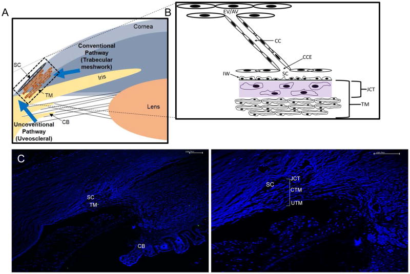
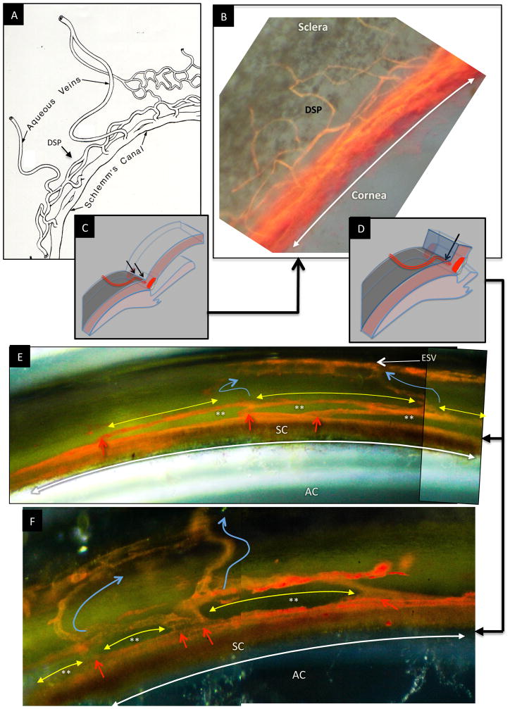
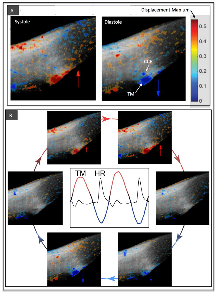
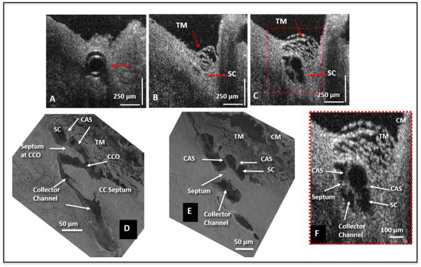
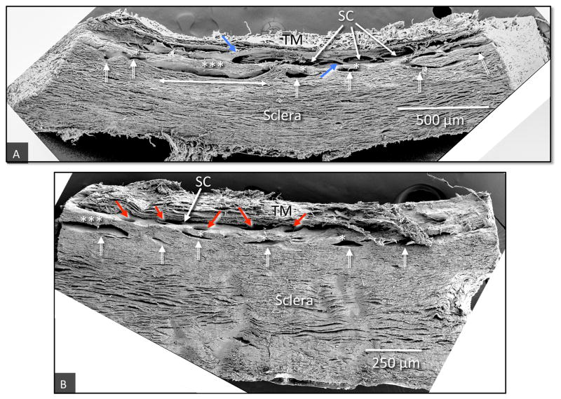
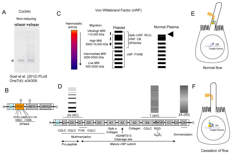
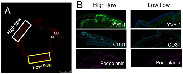
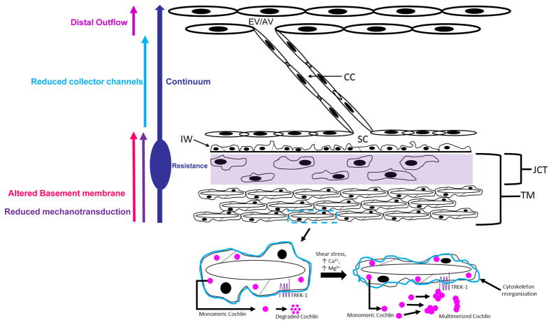
In glaucoma, lowered intraocular pressure (IOP) confers neuroprotection. Elevated IOP characterizes glaucoma and arises from impaired aqueous humor (AH) outflow. Increased resistance in the trabecular meshwork (TM), a filter-like structure essential to regulate AH outflow, may result in the impaired outflow. Flow through the 360° circumference of TM structures may be non-uniform, divided into high and low flow regions, termed as segmental. After flowing through the TM, AH enters Schlemm's canal (SC), which expresses both blood and lymphatic markers; AH then passes into collector channel entrances (CCE) along the SC external well. From the CCE, AH enters a deep scleral plexus (DSP) of vessels that typically run parallel to SC. From the DSP, intrascleral collector vessels run radially to the scleral surface to connect with AH containing vessels called aqueous veins to discharge AH to blood-containing episcleral veins. However, the molecular mechanisms that maintain homeostatic properties of endothelial cells along the pathways are not well understood. How these molecular events change during aging and in glaucoma pathology remain unresolved. In this review, we propose mechanistic possibilities to explain the continuum of AH outflow control, which originates at the TM and extends through collector channels to the episcleral veins.

Keywords: Basement membrane: turnover and stability; Collector channels; Continuum hypothesis; Deep scleral plexus; Distal outflow; Glaucoma; Mechanosensing; Segmental outflow; Trabecular meshwork; schlemm's canal.


Figures

Fig. 1
Fig. 1
Schematic diagram of outflow pathway and structures in the Trabecular Meshwork. (A) Schematic diagram depicting conventional and uveoscleral pathway in the anterior eye chamber. (B) A magnified view of trabecular meshwork (TM) depicting distal regions including collector channel entrances (CCE), collector channels (CC), episcleral vein (EV), and aqueous vein (AV). (C) A DAPI stained image of anterior chamber section showing ciliary body (CB), Schlemm’s canal (SC), TM (D) A magnified view of the same TM as in C, juxtacanalicular tissue (JCT), uveoscleral (UTM) and conventional (CTM) part of TM is as indicated.
Fig. 2
Fig. 2
Schematic diagram depicting anterior chamber regions (A) illustrating the relationship between Schlemm’s canal (SC), collector channel entrances (CCE) the deep intrascleral plexus (DSP) and the aqueous veins. (B) Microvascular cast of SC, the CCE, the (DSP) and aqueous veins that parallels the appearance of the schematic diagram in (A.) (C) Schematic illustration of the orientation of the microscope view from the corneoscleral surface of the image captured in (B). In views from the corneoscleral surface, collector channel entrances leave SC above and below the plane of section making it difficult to assess SC, CCE and DSP relationships. (D) Schematic illustration of the orientation of the microscope view taken through the cut corneal surface in images of (E) and (F). The view through the cut cornea captures the orientation of the CCE, and DSP adjacent to SC. (E, F) The red arrows identify locations where collector channel entrances arise from SC and connect with circularly oriented series of intrascleral channels coursing parallel to SC circumference (yellow arrows). The intrascleral channels communicate with one another providing a relatively continuous communicating ring. Blue arrows identify collecting vessels arising from the DSP that course radially through the sclera to the aqueous and episcleral veins on the surface of the eye. In the region between SC and the DSP running parallel to SC, a relatively thin layer of tissue is present (See Fig. 2, 8 & 9). The relatively long thin regions of tissue separating SC and the circularly oriented DSP are effectively hinged at their ends providing an anatomic relationship that ma provide the ability for the thin regions to move in response to IOP changes. (See Fig. 7–13) (A) is reproduced with permission from Hogan et al. (1971). [(B) non-human primate; m. nemestrina.] [(E) & (F); human.].
Fig. 3
Fig. 3
Data derived from imaging of cyclic, pulse-dependent tissue motion of the aqueous outflow system. (A) Live imaging of the aqueous outflow system in the temporal quadrant of the limbus of a normal human eye. The gray scale images are of structural details obtained by spectral domain optical coherence tomography (SD–OCT). Phase sensitive OCT (PhS–OCT) color displacement map information obtained from the same dataset is superimposed on the structural image and represents changes in displacement of trabecular meshwork (TM) and tissues surrounding collector channel entrances (CCE). The tissue motion results in lumen dimension changes of Schlemm’s canal, CCE and intrascleral collector channels. The increasing intensity in red represents the increase of displacement of tissue motion towards SC external wall and toward the sclera during systole when the pulse-induced IOP increases. Increasing intensity in blue indicates an increase of tissue displacement toward the anterior chamber during diastole when pulse-induced IOP decreases. (B) Arrows depict the image sequence around the periphery of the circle that encompasses one complete cardiac cycle. The central graph captures a tracing of trabecular meshwork (TM) bulk tissue motion over time using PhS-OCT. The heart rate (HR) tracing demonstrates that the TM motion is synchronous with the cardiac cycle but with a time delay. Reproduced with permission from Johnstone (2016).
Fig. 4
Fig. 4
Representative two-dimensional (2-D) structural OCT and scanning electron microscopy images from the limbal region of an eye. (A) shows the OCT image captured with the cannula inside Schlemm’s canal (SC) (arrow). The image location in (B) is ~150 μm away from the cannula tip and shows SC configuration before the infusion of perfusate. Arrows identify the TM and SC. In (C) the maximally dilated appearance of the same segment is visible after infusion of perfusate. (D) and (E) are representative SEM images from a radial limbal region, illustrating structural features of the outflow system that are mirrored in both the SEM and OCT images. (D) shows a collector channel entrance or ostia (CCO). A septum is present at the CCO that is attached to the TM by cylindrical attachment structures (CAS). (E) Is the adjacent section from the same segment showing the transition from the region of a CCO in (D) to a circumferentially oriented collector channel. (F) The 2X enlargement OCT image that is cropped from (C) permits a more detailed comparison of relationships. CM, ciliary muscle. Adopted from S. Hariri et al. Platform to investigate aqueous outflow system structure and pressure-dependent motion using high-resolution spectral domain optical coherence tomography. [non-human primate; M. nemestrina] Reproduced with permission from Hariri et al. (2014).
Fig. 5
Fig. 5
The 2-D OCT image from Fig. 11 and parameters derived from images. (A) SC and its adjacent collector channel (CC) are at their dilation maximum after a pulsed infusion. Parameters for quantification are shown: SC height, purple line; CC height, blue line; CAS height, red line; SC area, yellow line; CC area, green line. (B) Progressive increase in the height of SC (black curve), CC (red curve), and CAS (blue curve) with time. (C) The time-dependent change in SC lumen area. (D) The time-dependent change in CC lumen area. Height changes plateau in ~ 300 msec. Adopted from: S. Hariri et al. Platform to investigate aqueous outflow system structure and pressure-dependent motion using high-resolution spectral domain optical coherence tomography. J Biomed Opt 19(10) 106013. (2014) [non-human primate; m. nemestrina] Reproduced with permission from Hariri et al. (2014).
Fig. 6
Fig. 6
Pathway of Aqueous Humor: Scanning electron microscopy of aqueous outflow pathway from the anterior chamber (AC) through the trabecular meshwork (TM) to Schlemm’s canal (outlined in blue). Blue arrows denote further aqueous passage through the deep intrascleral plexus (DSP) into the more superficial plexus of intrascleral collector channels (ISCC) Aqueous finally enters the episcleral (AV-E) and conjunctival (AV-C) aqueous veins. CB = ciliary body. Red dashed rectangle identifies region shown in Fig. 7. [non-human primate; m. nemestrina] Reproduced with permission from Johnstone (2016).
Fig. 7
Fig. 7
Collector channel and hinged collagen flaps or leaflets in deep scleral plexus: From area of Fig. 7 outlined in red. Schlemm’s canal (SC) opens into a collector channel entrance (CCE). Collagen leaflets or flaps hinged at their scleral attachment (HCF) are present surrounding the convoluted pathway into the intrascleral collector channels (ISCC). Black T denotes the hinge locations. Area outlined in green denotes the juxtacanalicular space between the trabecular lamellae and SC inner wall. Cylindrical structures attach between SC inner wall and the HCF (*). If the TM moves, the HCF also will also move because of the connections between the structures. Reproduced with permission from Johnstone (2016).
Fig. 8
Fig. 8
The ultrastructure of region around Schlemm’s canal under various intraocular pressure (IOP). (A) IOP below episcleral venous pressure (EVP) during in vivo fixation. The trabecular meshwork (TM) is collapsed and far from the corneoscleral (CSW) of SC. Schlemm’s canal (SC) is dilated and filled with blood. A collagenous flap-like structure continuous with the collagen lamellae of the sclera protrudes into SC. The flap is only attached or anchored at one end with the other end unattached creating a hinged flap arrangement (HCF). The HCF is far from SC external wall. (B1) IOP of 25 mm Hg during in vivo fixation. (B2) In additional serial sections the HCF of (A) developed an attachment to the corneoscleral wall thus developing into a septum (SEP) that creates an intrascleral collector channel. (B3) In further serial sections, a more robust attachment developed between the SEP and the CSW. In each of the 3 images (B1–3), the TM is distended and SC is little more than a potential space with some areas of TM inner wall apposition to the corneoscleral wall of SC. A HCF at the entrance of the CCE was found in serial sections to be intermittently appositional (app) to SC external wall. Serial sections revealed that the SEP in (B) and (C) had intermittent areas of apposition to the CSW. At the hinge region (white arrows) a change in the orientation of the collagen lamellae of was regularly seen in serial sections. Reproduced with permission from Johnstone (2016). (non-human primate; M. mulatta).
Fig. 9
Fig. 9
Scanning electron microscopy of tilted frontal sections providing images of regions of the limbal circumference that include the trabecular meshwork, (TM), Schlemm’s canal (SC) and a series of intrascleral collector channels ISCC (white double bar arrows) in the deep scleral plexus. (A) The ISCC are distributed in a relatively narrow region close to SC. Rather than being round the ISCC have long dimensions’ parallel to and a short dimension perpendicular to SC. The ISCC configuration results in thin septa (*), attached at their ends to the sclera that are at times have a length many times their height (***). Blue arrows identify cylindrical structures crossing SC that connect the trabecular meshwork with regions of SC external wall near CCE. (B) The collector channel entrances (CCE) are depicted by red arrows (non-human primate, M. fascicularis).
Fig. 10
Fig. 10
Assessment of basement membrane (BM) proteins in the trabecular meshwork (TM) region. (A–B) Probing consecutive sections of TM with different collagen IV antibodies shows different distribution patterns. (C) DAPI stained image of the same TM region. (D) Western blot analysis for type IV collagen demonstrates more fragmentation and less intact collagen IV in normal controls compared to POAG TM. (E) Linear mode MALDI-TOF analyses for Wolframin (WFS1) shows more intact protein in the POAG compared to control TM. (F) The GAPDH normalized levels for all proteins as indicated suggest more intact BM and BM interactors in the POAD TM compared to controls [adopted from Goel et al. (2012) PLoS One7(4): e34309].
Fig. 11
Fig. 11
Basement membrane changes in ageing and glaucoma. (A–D) The basement membrane (BM) of inner limiting membrane (ILM) at the vitreoretinal surface of the retina undergoes a significant thickening due to ageing from fetal to 83-years-old as indicated. Adopted with permission from Candiello et al. (2010) Matrix Biology 29: 402–10. (E) The representative BM changes in the intra-trabecular space in transmission electron micrographs (TEM) en-bloc staining with uranyl acetate (Alcian Blue 8GX). As indicated, control (55-year-old) and primary open angle glaucoma (POAG; 55-year-old), both male cadaver donor eyes respectively. (F) Assessment of intra-trabecular BM thickness from 50 locations each from individual TEM images from 6 control and POAG donors each (age 55–59, equal distribution of genders). Asterisk indicate significance (p<0.05) using two-tailed equal variance t-test.
Fig. 12
Fig. 12
Potential in silico predicted interactors of cochlin including inner ear known basement membrane (BM) proteins. The balance of body depends on incremental movement of inner ear fluid. Such motions are often infinitesimally small in magnitude. In silico cochlin is predicted to interact with α-tectorin (TECTA), Wolframin 1 (WFS1), Diaphanous like formin-1 (DIAPH1 or DRF-1) as indicated by arrow. The α-tectorin is a bonafide BM protein of inner ear tectorial membrane some of its interactors have been recently predicted to be part of BM such as ruffle membranes. Mutation in WFS1, Gasdermin and DIAPH1 have been found to be associated with eye diseases.
Fig. 13
Fig. 13
Solution phase mechanosensing in the Trabecular meshwork. (A) Multimerization of cochlin under shear stress and under non-reducing conditions [adopted from Goel et al. (2012) PLoS One7(4): e34309]. (B) The schematic depiction of domains in cochlin protein. The factor C homology domain (FCH) identified mutations have been shown to play in progressive hearing disease DFNA9, which is associated with inner ear fluid flow homeostasis. Two von willebrand factor A domains (vWFA1 &2) and glycosylation sites (G) are as indicated. (C) Schematic depiction of different von Willebrand factor (vWF) multimer sizes and their known role in hemostatic activity (the spectrum has been indicated; arrow indicates decreased activity). Ultralarge MW (ULMW) forms are found in platelet or endothelial cells but are absent in normal plasma (arrow head). Dashed arrow indicates decreased binding to proteins as indicated such as ricosetin (vWF: Rco) or collagen (vWF: CB). (D) The different vWF domains, their binding propensity with proteins and role in multimer formation. ADAMTS13 cleavage site as indicated. The multimers for three vWF disease conditions have been shown (1, 2A IIC and 2A IID). (E, F) Mechanosensing by polycystein1 (PC1) in kidney. (F) The truncated PC1enters nucleus and stops gene expression as a consequence of flow cessation.
Fig. 14
Fig. 14
A representative optical coherence tomography image of distal outflow region in a human eye. Circumferential limbal scans in the original study were processed and assembled manually in 3D space to yield a full casting of the episcleral and intrascleral venous plexus throughout the limbus in-situ during active perfusion. Adopted with permission from Kagemann et al. Exp. Eye Res. (2011) 93 308–315.
Fig. 15
Fig. 15
Distal outflow is likely to be impaired in pathologic state (glaucoma). (A) The DBA/2J hypertensive mouse shows regions of high and low flow, which is found to be associated with low and high elastic modulus respectively. Trabecular meshwork (TM) and Schlemm’s canal (SC) located as indicated. (B) The adjacent regions of TM in DBA/2J hypertensive mouse demonstrates presence of elevated levels of vessel markers LYVE-1, CD31 and Podoplanin in high flow regions contrast to low flow as indicated. The distal flow regions in DBA/2J mouse are non-uniform. These regions have been shown to have high non-uniformity and abnormalities in human glaucoma subjects compared to controls.
Fig. 16
Fig. 16
The proposed model of continuum. Reduced or altered mechanotransduction in the TM due to alteration of soluble mechanosensing molecules or their deposition results in pathology in the TM. At all levels of TM including Schlemm’s canal (SC), the basement membrane degradation is impaired resulting in lack of generation of pro- and anti-angiogenic molecules such as endostatin, canstatin, tumstatin fragments of type IV collagen. There is observed reduced collector channel (CC) frequency and/or dimension in the surrounding region of TM. The fine regulation of degraded protein fragments of BM may be involved in regulation of CCs and distal flow regions. The model supports an integrated pathology encompassing TM, SC, CCs and distal outflow regions. JCT= juxtacanalicular tissue, IW inner wall of SC, AV = aqueous veins, EV= episcleral vein.

Similar articles

Cited by

References

    1. Anderson DR. Glaucoma: the damage caused by pressure. XLVI Edward Jackson memorial lecture. Am J Ophthalmol. 1989;108:485–495. - PubMed
    1. Anderson DR. Collaborative normal tension glaucoma study. Curr Opin Ophthalmol. 2003;14:86–90. - PubMed
    1. Ascher KW. Physiologic importance of the visible elimination of intraocular fluid. Am J Ophth. 1942;25:1174–1209. - PubMed
    1. Ascher KW. Backflow phenomena in aqueous veins. Am J Ophth. 1944;27:1074–1076.
    1. Ascher KW. Aqueous veins and their significance for pathogenesis of glaucoma. Arch Ophthal. 1949;42:66–76. - PubMed

Publication types