. 2016 Dec 16:9:90.
doi: 10.1186/s13039-016-0297-x. eCollection 2016.

Inherent variability of cancer-specific aneuploidy generates metastases

Affiliations

Inherent variability of cancer-specific aneuploidy generates metastases

Mathew Bloomfield et al. Mol Cytogenet. .

Abstract

Background: The genetic basis of metastasis is still unclear because metastases carry individual karyotypes and phenotypes, rather than consistent mutations, and are rare compared to conventional mutation. There is however correlative evidence that metastasis depends on cancer-specific aneuploidy, and that metastases are karyotypically related to parental cancers. Accordingly we propose that metastasis is a speciation event. This theory holds that cancer-specific aneuploidy varies the clonal karyotypes of cancers automatically by unbalancing thousands of genes, and that rare variants form new autonomous subspecies with metastatic or other non-parental phenotypes like drug-resistance - similar to conventional subspeciation.

Results: To test this theory, we analyzed the karyotypic and morphological relationships between seven cancers and corresponding metastases. We found (1) that the cellular phenotypes of metastases were closely related to those of parental cancers, (2) that metastases shared 29 to 96% of their clonal karyotypic elements or aneusomies with the clonal karyotypes of parental cancers and (3) that, unexpectedly, the karyotypic complexity of metastases was very similar to that of the parental cancer. This suggests that metastases derive cancer-specific autonomy by conserving the overall complexity of the parental karyotype. We deduced from these results that cancers cause metastases by karyotypic variations and selection for rare metastatic subspecies. Further we asked whether metastases with multiple metastasis-specific aneusomies are assembled in one or multiple, sequential steps. Since (1) no stable karyotypic intermediates of metastases were observed in cancers here and previously by others, and (2) the karyotypic complexities of cancers are conserved in metastases, we concluded that metastases are generated from cancers in one step - like subspecies in conventional speciation.

Conclusions: We conclude that the risk of cancers to metastasize is proportional to the degree of cancer-specific aneuploidy, because aneuploidy catalyzes the generation of subspecies, including metastases, at aneuploidy-dependent rates. Since speciation by random chromosomal rearrangements and selection is unpredictable, the theory that metastases are karyotypic subspecies of cancers also explains Foulds' rules, which hold that the origins of metastases are "abrupt" and that their phenotypes are "unpredictable."


Figures
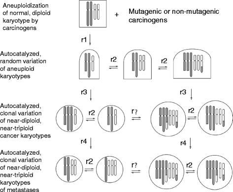
Fig. 1
Fig. 1
Karyotypic theory of metastasis. According to the karyotypic theory mutagenic and non-mutagenic carcinogens induce random aneuploidy in normal cells at carcinogen-dependent rates, r1. Aneuploidy then auto-catalyzes further random karyotypic variation, because it unbalances thousands of genes including mitosis-genes at aneuploidy-dependent rates, r2. Most such random variants perish. Rare karyotypic variants, however, form new autonomous cancer species with near-diploid, hypo-diploid or hyper-diploid karyotypes at very low rates, r3, because the probability to form a new autonomous cell is very low [–77, 104, 105, 109, 135]. Owing to the inherent instability of aneuploidy, the karyotypes of new cancer species vary at aneuploidy-dependent rates, r2, within margins that are defined by selection for cancer-specific autonomy. As a result of this inherent variability of cancer karyotypes, new autonomous karyotypic subspecies with metastatic phenotypes arise stochastically. Since the probability of forming new autonomous subspecies by random variation of cancer karyotypes is very low – as in conventional subspeciation – metastases would occur typically at low rates, r4, such as the 10^-8 per-cell rate described in the Background in reference [9]. These rates are still higher than those of carcinogenesis from normal cells, r3 [136]. The Figure also records graphically our results that the karyotypic complexity of cancers is highly conserved in metastases. Thus hyper-triploid cancers formed hyper-triploid metastases and near-diploid cancers formed near-diploid metastases. There was no evidence of spontaneous alterations of the clonal karyotypic complexity of cancers or metastases; hence ‘r?’ in Fig. 1
Fig. 2
Fig. 2
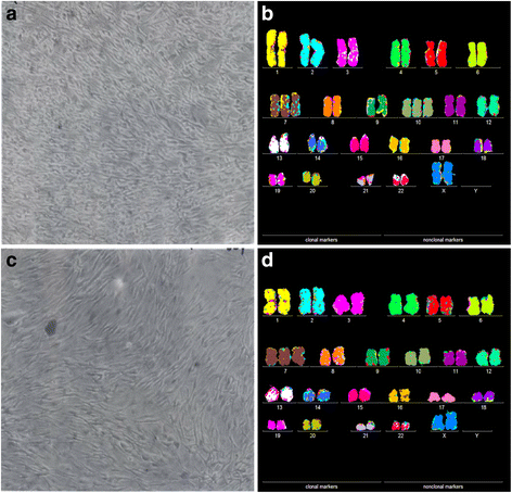
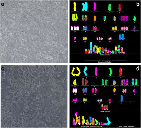
Cellular morphologies and karyotypes of breast cancer HIM-2 (a, b) and of a corresponding brain metastasis HIM-5 (c, d). The comparisons show that the primary cancer HIM-2 and corresponding metastasis HIM-5 have very similar but distinct cell morphologies (a, c) and karyotypes (b, d). Both primary cancer and metastasis have near diploid karyotypes with the same numbers and structures of chromosomes, except for a trisomy 10 that is missing in the metastasis HIM-5 (d). The cells were photographed at 120X in cell culture dishes (Methods). The cells of the primary cancer were found to be more refractive and growing at modestly higher rates than the metastasis. The chromosomes were prepared from metaphase-cells and color-coded as described in Methods
Fig. 3
Fig. 3
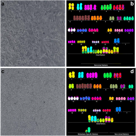
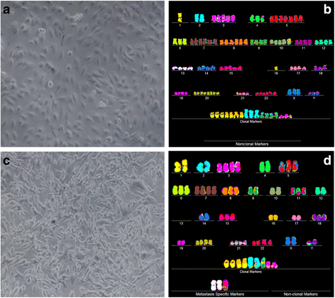
Cellular morphologies and karyotypes of melanoma WM-115 (a, b) and a corresponding metastasis WM-266-4 (c, d). The comparisons show that the primary cancer WM-115 and the corresponding metastasis WM-266-4 have similar, but distinct cell morphologies (a, c) and karyotypes (b, d). Both primary cancer and metastasis have hyper-triploid karyotypes with similar numbers of chromosomes and aneusomies and both lack intact chromosome 9. They also differ from each other in the total numbers of chromosomes and in the structures of some marker chromosomes (see Tables 1 and 2). The absence of normal chromosomes 6 from the presumed primary and the presence of "primary specific" marker chromosomes 6 indicate that both the presumed primary and the metastasis derived from an unknown primary with normal chromosomes 6. The cells were propagated and karyotyped as described for Fig. 2
Fig. 4
Fig. 4
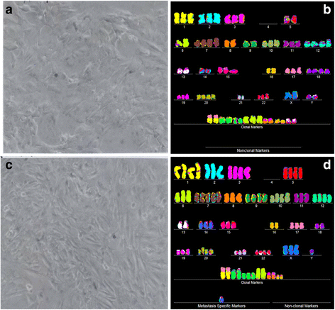
Cellular morphologies and karyotypes of liver cancer H2P (a, b) and a corresponding portal vein metastasis H2M (c, d). The comparisons show that the primary cancer H2P and the corresponding metastasis H2M have similar, but distinct cell morphologies (a, c) and karyotypes (b, d). Both primary cancer and metastasis have hyper-triploid karyotypes with similar numbers of chromosomes and aneusomies, and both lack intact chromosome 4. They differ from each other in the total numbers of chromosomes and in the structures of some marker chromosomes (see Tables 1 and 2). The cells were propagated and karyotyped as described for Fig. 2
Fig. 5
Fig. 5
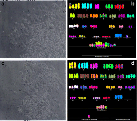
Cellular morphologies and karyotypes of medulloblastoma M-458 (a, b) and a corresponding metastasis M-425 (c, d). The comparisons show again that the primary cancer M-458 and the corresponding metastasis M-425 have similar, but distinct cell morphologies (a, c) and karyotypes (b, d). Some of the metastatic M-425 cells grew in suspension (One reason, why M-425 was named the metatasis. See note above in this section.), while the rest was attached to the culture dish. The karyotypes of both the primary cancer and the metastasis are hyper-triploid and have similar numbers of chromosomes and of aneusomies. But they also differ in the total numbers of chromosomes and in the structures of some individual marker chromosomes (see Tables 1 and 2). The cells were propagated and karyotyped as described for Fig. 2
Fig. 6
Fig. 6
Cellular morphologies and karyotypes of colon cancer SW-480 (a, b) and of a corresponding metastasis SW-620 (c, d). The comparisons show once more that the primary cancer SW-480 and the corresponding metastasis SW-620 have similar, but distinct cell morphologies (a, c) and karyotypes (b, d). Both primary cancer and metastasis have hyper-diploid karyotypes with similar numbers of chromosomes and of aneusomies. They also differ from each other in the total numbers of chromosomes and in the structures of some marker chromosomes (see Tables 1 and 2). We adduce evidence in the text that both SW-480 and SW-620 are probably both metastases of an unknown primary. The cells were propagated and karyotyped as described for Fig. 2
Fig. 7
Fig. 7
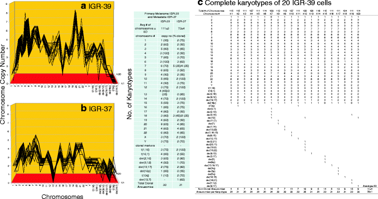
Cellular morphologies and karyotypes of melanoma IGR-39 (a, b) and of a corresponding metastasis IGR-37 (c, d). The comparisons show once more that the primary melanoma IGR-39 and the corresponding metastasis IGR-37 have similar, but distinct cell morphologies (a, c) and that their karyotypes are closely related but differ in ploidy number (b, d). The primary cancer has a hyper-tetraploid karyotype and the metastasis a closely related, but hyper-triploid karyotype. The relationship is based on chromosome copy numbers that differ from each other by the ploidy factor that sets apart the two karyotypes. The karyotypes also differ from each other in several individual chromosome numbers and in several individual marker chromosomes (see Tables 1 and 2). The cells were propagated and karyotyped as described for Fig. 2 and in the text
Fig. 8
Fig. 8
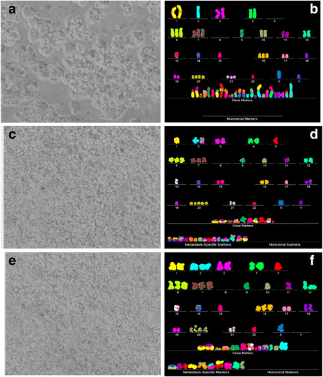
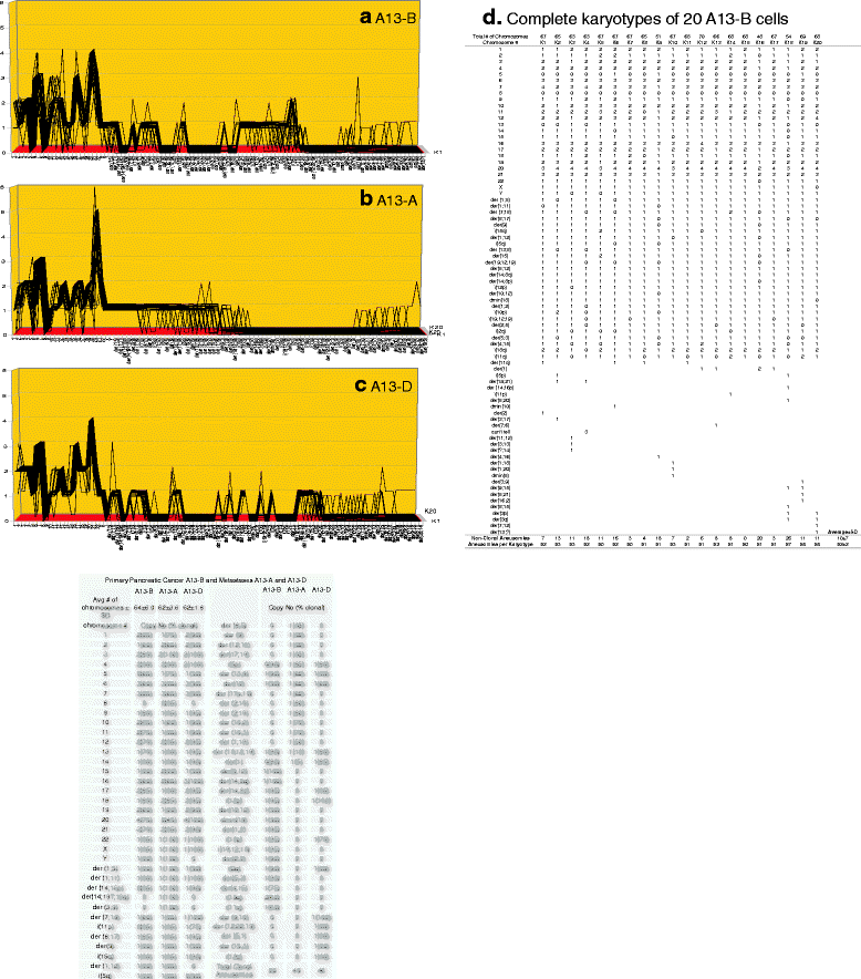
Cellular morphologies and karyotypes of pancreatic cancer A13-B (a, b) and of two corresponding metastases, a pancreatic metastasis A13-A (c, d) and a liver metastasis A13-D (e, f). The comparisons show once more that the primary cancer A13-B and the corresponding metastases A13-A and A13-D have similar, but distinct cell morphologies (a, c, e) and karyotypes (b, d, f). The primary cancer and both metastases have hyper-diploid karyotypes with similar numbers of chromosomes and of aneusomies. The primary and the two metastases differ from each other in the total numbers of chromosomes and in the structures of some individual marker chromosomes see Tables 1 and 2). The cells were propagated and karyotyped as described for Fig. 2 [103]
Fig. 9
Fig. 9
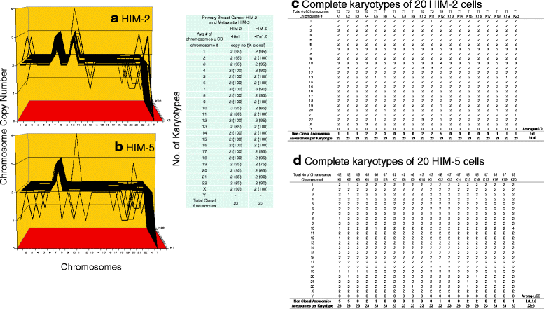
a, b, c, d Karyotypic evidence that the brain metastasis HIM-5 is an individual subspecies of the breast cancer HIM-2. The karyotypic theory of metastasis predicts that metastases have individual clonal karyotypes that differ from those of parental cancers in individual metastasis-specific aneusomies. To test this theory we have compared karyotype-arrays of the brain metastasis HIM-5 to that of the primary cancer HIM-2. Karyotype arrays are three-dimensional tables of 20 karyotypes, which list the chromosome numbers of arrayed karyotypes on the x-axis, the copy numbers of each chromosome on the y-axis, and the number of karyotypes arrayed on the z-axis, as detailed in Results (Section, Metastases are karyotypic subspecies of cancers). Figure 9 a, b and the attached table show that 85 to 100% of the chromosomes of the cancer HIM-2 and the metastasis HIM-5 were clonal, and that cancer and metastasis formed very similar clonal patterns. The karyotype of the metastasis differed from that of the primary only in the loss of trisomy 10. The copy numbers of the non-clonal chromosomes differed from the clonal averages typically  ± 1 (see Fig. 9 a,b and specifically Fig. 9 c,d). These non-clonal copy numbers represent the ongoing karyotypic variation predicted by the inherent variability of cancer-specific aneuploidy (Background). We conclude that the brain metastasis HIM-5 is a subspecies of the parental breast cancer HIM-2
Fig. 10
Fig. 10
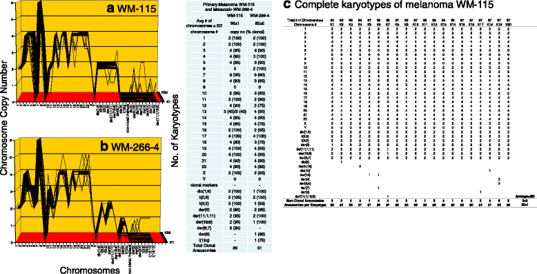
a, b, c Karyotypic evidence that the metastasis WM-266-4 is an individual subspecies of melanoma WM-115. The karyotypic theory of metastasis predicts that metastases have individual clonal karyotypes that differ from those of parental cancers in individual metastasis-specific aneusomies. To test this theory we have compared karyotype-arrays of the melanoma metastasis WM-266-4 to that of the primary cancer WM-115 prepared as described for Fig. 9. Figure 10 a, b and the attached table show that 80 to 100% of the chromosomes of the metastasis WM-266-4 and of the cancer WM-115 were clonal, and that cancer and metastasis both formed very similar clonal patterns. The karyotype of the metastasis differed from that of the primary cancer in about 13 of an average of 31 aneusomies (Fig. 10 a, b, c and Table 1). The copy numbers of the non-clonal chromosomes differed from clonal averages ± 1; there were also several non-clonal marker chromosomes (Fig. 10 a, b, c). These non-clonal chromosomes represent the ongoing karyotypic variation predicted by the inherent variability of cancer-specific aneuploidy (see Fig. 10 a, b and specifically Fig. 10c, and Background). We conclude that the melanoma metastasis WM-266-4 is a subspecies of the parental melanoma WM-115
Fig. 11
Fig. 11
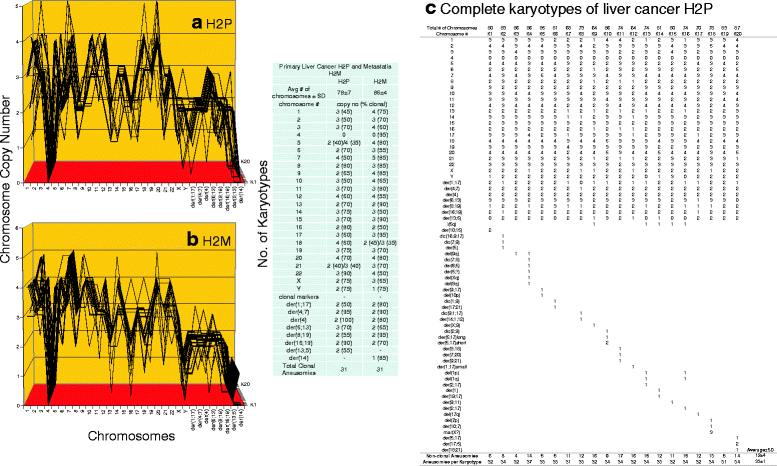
a, b, c Karyotypic evidence that the metastasis H2M is an individual subspecies of liver cancer H2P. The karyotypic theory of metastasis predicts that metastases have individual clonal karyotypes that differ from those of parental cancers in individual metastasis-specific aneusomies. To test this theory we have compared karyotype-arrays of the metastasis H2M to that of the primary cancer H2P prepared as described for Fig. 9. Figure 11 a, b and the attached table show that 45–80% of the chromosomes of cancer H2P and 50–90% of the chromosomes of metastasis H2M were clonal, and that cancer and metastasis formed similar clonal patterns. The karyotype of the metastasis differed from that of the primary cancer in about 15 of an average of 31 H2M aneusomies (Fig. 11 a, b, c and Table 1). The copy numbers of non-clonal chromosomes including marker chromosomes differed from clonal averages ± 1 (see Fig. 11 a, b and specifically Fig. 11c). The chromosomes with non-clonal copy numbers represent the ongoing karyotypic variation predicted by the inherent variability of cancer-specific aneuploidy (See Fig. 11c and Background). We conclude that the metastasis H2M is a subspecies of the parental liver cancer H2P
Fig. 12
Fig. 12
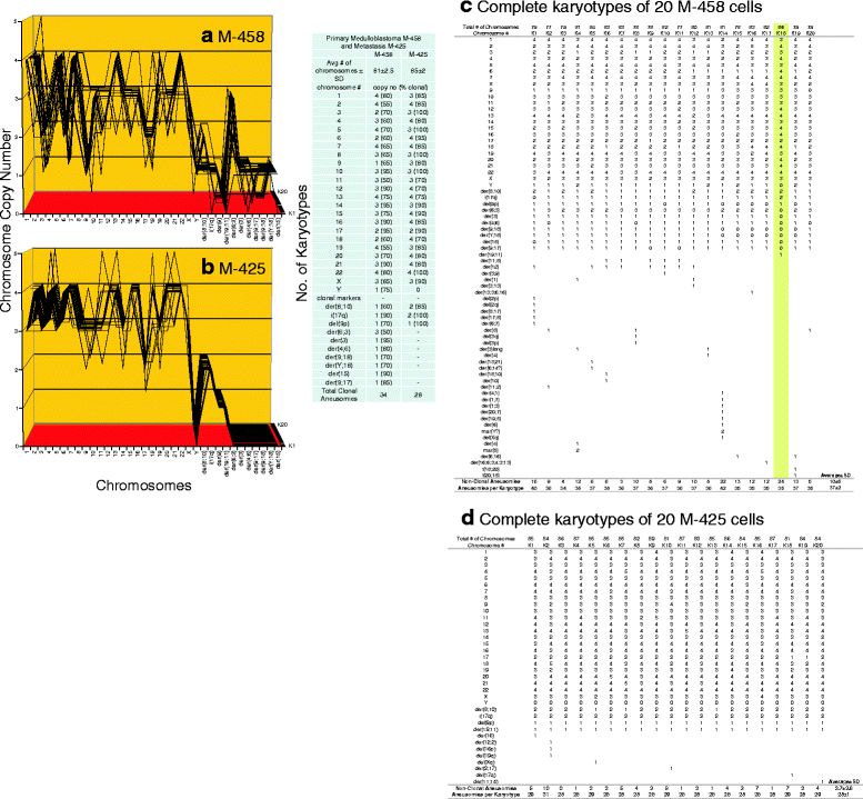
a, b, c, d Karyotypic evidence that the metastasis M-425 is an individual subspecies of medulloblastoma M-458. The karyotypic theory of metastasis predicts that metastases have individual clonal karyotypes that differ from those of parental cancers in individual metastasis-specific aneusomies. To test this theory we have compared karyotype-arrays of the metastasis M-425 to that of the primary cancer M-458 prepared as described for Fig. 9. Figure 12 a, b and the attached table show that 55–90% of the chromosomes of the cancer M-458 and 70–100% of the chromosomes of the metastasis M-425 were clonal, and that cancer and metastasis formed similar clonal patterns. The fact that the karyotype of M-425 was more clonal than that of M-458, again supports the view that M-425 is the metastasis and M-458 the original cancer (See comment regarding this question in section "Karyotypic and phenotypic relationships between metastases and parental cancers"). The karyotype of the metastasis differed from that of the primary cancer in about 17 of an average of 28 M-425 aneusomies (Fig. 12 a, b, c, d and Table 1). The copy numbers of most non-clonal chromosomes including marker chromosomes differed from clonal averages ± 1 (see Fig. 12 a, b and specifically Fig. 12 c, d). The chromosomes with non-clonal copy numbers represent the ongoing karyotypic variation predicted by the inherent variability of cancer-specific aneuploidy (See Fig. 11c and Background). We conclude that the metastasis M-425 is subspecies of the parental medulloblastoma M-458
Fig. 13
Fig. 13
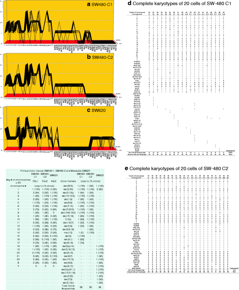
a, b, c, d, e Karyotypic evidence that the metastasis SW-620 is an individual subspecies of SW-480 or of an unknown common precursor. The karyotypic theory of metastasis predicts that metastases have individual clonal karyotypes that differ from those of parental cancers in individual metastasis-specific aneusomies. To test this theory we have compared karyotype-arrays of the metastasis SW-620 to that of two clones of the presumed primary SW-480, prepared as described for Fig. 9. Figure 13 a, b, c and the attached table show that 75 - 100% of the chromosomes of SW-620 and 53–100% of the chromosomes of SW-480 C1 and 75-100% of the chromosomes of SW-480 C2 were clonal, and that the two cancer clones and the metastasis formed similar clonal patterns. The karyotype of the metastasis differed from that of the primary cancer SW-480 C1 in 25 of 38 average SW-480 C1 aneusomies and differed from SW-480 C2 in 27 of 38 average aneusomies (Fig. 13a, b, c, d, e and Table 1). The copy numbers of most non-clonal chromosomes including marker chromosomes differed from clonal averages ± 1 (Fig. 13a, b, c and specifically Fig. 13d, e). The chromosomes with non-clonal copy numbers represent the ongoing karyotypic variation predicted by the inherent variability of cancer-specific aneuploidy (See Fig. 11d, e and Background). We conclude that the metastasis SW-620 is a subspecies of the parental colon cancer SW-480 or of a common unknown precursor
Fig. 14
Fig. 14
a, b, c Karyotypic evidence that the metastasis IGR-37 is an individual subspecies of melanoma IGR-39. The karyotypic theory of metastasis predicts that metastases have individual clonal karyotypes that differ from those of parental cancers in individual metastasis-specific aneusomies. To test this theory we have compared karyotype-arrays of the metastasis IGR-37 to that of the primary cancer IGR-39 prepared as described for Fig. 9. Figure 14 a, b, c and the attached table show that 55–100% of the chromosomes of the parental cancer IGR-39 and 50–100% of the chromosomes of the metastasis IGR-37 were clonal, and that cancer and metastasis formed similar clonal patterns. These patterns show, however, that metastasis coincided with a reduction in the ploidy of the parental cancer from hyper-tetraploid to hyper-triploid. Moreover, the karyotype of the metastasis differed from that of the primary cancer in about 27 of an average of 31 metastasis-specific aneusomies (Fig. 14 a, b, c and Table 1). Since the ploidy-shift changed the relative chromosome copy numbers of many aneusomies, the percentage of metastasis-specific aneusomies is, however, larger than if it were based on qualitative differences only (see Table 1). As in all other hyper-diploid cancers, the copy numbers of most non-clonal chromosomes including marker chromosomes differed from clonal averages ± 1 (Fig. 14 a, b and specifically Fig. 14c). Again, the chromosomes with non-clonal copy numbers represent the ongoing karyotypic variation predicted by the inherent variability of cancer-specific aneuploidy (See Background). We conclude that the metastasis IGR-37 is a subspecies of the parental melanoma IGR-39
Fig. 15
Fig. 15
a, b, c, d Karyotypic evidence that the metastases A13-A and A13-D are individual subspecies of the pancreatic cancer A-13B. The karyotypic theory of metastasis predicts that metastases have individual clonal karyotypes that differ from those of parental cancers in individual metastasis-specific aneusomies. To test this theory we have compared the karyotype-arrays of the metastases A13-A and A13-D to that of the primary cancer A-3B. The karyotype arrays were again prepared as described for Fig. 9. Figure 15 a, b, c and the attached table show that the chromosomes of the cancer were 55–95% clonal and that of the chromosomes of metastasis A13-A were 75–100% and those of metastasis A13-D were 75–100% clonal, and that all three cancers formed related clonal patterns. As shown in Table 1, the karyotype of the metastasis A13-A differed from that of the primary cancer A13-B in 27 of 49 aneusomies and metastasis A13-D differed from that of the primary in 16 of 49 aneusomies (Fig. 15 a, b, c, d). The copy numbers of most non-clonal chromosomes including marker chromosomes differed from clonal averages ± 1 (Fig. 15 a, b, c and specifically Fig. 15d). The chromosomes with non-clonal copy numbers represent the ongoing karyotypic variation predicted by the inherent variability of cancer-specific aneuploidy (See Background). We conclude that the metastases A13-A and D are subspecies of the parental pancreatic cancer A13-B

Similar articles

Cited by

References

    1. New Oxford American Dictionary: 2010, 2013 by Oxford University Press, Inc
    1. Foulds L. Multiple etiologic factors in neoplastic development. Cancer Res. 1965;25(8):1339–1347. - PubMed
    1. Weinberg RA. The biology of cancer, Second edition. New York; London: Garland Science; 2014.
    1. Alberts B, Johnson A, Lewis J, Raff M, Roberts K, Walter P. Molecular Biology of the Cell. New York: Garland; 2014.
    1. Foulds L. Tumor progression: a review. Cancer Res. 1954;14:327–339. - PubMed

LinkOut - more resources