Review
. 2017 Jan;97(1):411-463.
doi: 10.1152/physrev.00031.2014.

Ghrelin, CCK, GLP-1, and PYY(3-36): Secretory Controls and Physiological Roles in Eating and Glycemia in Health, Obesity, and After RYGB

Affiliations
Review

Ghrelin, CCK, GLP-1, and PYY(3-36): Secretory Controls and Physiological Roles in Eating and Glycemia in Health, Obesity, and After RYGB

Robert E Steinert et al. Physiol Rev. 2017 Jan.

Erratum in

  • Corrigendum.
    [No authors listed] [No authors listed] Physiol Rev. 2017 Jul 1;97(3):1229. doi: 10.1152/physrev.z9j-2792-corr.2011. Physiol Rev. 2017. PMID: 28615464 Free PMC article. No abstract available.

Abstract

The efficacy of Roux-en-Y gastric-bypass (RYGB) and other bariatric surgeries in the management of obesity and type 2 diabetes mellitus and novel developments in gastrointestinal (GI) endocrinology have renewed interest in the roles of GI hormones in the control of eating, meal-related glycemia, and obesity. Here we review the nutrient-sensing mechanisms that control the secretion of four of these hormones, ghrelin, cholecystokinin (CCK), glucagon-like peptide-1 (GLP-1), and peptide tyrosine tyrosine [PYY(3-36)], and their contributions to the controls of GI motor function, food intake, and meal-related increases in glycemia in healthy-weight and obese persons, as well as in RYGB patients. Their physiological roles as classical endocrine and as locally acting signals are discussed. Gastric emptying, the detection of specific digestive products by small intestinal enteroendocrine cells, and synergistic interactions among different GI loci all contribute to the secretion of ghrelin, CCK, GLP-1, and PYY(3-36). While CCK has been fully established as an endogenous endocrine control of eating in healthy-weight persons, the roles of all four hormones in eating in obese persons and following RYGB are uncertain. Similarly, only GLP-1 clearly contributes to the endocrine control of meal-related glycemia. It is likely that local signaling is involved in these hormones' actions, but methods to determine the physiological status of local signaling effects are lacking. Further research and fresh approaches are required to better understand ghrelin, CCK, GLP-1, and PYY(3-36) physiology; their roles in obesity and bariatric surgery; and their therapeutic potentials.


Figures

FIGURE 1.
FIGURE 1.
Schematic depictions of the localization of enteroendocrine cells and changes after RYGB. A: distribution of enteroendocrine cells secreting ghrelin, CCK, GLP-1, and PYY in the stomach (pink), duodenum (yellow), jejunum (green), and ileum (violet). Black areas indicate the relative densities of expression of enteroendocrine cells producing the hormones indicated. Enteroendocrine cells secreting particular hormones were initially categorized histologically, e.g., I cells for CCK, L cells for enteroglucagons and PYY, etc. (166, 567, 591). It is now clear, however, that this categorization is not a reliable guide to hormone secretion. Rather, individual enteroendocrine cells secrete variable mixtures of hormones (231, 303, 597, 738). Bottom salmon rectangle, proximal large intestine. B: intact gastrointestinal tract (left) and gastrointestinal rearrangement after RYGB (right). Pink areas are stomach, salmon areas are large intestine (∼1.5 m long in healthy adults), yellow is duodenum (typically ∼25 cm long), green is jejunum (∼2–3 m), and violet is ileum (∼3–4 m). For RYGB, the stomach is divided into a small upper pouch with a volume of ∼25 ml and an isolated gastric remnant, the small intestine is divided ∼50 cm from the pylorus, and the distal limb of the small intestine (Roux or alimentary limb) is brought up to the gastric pouch and connected to it by an end-to-side gastroenterostomy. As a result, ingested food enters the small gastric pouch and empties directly into the jejunum. The gastric remnant and isolated ∼50 cm of small intestine (“biliopancreatic limb”) is connected to the jejunum ∼150 cm distal to the gastroenterostomy. The small intestine distal to the anastomosis is called the common channel.
FIGURE 2.
FIGURE 2.
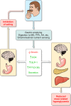
Overview of the hypothesized physiological roles of ghrelin, CCK, GLP-1, and PYY(3–36) in the control of eating and of meal-related glycemia. Gastric emptying, which controls the rate of appearance of ingested food in the small intestine, intestinal transit, rate of digestion, and small intestinal nutrient sensing are the major determinants of the inhibition of ghrelin secretion and the stimulation of CCK, GLP-1, and PYY(3–36) secretion during and after meals. Left: changes in hormone levels lead to GI and central nervous system events whose outcome is to inhibit eating. Right: changes in hormone levels lead GI, pancreatic, hepatic, and central nervous system events whose outcome is to dampen postprandial increases in blood glucose. All four hormones have been hypothesized to contribute to each type of outcome. MS, monosaccharides; FFA, free fatty acids; AA, amino acids.
FIGURE 3.
FIGURE 3.
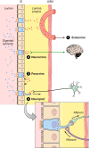
Schematic of the small intestinal mucosa showing potential modes of action of CCK, GLP-1, and PYY. The mucosa includes the epithelial cell layer (IE) on the luminal side, the lamina propria, and the lamina muscularis mucosae (LMM), which limns the submucosa and underlying serosa (not shown). The epithelium consists of enterocytes (tan), which are specialized for nutrient absorption, enteroendocrine cells (blue, villi not shown), which secrete GI hormones, and other cell types (not shown). Digested nutrients activate specific nutrient receptors and transporters (orange <) expressed on the apical surface of enteroendocrine cells, leading to secretion of CCK, GLP-1, and PYY from the basolateral side of enteroendocrine cells. Four modes of action are diagrammed. Mode 1 is the classical endocrine mode, in which hormones diffuse from the lamina propria into mesenteric capillaries (salmon), which drain into the hepatic-portal vein and finally the systemic circulation, allowing hormones to act on distant targets. Modes 2–4 show variations of local actions. Mode 2 is a neuroendocrine mode, in which hormones in the lamina propria activate vagal afferents (green arrow), which in turn stimulate brain-mediated responses. Mode 3 is the paracrine mode, in which hormones in the lamina propria act on receptors (black <) on nearby cells, either neuroendocrine cells or other cell types. Mode 4 shows the anatomical basis for a neuropod mode of action, which has been described for enteroendocrine CCK and PYY cells, and may exist for other GI hormones. This involves hormone release from enteroendocrine-cell neuropods that end in synapse-like appositions to glial cells of the enteric nervous system and other cell types. Note that the hormone concentrations involved in these different modes vary: hormone concentrations in the small gap between neuropods and adjacent cells are likely to be highest, paracrine and vagal neuroendocrine signaling may be the next highest hormone concentrations, endocrine signaling in the liver involves moderate hormone concentrations, and endocrine signaling in which hormones reach their receptors via the systemic circulation involves relatively low hormone concentrations. Hormones also enter the lymph from the lamina propria via bulk flow (not shown), but this is not known to be functionally relevant. Although ghrelin secretion is not stimulated directly by nutrients, secreted ghrelin may act in the modes shown here.
FIGURE 4.
FIGURE 4.
Typical patterns of gastric emptying of solid (green) and liquid (red) foods in relation to meals and intermeal intervals. Depending on the physical digestibility of solid foods, emptying during the first several minutes is very slow (the lag phase), whereas it is uncontrolled and rapid for liquids. The overall shapes of the gastric emptying curves for solid food after the lag phase and for liquid foods are exponential, although significant extents of each approximate linear functions. As described in the text, gastric emptying plays important roles in the control of eating and meal-related glycemia.
FIGURE 5.
FIGURE 5.
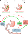
Gastric volume, gastric emptying, and ghrelin, CCK, GLP-1, and PYY(3–36) secretion in relation to meals. A: eating a meal increases gastric volume-related mechanoreception (bold green arrows), which increases satiation signaling via neural afferents, and increases gastric emptying and the delivery of ingested nutrients into the small intestine (bold red arrow), which increases satiation and satiety signaling and decreases hunger signaling. As the intermeal interval (IMI) progresses, volume sensing and gastric emptying progressively decrease (thin red and green arrows). B: gastric emptying determines the rate of appearance of nutrients into the small intestine and, together with the rate of digestion and small-intestinal motility, controls small intestinal-nutrient sensing. For most meals, small intestinal-nutrient sensing inhibits ghrelin secretion (red arrow, −) and stimulates CCK, GLP-1, and PYY secretion (green arrows, +). In turn, ghrelin stimulates (green arrow, +) and CCK, GLP-1, and PYY(3–36) inhibit (red arrows, −) gastric emptying. Note that each feedback loop is negative, as indicated by the change in sign (e.g., red to green) between (small intestinal-nutrient sensing)–(hormone secretion) and (hormone secretion)–(gastric emptying).
FIGURE 6.
FIGURE 6.
Schematic of the organization of ghrelin, CCK, GLP-1, and PYY entroendocrine cells. A: gastric ghrelin cells (blue) are closed-type. Their apical aspects are enclosed by epithelial cells (tan) so that they have no direct contact with the gastric lumen. 1) Neural signals provide the major stimulatory control of ghrelin secretion. 2) Secreted ghrelin (red dots) diffuses through the lamina propria (yellow) into gastric capillaries (salmon) and is transported into the hepatic-portal vein and systemic circulation. 3) Ghrelin cells express a number of nutrient receptors, mainly on the basal and lateral aspects (orange <). These are probably stimulated mainly by metabolites reaching them by diffusion from the gastric capillaries through the lamina propria, although some nutrients may reach them directly from the stomach. 4) CCK, PYY(3–36), perhaps other small intestinal hormones, and other humoral stimuli reach ghrelin cells via the circulation and inhibit ghrelin secretion. Paracrine signals (not shown) may also be involved. B: CCK, GLP-1, and PYY cells (blue) are open-type, with direct contact with the small intestinal lumen. 1) Each expresses a number of nutrient receptors, mainly on the apical and lateral aspects (orange <). These are probably the major controls of secretion of these hormones. The nutrient receptors expressed by ghrelin, CCK, GLP-1, and PYY cells are listed in Table 4, which also indicates the extensive overlap in the nutrient receptors expressed by the these cell types. 2) Secreted hormones (red dots) diffuse through the lamina propria (yellow) into small-intestinal capillaries (salmon) and are transported into the hepatic-portal vein and systemic circulation. 3) Metabolites, hormones, and other humoral factors reach CCK, GLP-1, and PYY cells by diffusion from mesenteric capillaries through the lamina propria (yellow) or from nearby small-intestinal epithelial cells (tan). 4) Neural inputs also control CCK, GLP-1, and PYY secretion.
FIGURE 7.
FIGURE 7.
Some features of ghrelin physiology. Ghrelin is secreted from closed-type enteroendocrine cells (blue) dispersed in the epithelial layer (tan) of the gastric mucosa. Ghrelin diffuses through the lamina propria (yellow) and into gastric capillaries (salmon). 1) Ghrelin's potential physiological effects include acting in the brain to stimulate eating, acting in the stomach to stimulate gastric emptying, and acting on the pancreatic β-cells to inhibit insulin secretion. 2) Ghrelin secretion is stimulated mainly by neural controls. 3) Feedback from small-intestinal nutrient sensing, mediated in part by open-type CCK and PYY(3–36) cells, inhibits ghrelin secretion during and after meals.
FIGURE 8.
FIGURE 8.
Evidence that endogenous CCK signals satiation in healthy humans. A: intravenous infusion of a physiological dose of CCK inhibited eating. Ten healthy-weight women [body mass index (BMI) 22 ± 3 kg/m2] and 8 obese women (BMI 39 ± 2 kg/m2) received 60 min intravenous (IV) infusions of 0.24 pmol·kg ideal body weight−1·min−1 CCK-33 or saline (SAL) beginning at 0800 after an overnight fast. At 0900, a 132 kcal preload of bananas was served, and at 0915, a banana-shake meal was served in excess; bananas were used because they did not elicit CCK secretion. CCK significantly reduced meal size (*) without physical or subjective side effects. [From Lieverse et al. (438), with permission from BMJ Publishing Group Ltd.] B: the CCKA receptor antagonist loxiglumide (LOX) antagonized the satiating action of endogenous CCK stimulated by intraduodenal (ID) infusion of a fat emulsion. Healthy-weight adult males began a midday lunch buffet 4 h after a standard breakfast, 90 min after onset of an IV infusion of 10 μmol·kg−1·h−1 LOX or SAL, 60 min after an ID infusion of 0.4 ml/min corn oil (FAT) or SAL, and 20 min after an oral preload of 400 ml of a low-fat banana milkshake. Infusions were continued throughout the meal. ID fat infusion significantly reduced the size of the lunch meal (+), and that this was reversed by LOX (*); no physical or subjective side effects occurred in any condition. [From Matzinger et al. (480).] C: antagonism of CCK signaling with the CCKA receptor antagonist LOX stimulated eating. Healthy-weight adult males began a midday lunch buffet 4 h after a standard breakfast and 60 min after beginning an IV infusion of 22 μmol·kg−1·h−1 LOX or SAL. Infusions were continued throughout the meal. LOX significantly increased meal size (*) without physical or subjective side effects. [From Beglinger et al. (70).]
FIGURE 9.
FIGURE 9.
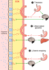
Some features of CCK physiology. CCK (red dots) secretion is stimulated by the digestive products of all three macronutrients acting on nutrient receptors on the apical aspects of enteroendocrine CCK cells (blue) dispersed in the epithelial layer (tan) of the small intestinal mucosa. CCK acts in an endocrine mode by diffusing through the lamina propria (yellow) and into intestinal capillaries (salmon) to reach distant target organs (red arrows), or acts locally. 1) CCK's physiological effects include stimulating satiation. This may occur via endocrine actions in the pyloric area of the stomach that produce signals relayed to the brain via vagal afferents (green arrow) or via local actions on vagal afferents in the lamina propria. An endocrine action in the brain may also contribute. 2) CCK lowers meal-related glycemia via an endocrine effect on gastric emptying and perhaps via a vagal-vagal reflex. 3) Similarly, CCK slows gastric emptying via a direct endocrine effect and perhaps via a vagal-vagal reflex. Solid lines indicate well-established effects, and dashed lines indicate less well established effects.
FIGURE 10.
FIGURE 10.
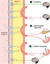
Some features of GLP-1 physiology. GLP-1 secretion is stimulated by the digestive products of all three macronutrients acting on nutrient receptors on the apical aspects of enteroendocrine GLP-1 cells (blue) dispersed in the epithelial layer (tan) of the small intestinal mucosa. GLP-1 acts in an endocrine mode by diffusing through the lamina propria (yellow) and into intestinal capillaries (salmon) to reach distant target organs (red arrows), or acts locally. 1) GLP-1 stimulates satiation. Data in rats indicate GLP-1 signals satiation via a local action on vagal afferents (green arrow) in the lamina propria. GLP-1 may also act in the brain to affect satiation or postprandial satiety. 2) GLP-1 improves meal-related glycemia by increasing pancreatic β-cell insulin secretion in a glucose-dependent manner, by inhibiting pancreatic α-cell glucagon secretion, and by inhibiting gastric emptying; all three appear to be endocrine effects of GLP-1. 3) GLP-1 slows gastric emptying via a direct endocrine effect and perhaps via a vagal-vagal reflex. Solid lines indicate well established effects, and dashed lines indicate less well established effects.
FIGURE 11.
FIGURE 11.
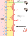
Some features of PYY(3–36) physiology. PYY secretion is stimulated by the digestive products of all three macronutrients acting on nutrient receptors on the apical aspects of enteroendocrine PYY cells (blue) dispersed in the epithelial layer (tan) of the small-intestinal mucosa. PYY is transformed into PYY(3–36) beginning in the lamina propria. PYY(3–36) acts in an endocrine mode by diffusing through the lamina propria (yellow) and into intestinal capillaries (salmon) to reach distant target organs (red arrows), or acts locally. 1) The role of PYY(3–36) in eating is uncertain. It may inhibit eating via a local action on vagal afferents (green arrow) in the lamina propria or by acting directly in the brain. 2) Whether PYY(3–36) improves meal-related glycemic control is uncertain. 3) PYY(3–36) appears to slow gastric emptying via a direct endocrine effect on the stomach; whether vagal-vagal reflexes contribute is unknown. Solid lines indicate well established effects, and dashed lines indicate less well established effects.
FIGURE 12.
FIGURE 12.
A thought experiment depicting how learning may influence the control of eating by GI hormones. Left: when an individual is served a palatable but unfamiliar food, meal size is determined mainly by unconditioned satiation signals related to gastric volume, CCK and GLP-1 secretion, etc., as discussed in the review. Under these conditions, CCK infusion during the meal might exert its full unconditioned effect, indicated by the 40% reduction in meal size. Right: if the same individual is tested after extensive experience eating the test food, meal size might be the same as initially, but will now be under the control of conditioned responses such as expected satiation (111), portion-size estimation (629), etc., that override unconditioned signals, and because conditioned eating controls are resistant to physiological feedback, the same CCK infusion might now reduce meal size less, indicated by the 20% effect.

Similar articles

Cited by

References

    1. Abbott CR, Monteiro M, Small CJ, Sajedi A, Smith KL, Parkinson JR, Ghatei MA, Bloom SR. The inhibitory effects of peripheral administration of peptide YY(3–36) and glucagon-like peptide-1 on food intake are attenuated by ablation of the vagal-brainstem-hypothalamic pathway. Brain Res : 127–131, 2005. - PubMed
    1. Abbott CR, Small CJ, Kennedy AR, Neary NM, Sajedi A, Ghatei MA, Bloom SR. Blockade of the neuropeptide Y Y2 receptor with the specific antagonist BIIE0246 attenuates the effect of endogenous and exogenous peptide YY(3–36) on food intake. Brain Res : 139–144, 2005. - PubMed
    1. Abegg K, Schiesser M, Lutz TA, Bueter M. Acute peripheral GLP-1 receptor agonism or antagonism does not alter energy expenditure in rats after Roux-en-Y gastric bypass. Physiol Behav : 70–78, 2013. - PMC - PubMed
    1. Abizaid A, Liu ZW, Andrews ZB, Shanabrough M, Borok E, Elsworth JD, Roth RH, Sleeman MW, Picciotto MR, Tschop MH, Gao XB, Horvath TL. Ghrelin modulates the activity and synaptic input organization of midbrain dopamine neurons while promoting appetite. J Clin Invest : 3229–3239, 2006. - PMC - PubMed
    1. Acosta A, Camilleri M, Shin A, Vazquez-Roque MI, Iturrino J, Burton D, O'Neill J, Eckert D, Zinsmeister AR. Quantitative gastrointestinal and psychological traits associated with obesity and response to weight-loss therapy. Gastroenterology : 537–546, 2015. - PMC - PubMed

Publication types

LinkOut - more resources