

Upregulated inflammatory associated factors and blood-retinal barrier changes in the retina of type 2 diabetes mellitus model
- PMID: 27990361
- PMCID: PMC5145086
- DOI: 10.18240/ijo.2016.11.09
Upregulated inflammatory associated factors and blood-retinal barrier changes in the retina of type 2 diabetes mellitus model
Abstract
Aim: To examine the expression of high mobility group box-1 (HMGB-1) and intercellular adhesion molecule-1 (ICAM-1) in the retina and the hippocampal tissues; and further to evaluate the association of these two molecules with the alterations of blood-retinal barrier (BRB) and blood-brain barrier (BBB) in a rat model of type 2 diabetes.
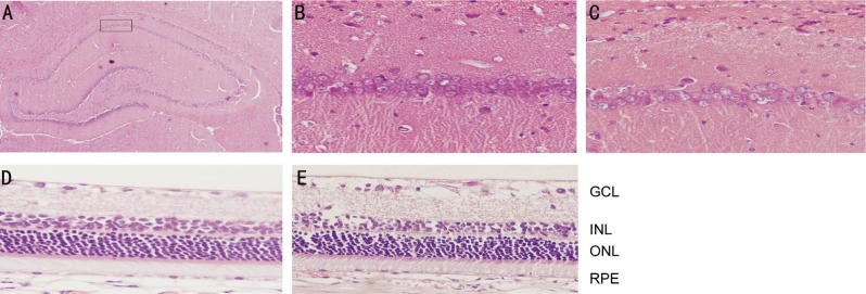
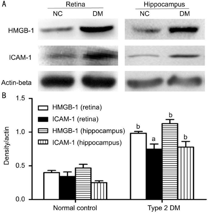
Methods: The type-2 diabetes mellitus (DM) model was established with a high-fat and high-glucose diet combined with streptozotocin (STZ). Sixteen weeks after DM induction, morphological changes of retina and hippocampus were observed with hematoxylin-eosin staining, and alternations of BRB and BBB permeability were measured using Evans blue method. Levels of HMGB-1 and ICAM-1 in retina and hippocampus were detected by Western blot. Serum HMGB-1 levels were determined by enzyme-linked immunosorbent assay (ELISA).
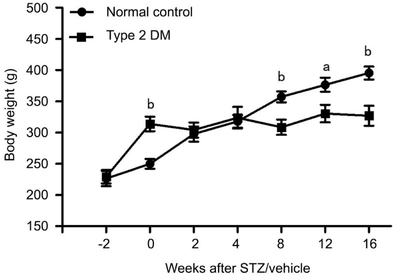
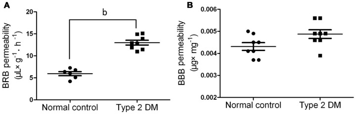
Results: A significantly higher serum fasting blood glucose level in DM rats was observed 2wk after STZ injection (P<0.01). The serum levels of fasting insulin, Insulin resistance homeostatic model assessment (IRHOMA), total cholesterol (TC), total triglycerides (TG) and low density lipoprotein cholesterol (LDL-C) in the DM rats significantly higher than those in the controls (all P<0.01). HMGB-1 (0.96±0.03, P<0.01) and ICAM-1 (0.76±0.12, P<0.05) levels in the retina in the DM rats were significantly higher than those in the controls. HMGB-1 (0.83±0.13, P<0.01) and ICAM-1 (1.15±0.08, P<0.01) levels in the hippocampal tissues in the DM rats were also significantly higher than those in the controls. Sixteen weeks after induction of DM, the BRB permeability to albumin-bound Evans blue dye in the DM rats was significantly higher than that in the controls (P<0.01). However, there was no difference of BBB permeability between the DM rats and controls. When compared to the controls, hematoxylin and eosin staining showed obvious irregularities in the DM rats.
Conclusion: BRB permeability increases significantly in rats with type-2 DM, which may be associated with the up-regulated retinal expression of HMGB-1 and ICAM-1.
Keywords: blood-brain barrier; blood-retinal barrier; diabetes mellitus; high mobility group box-1 protein; permeability; rats.
Figures
Similar articles
-
High-mobility group box-1 protein activates inflammatory signaling pathway components and disrupts retinal vascular-barrier in the diabetic retina.Exp Eye Res. 2013 Feb;107:101-9. doi: 10.1016/j.exer.2012.12.009. Epub 2012 Dec 21. Exp Eye Res. 2013. PMID: 23261684
-
Disruption of the hippocampal and hypothalamic blood-brain barrier in a diet-induced obese model of type II diabetes: prevention and treatment by the mitochondrial carbonic anhydrase inhibitor, topiramate.Fluids Barriers CNS. 2019 Jan 8;16(1):1. doi: 10.1186/s12987-018-0121-6. Fluids Barriers CNS. 2019. PMID: 30616618 Free PMC article.
-
An endothelin type A receptor antagonist reverses upregulated VEGF and ICAM-1 levels in streptozotocin-induced diabetic rat retina.Curr Eye Res. 2006 Jan;31(1):79-89. doi: 10.1080/02713680500478923. Curr Eye Res. 2006. PMID: 16421022
-
Protective effects of a novel drug RC28-E blocking both VEGF and FGF2 on early diabetic rat retina.Int J Ophthalmol. 2018 Jun 18;11(6):935-944. doi: 10.18240/ijo.2018.06.07. eCollection 2018. Int J Ophthalmol. 2018. PMID: 29977804 Free PMC article.
-
Α-Melanocyte-Stimulating Hormone Protects Early Diabetic Retina from Blood-Retinal Barrier Breakdown and Vascular Leakage via MC4R.Cell Physiol Biochem. 2018;45(2):505-522. doi: 10.1159/000487029. Epub 2018 Jan 25. Cell Physiol Biochem. 2018. PMID: 29402864
Cited by
-
Time dependent effects of prolonged hyperglycemia in zebrafish brain and retina.Front Ophthalmol (Lausanne). 2022 Aug 25;2:947571. doi: 10.3389/fopht.2022.947571. eCollection 2022. Front Ophthalmol (Lausanne). 2022. PMID: 38983568 Free PMC article.
-
A Novel Pathological Scoring System for Hepatic Cirrhosis with Hepatocellular Carcinoma.Cancer Manag Res. 2020 Jul 8;12:5537-5547. doi: 10.2147/CMAR.S223417. eCollection 2020. Cancer Manag Res. 2020. PMID: 32753967 Free PMC article.
-
Roles of Drug Transporters in Blood-Retinal Barrier.Adv Exp Med Biol. 2019;1141:467-504. doi: 10.1007/978-981-13-7647-4_10. Adv Exp Med Biol. 2019. PMID: 31571172 Free PMC article. Review.
-
Serum Low-Density Lipoprotein Cholesterol Levels are Associated with Relapse in Neuromyelitis Optica Spectrum Disorder.J Inflamm Res. 2024 Nov 5;17:8227-8240. doi: 10.2147/JIR.S489723. eCollection 2024. J Inflamm Res. 2024. PMID: 39525310 Free PMC article.
-
miR-Let7A Controls the Cell Death and Tight Junction Density of Brain Endothelial Cells under High Glucose Condition.Oxid Med Cell Longev. 2017;2017:6051874. doi: 10.1155/2017/6051874. Epub 2017 Jun 7. Oxid Med Cell Longev. 2017. PMID: 28680530 Free PMC article.
References
-
- Xu Y, Wang L, He J, Bi Y, Li M, Wang T, Wang L, Jiang Y, Dai M, Lu J, Xu M, Li Y, Hu N, Li J, Mi S, Chen CS, Li G, Mu Y, Zhao J, Kong L, Chen J, Lai S, Wang W, Zhao W, Ning G, 2010 China Noncommunicable Disease Surveillance Group Prevalence and control of diabetes in Chinese adults. JAMA. 2013;310(9):948–959. - PubMed
-
- Ozkan E, Gocmen R, Topcuoglu MA, Arsava EM. Blood-retina-barrier disruption accompanying blood-brain-barrier dysfunction in posterior reversible encephalopathy syndrome. J Neurol Sci. 2014;346(1–2):315–317. - PubMed
LinkOut - more resources
Full Text Sources
Other Literature Sources
Miscellaneous