. 2017 May;24(4):10.1111/micc.12341.
doi: 10.1111/micc.12341.

Arteriogenesis in murine adipose tissue is contingent on CD68+ /CD206+ macrophages

Affiliations

Arteriogenesis in murine adipose tissue is contingent on CD68+ /CD206+ macrophages

Scott A Seaman et al. Microcirculation. 2017 May.

Abstract

Objective: The surgical transfer of skin, fat, and/or muscle from a donor site to a recipient site within the same patient is a widely performed procedure in reconstructive surgeries. A surgical pretreatment strategy that is intended to increase perfusion in the flap, termed "flap delay," is a commonly employed technique by plastic surgeons prior to flap transplantation. Here, we explored whether CD68+ /CD206+ macrophages are required for arteriogenesis within the flap by performing gain-of-function and loss-of-function studies in a previously published flap delay murine model.

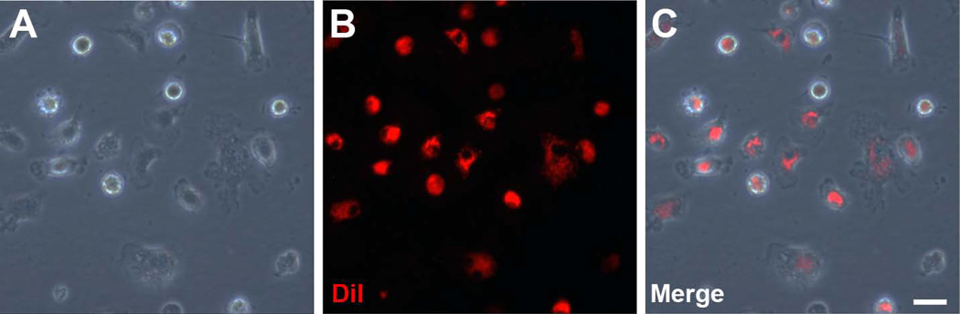
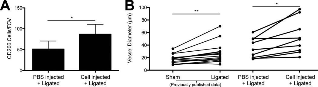
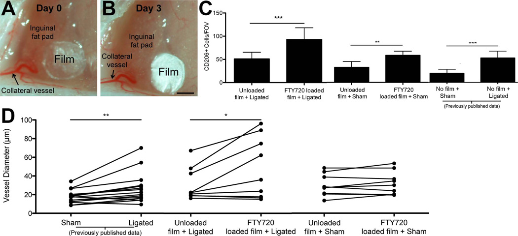
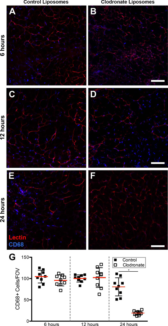
Methods and results: Local injection of M2-polarized macrophages into the flap resulted in an increase in collateral vessel diameter. Application of a thin biomaterial film loaded with a pharmacological agent (FTY720), which has been previously shown to recruit CD68+ /CD206+ macrophages to remodeling tissue, increased CD68+ /CD206+ cell recruitment and collateral vessel enlargement. Conversely, when local macrophage populations were depleted within the inguinal fat pad via clodronate liposome delivery, we observed fewer CD68+ cells accompanied by diminished collateral vessel enlargement.

Conclusions: Our study underscores the importance of macrophages during microvascular adaptations that are induced by flap delay. These studies suggest a mechanism for a translatable therapeutic target that may be used to enhance the clinical flap delay procedure.

Keywords: adipose; arteriogenesis; collateral vessel; macrophage.


Figures

Figure 1
Figure 1. Fluorescent labeling of bone marrow-derived macrophages
In vitro culture of bone marrow-derived macrophages can be fluorescently labeled with DiI. A.) Bone marrow-derived macrophages display prototypical “fried egg” morphology in culture. B.) DiI, a membrane dye, labels macrophages prior to injection. C.) Nearly all bone marrow-derived macrophages are labeled with DiI. Scale bar = 50 µm.
Figure 2
Figure 2. In vivo injection of bone marrow-derived macrophages
Fluorescent immunohistochemical labeling reveals an increase in CD68+ and CD206+ cells in inguinal fat pads injected with DiI labeled, M2-polarized macrophages. A.) No fluorescent signal was detectable in inguinal fat pads that were injected with unlabeled cells (scale bar = 50 µm), whereas (B.) DiI labeled cells were visible in inguinal fat pads that were injected with DiI labeled, M2-polarized macrophages (scale bar = 50 µm). C.,D.) Fluorescent immunohistochemical labeling of inguinal fat pad revealed increase in CD68+ cells in (C) DiI+ cell injected tissue when compared to (D) uninjected tissue (scale bar = 20 µm). E.,F.) Fluorescent immunohistochemical labeling of inguinal fat pad revealed increase of CD206+ cells in (E) DiI+ cell injected tissue when compared to (F) uninjected tissue (scale bar = 20 µm) G.) Number of CD68+ cells in injected inguinal fat pads was increased compared to uninjected inguinal fat pads. n = 6 mice. Student’s t-test, * = p-value < 0.05. H.) Number of CD206+ cells was increased in injected inguinal fat pads when compared to uninjected inguinal fat pads. n = 5 mice. Student’s t-test, * = p-value < 0.05. Data are mean ± standard deviation.
Figure 3
Figure 3. Injection of bone marrow-derived macrophages into inguinal fat pad
Injection of bone marrow-derived macrophages increased the number of CD206+ cells and increased collateral vessel diameters when compared to PBS-injected inguinal fat pads. A.) Quantification of the number of CD206+ cells/FOV. Student’s t-test, * = p-value < 0.05. Data are mean + standard deviation. B.) Individual diameter measurements for collateral vessels in PBS-injected tissues were paired with corresponding collateral branch diameter measurements in cell-injected tissues (in the same mouse). Collateral vessels in fat pads receiving cell injections had increased diameters when compared to corresponding collateral vessels in PBS-injected fat pads. Data from fat pads experiencing either sham surgery or ligation surgery (without PBS or cell injection) from a previously published study are presented for comparison purposes. Paired t-test, ** = p-value < 0.01, * = p-value < 0.05. n = 3 mice for all experiments, 4 unique FOVs for CD206+ cell quantification.
Figure 4
Figure 4. FTY720 delivery to inguinal fat pad
FTY720-loaded PLAGA films were applied to inguinal fat pads, resulting in enhanced arteriogenesis. A.) Representative images of PLAGA film placement during ligation surgery and (B.) three days post-surgery. Scale bar = 500 µm C.) Quantification of CD206+ cells in unloaded and FTY720-loaded PLAGA films three days after sham or ligation surgery. Quantification reveals a significant increase in the number of CD206+ cells in ligated tissue treated with FTY720-loaded films vs. unloaded films. After sham surgery, a significant increase in the number of CD206+ cells was observed in FTY720 treated tissue. Student’s t-test, * = p-value < 0.05, ** = p-value < 0.01. *** = p-value < 0.001 D.) Individual diameter measurements for collateral vessels in inguinal fat pads treated with FTY720-loaded film were paired with corresponding branch diameter measurements for collateral vessels in fat pads treated with unloaded film (in the same mouse). A significant increase in the diameters of collateral vessels was observed in fat pads treated with FTY720-loaded film when compared to fat pads treated with unloaded films. No significant increase was seen in the sham surgery trials between FTY720-loaded and unloaded films. Sham and ligated data from a previously published study are presented for comparison purposes. Paired t-test, * = p-value < 0.05. n = 3 mice for all experiments, 4 unique FOVs for CD206+ cell quantification.
Figure 5
Figure 5. Clodronate depletion of CD68+ macrophages within inguinal fat pad
Direct injection of clodronate liposomes into the inguinal fat pad significantly depleted local CD68+ macrophage populations after 24 hours. A.,B.) Representative image of control liposome-injected inguinal fat pad (A) and clodronate liposome-injected inguinal fat pad (B) at 6 hours. C.,D.) Representative image of control liposome-injected inguinal fat pad (C) and clodronate liposome-injected inguinal fat pad (D) at 12 hours. E.,F.) Representative image of control liposome-injected inguinal fat pad (E) and clodronate liposome-injected inguinal fat pad (F) at 6 hours. G.) Quantification of CD68+ cells at 6 hours, 12 hours, and 24 hours following either control liposome or clodronate liposome injection. A significant decrease in the number of CD68+ cells was observed 24 hours post-injection in clodronate liposome-injected inguinal fat pads (Student’s t-test, * = p-value < 0.05) relative to control liposome-injected fat pads. Scale bar = 100 µm, n = 3 mice for each time point, 3 unique FOVs for CD68+ cell quantification.
Figure 6
Figure 6. Injection of clodronate liposomes within remodeling inguinal fat pad
Clodronate liposomes depleted the local macrophage population after ligation surgery and affected the arteriogenic response of collateral vessels. A.) Quantification of CD68+ cells reveals a significant decrease in the number of CD68+ cells within clodronate liposome-treated tissue compared to control liposome-treated tissue. Student’s t-test, ** = p-value < 0.01 B.) Collateral vessel diameters in the clodronate liposome-injected tissues were paired with the diameter measurements from the corresponding collateral vessel branches in the control liposome-injected tissues in the same mouse. Collateral vessels in control liposome-injected fat pads had larger diameters compared to corresponding collateral vessels in clodronate liposome-injected fat pads. Sham and ligated data from a previously published study is presented for comparison purposes. Paired t-test, ** = p-value < 0.01. n = 3 mice for all experiments, 4 unique FOVs for CD68+ cell quantification.

Similar articles

Cited by

References

    1. Ashley JW, Shi Z, Zhao H, Li X, Kesterson RA, Feng X. Genetic Ablation of CD68 Results in Mice with Increased Bone and Dysfunctional Osteoclasts. PLoS One. 2011;6 - PMC - PubMed
    1. Awojoodu AO, Ogle ME, Sefcik LS, Bowers DT, Martin K, Brayman KL, Lynch KR, Peirce-Cottler SM, Botchwey E. Sphingosine 1-phosphate receptor 3 regulates recruitment of anti-inflammatory monocytes to microvessels during implant arteriogenesis. Proc. Natl. Acad. Sci. U. S. A. 2013;110:13785–13790. - PMC - PubMed
    1. Bain CC, Bravo-blas A, Scott CL, Perdiguero EG, Geissmann F, Henri S, Malissen B, Osborne LC, Artis D, Mowat AM. Constant replenishment from circulating monocytes maintains the macrophage pool in the intestine of adult mice. Nat. Imm. 2014;15 - PMC - PubMed
    1. Biewenga J, van der Ende MB, Krist LF, Borst A, Ghufron M, van Rooijen N. Macrophage depletion in the rat after intraperitoneal administration of liposome-encapsulated clodronate: depletion kinetics and accelerated repopulation of peritoneal and omental macrophages by administration of Freund’s adjuvant. Cell Tissue Res. 1995;280:189–196. - PubMed
    1. Bruce AC, Kelly-Goss MR, Heuslein JL, Meisner JK, Price RJ, Peirce SM. Monocytes are recruited from venules during arteriogenesis in the murine spinotrapezius ligation model. Arterioscler. Thromb. Vasc. Biol. 2014:1–11. - PMC - PubMed

MeSH terms

LinkOut - more resources