Characterization of an Immunodominant Epitope in the Endodomain of the Coronavirus Membrane Protein
- PMID: 27973413
- PMCID: PMC5192388
- DOI: 10.3390/v8120327
Characterization of an Immunodominant Epitope in the Endodomain of the Coronavirus Membrane Protein
Abstract
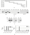
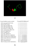
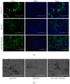
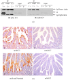
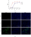
The coronavirus membrane (M) protein acts as a dominant immunogen and is a major player in virus assembly. In this study, we prepared two monoclonal antibodies (mAbs; 1C3 and 4C7) directed against the transmissible gastroenteritis virus (TGEV) M protein. The 1C3 and 4C7 mAbs both reacted with the native TGEV M protein in western blotting and immunofluorescence (IFA) assays. Two linear epitopes, 243YSTEART249 (1C3) and 243YSTEARTDNLSEQEKLLHMV262 (4C7), were identified in the endodomain of the TGEV M protein. The 1C3 mAb can be used for the detection of the TGEV M protein in different assays. An IFA method for the detection of TGEV M protein was optimized using mAb 1C3. Furthermore, the ability of the epitope identified in this study to stimulate antibody production was also evaluated. An immunodominant epitope in the TGEV membrane protein endodomain was identified. The results of this study have implications for further research on TGEV replication.
Keywords: coronavirus; endodomain; immunodominant epitope; membrane protein.
Conflict of interest statement
The authors declare no conflict of interest.
Figures



Similar articles
-
Immunogenic characterization and epitope mapping of transmissible gastroenteritis virus RNA dependent RNA polymerase.J Virol Methods. 2011 Jul;175(1):7-13. doi: 10.1016/j.jviromet.2011.04.007. Epub 2011 Apr 12. J Virol Methods. 2011. PMID: 21513742 Free PMC article.
-
Production and characterization of a monoclonal antibody against spike protein of transmissible gastroenteritis virus.Hybridoma (Larchmt). 2010 Aug;29(4):345-50. doi: 10.1089/hyb.2010.0009. Hybridoma (Larchmt). 2010. PMID: 20715993
-
Monoclonal antibody against membrane protein of transmissible gastroenteritis virus.Monoclon Antib Immunodiagn Immunother. 2013 Feb;32(1):36-40. doi: 10.1089/mab.2012.0065. Monoclon Antib Immunodiagn Immunother. 2013. PMID: 23600504
-
An overview of immunological and genetic methods for detecting swine coronaviruses, transmissible gastroenteritis virus, and porcine respiratory coronavirus in tissues.Adv Exp Med Biol. 1997;412:37-46. doi: 10.1007/978-1-4899-1828-4_4. Adv Exp Med Biol. 1997. PMID: 9191988 Review.
-
Transmissible gastroenteritis virus infection: a vanishing specter.Dtsch Tierarztl Wochenschr. 2006 Apr;113(4):157-9. Dtsch Tierarztl Wochenschr. 2006. PMID: 16716052 Review.
Cited by
-
Development and Application of an Indirect Enzyme-Linked Immunosorbent Assay Based on a Recombinant Matrix Protein for the Serological Study of Porcine Deltacoronavirus in Mexican Pigs.Vet Med Sci. 2024 Nov;10(6):e70108. doi: 10.1002/vms3.70108. Vet Med Sci. 2024. PMID: 39494986 Free PMC article.
-
A Candidate Antigen of the Recombinant Membrane Protein Derived from the Porcine Deltacoronavirus Synthetic Gene to Detect Seropositive Pigs.Viruses. 2023 Apr 25;15(5):1049. doi: 10.3390/v15051049. Viruses. 2023. PMID: 37243136 Free PMC article.
References
-
- De Groot R.J., Baker S.G., Baric R.S., Enjuanes L., Gorbalenya A.E. Coronaviridae. In: King A.M.Q., Adams M.J., Carstens E.B., Lefkowitz E.J., editors. Virus Taxonomy: Ninth Report of the International Committee on Taxonomy of Viruses. Elsevier Academic Press; San Diego, CA, USA: 2011. pp. 774–796.
MeSH terms
Substances
LinkOut - more resources
Full Text Sources
Other Literature Sources

