. 2017 Feb;23(2):162-173.
doi: 10.1111/cns.12660. Epub 2016 Dec 9.

Effects of LRRK2 Inhibitors on Nigrostriatal Dopaminergic Neurotransmission

Affiliations

Effects of LRRK2 Inhibitors on Nigrostriatal Dopaminergic Neurotransmission

Qi Qin et al. CNS Neurosci Ther. 2017 Feb.

Abstract

Introduction: Mutations in leucine-rich repeat kinase 2 (LRRK2) are the most prevalent cause of familial and sporadic Parkinson's disease (PD). Because most pathogenic LRRK2 mutations result in enhanced kinase activity, it suggests that LRRK2 inhibitors may serve as a potential treatment for PD. To evaluate whether LRRK2 inhibitors are effective therapies for PD, it is crucial to know whether LRRK2 inhibitors will affect dopaminergic (DAergic) neurotransmission. However, to date, there is no study to investigate the impact of LRRK2 inhibitors on DAergic neurotransmission.

Aims: To address this gap in knowledge, we examined the effects of three types of LRRK2 inhibitors (LRRK2-IN-1, GSK2578215A, and GNE-7915) on dopamine (DA) release in the dorsal striatum using fast-scan cyclic voltammetry and DA neuron firing in the substantia nigra pars compacta (SNpc) using patch clamp in mouse brain slices.

Results: We found that LRRK2-IN-1 at a concentration higher than 1 μM causes off-target effects and decreases DA release, whereas GSK2578215A and GNE-7915 do not. All three inhibitors at 1 μM have no effect on DA release and DA neuron firing rate. We have further assessed the effects of the inhibitors in two preclinical LRRK2 mouse models (i.e., BAC transgenic hG2019S and hR1441G) and demonstrated that GNE-7915 enhances DA release and synaptic vesicle mobilization/recycling.

Conclusion: GNE-7915 can be validated for further therapeutic development for PD.

Keywords: Dopamine; Fast-scan cyclic voltammetry; Inhibitor; LRRK2; Parkinson's disease.


Conflict of interest statement

The authors declare no conflict of interest.

Figures

Figure 1
Figure 1
Loss of LRRK2 does not alter DA release and synaptic vesicle replenishment /recycling. (A) Representative voltammetric traces of evoked DA release with different stimulations (one pulse (1p), two pulses at 100 Hz (2p@100 Hz), four pulses at 20 Hz (4p@20 Hz), paired stimuli at variable interpulse intervals) in WT and LRRK2 KO mice. (B) Bar graphs showing no alteration of DA release evoked by 1p or 4p@20 Hz, n = 11. (C) Bar graphs showing no alteration of DA release and PPR by LRRK2 deletion. 4p@20 Hz/1p: 4p@20 Hz train stimuli evoked DA release normalized to 1‐p‐evoked DA release, n = 11; 2p@100 Hz/1p: 2p@100 Hz stimuli evoked DA release normalized to 1p‐evoked DA release, n = 13; PPR/5s, PPR/10s, and PPR/20s: paired‐pulse stimulation at 5‐, 10‐, and 20‐seconds interval, n = 19.
Figure 2
Figure 2
Effects of different concentrations of LRRK2 inhibitors on DA release in WT mice. (A) Representative voltammetric traces of evoked DA release with different stimulations before and after LRRK2‐IN‐1 (3 μM, 2‐h incubation) treatment. (B) Effects of LRRK2‐IN‐1 (1 and 3 μM, 2‐h incubation) on evoked DA release and PPR. DMSO, n = 14; 1 μM, n = 10; 3 μM, n = 7. LRRK2‐IN‐1 at 1 μM had no effect on evoked DA release and PPR. However, LRRK2‐IN‐1 at a concentration of 3 μM decreased 1p‐evoked DA release (treated group: 1.57 ± 0.21 μM, n = 7; control group: 2.19 ± 0.08 μM, n = 7, P < 0.05) and 4p@20 Hz‐evoked DA release (treated group: 2.01 ± 0.13 μM, n = 7; control group: 2.67 ± 0.13 μM, n = 7, P < 0.05) and PPR with 5‐s interval (treated group: 0.40 ± 0.02, n = 7; control group: 0.46 ± 0.02, n = 7, P < 0.05), 10‐s interval (treated group: 0.54 ± 0.02, n = 7; control group: 0.64 ± 0.02, n = 7, P < 0.05), and 20‐s interval (treated group: 0.73 ± 0.02, n = 7; control group: 0.83 ± 0.03, n = 7, P < 0.05). (C) GSK2578215A (1 and 3 μM) had no effect on evoked DA release and PPR. DMSO, n = 14; 1 μM, n = 10; 3 μM, n = 10. (D) GNE‐7915 (1 and 3 μM) had no effect on evoked DA release and PPR. DMSO, n = 14; 1 μM, n = 11; 3 μM, n = 9. *P < 0.05, paired t‐test. All the three inhibitors at 0.1 or 0.3 μM had no effect on evoked DA release (data not shown here, but summarized in Figure 3B).
Figure 3
Figure 3
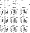
LRRK2‐IN‐1 at 3 μM shows off‐target effects on evoked DA release in KO mice, whereas GSK2578215A and GNE‐7915 do not. (A) Upper panels show no effect of the three LRRK2 inhibitors (1 μM, 2‐h incubation) on DA release and PPR. LRRK2‐IN‐1, n = 12 for each condition; GSK, n = 8 for each condition; GNE, n = 10 for each condition. Lower panels show that LRRK2‐IN‐1 at a concentration of 3 μM decreased 1‐p‐evoked DA release (treated group: 1.49 ± 0.22 μM, n = 14; control group: 1.88 ± 0.20 μM, n = 4, P < 0.01), 4p@20 Hz‐evoked DA release (treated group: 2.07 ± 0.26 μM, n = 14; control group: 2.65 ± 0.26 μM, n = 4, P < 0.01) and PPR with 5‐s interval (treated group: 0.37 ± 0.02, n = 9; control group: 0.43 ± 0.04, n = 9, P < 0.01), 10‐s interval (treated group: 0.52 ± 0.02, n = 9; control group: 0.61 ± 0.04, n = 9, P < 0.01), and 20‐s interval (treated group: 0.74 ± 0.02, n = 9; control group: 0.82 ± 0.02, n = 9, P < 0.01). GSK, n = 8 for each condition; GNE, n = 10 for each condition. *P < 0.05, **P < 0.01, paired t‐test. (B) Dose–response curves for each LRRK2 inhibitor, respectively, in WT mice (left panel) and KO mice (right panel). The amplitude of 1p‐evoked DA release in the treated group was normalized to the control group (DMSO‐treated group). 3 μM LRRK2‐IN‐1 decreased 1p‐evoked DA release in both WT and KO mice suggesting off‐target effects. *P < 0.05, **P < 0.01, One‐way ANOVA followed by Bonferroni test.
Figure 4
Figure 4
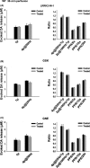
None of the three LRRK2 inhibitors have any significant effect on DA release or synaptic vesicle replenishment/recycling with 30‐min perfusion. Evoked DA release was recorded by FSCV at one site within a slice, and 1 μM LRRK2 inhibitor was perfused for 30 min. There was no significant difference of DA release and PPR before and after 30‐min perfusion. (A) LRRK2‐IN‐1, n = 6 for each condition. (B) GSK, n = 6 for each condition; (C) GNE, n = 6 for each condition.
Figure 5
Figure 5
None of the three LRRK2 inhibitors have any significant effect on DA release or synaptic vesicle replenishment/recycling in the G2019S Tg mice with 2‐h incubation or 30‐min perfusion. (A) Representative voltammetric traces of 1‐p‐evoked DA release under control condition and with LRRK2 inhibitor (1 μM, 2 hr) treatment. (B) No inhibitor (1 μM, 2‐h incubation) produced a significant effect on DA release and recovery. LRRK2‐IN‐1, n = 10 for each condition; GSK, n = 11 for each condition; GNE, n = 11 for each condition. (C) No inhibitor (1 μM, 30‐min perfusion) produced a significant effect on DA release and recovery. LRRK2‐IN‐1, n = 7 for each condition; GSK, n = 7 for each condition; GNE, n = 7 for each condition.
Figure 6
Figure 6
GNE‐7915 enhances DA release and synaptic vesicle replenishment/recycling in the R1441G Tg mice. (A) Representative voltammetric traces of 1‐p‐evoked DA release under control condition and with LRRK2 inhibitor (1 μM, 2‐h incubation) treatment. LRRK2‐IN‐1 and GSK had no significant effect on DA release, whereas GNE enhanced DA release. (B) Neither LRRK2‐IN‐1 (1 μM, 2‐h incubation, n = 10 for each condition) nor GSK (1 μM, 2‐h incubation, n = 10 for each condition) had any significant effect on DA release and recovery. In contrast, GNE‐7915 (1 μM, 2‐h incubation) increased 1‐p and 4p@20 Hz‐evoked DA release (n = 10, P < 0.05) and PPR at 5s (n = 10, P < 0.05), 10s (n = 10, P < 0.01) and PPR at 20s (n = 10, P < 0.001). 4p@20 Hz/1p, n = 9; 2p@100 Hz/1p, n = 9. (C) Neither LRRK2‐IN‐1 (1 μM, 30‐min perfusion, n = 7 for each condition) nor GSK (1 μM, 30‐min perfusion, n = 7 for each condition) had any effect on DA release and kinetics. In contrast, GNE‐7915 (1 μM, 30‐min perfusion) increased single‐pulse and 4p@20 Hz‐evoked DA release (n = 6, P < 0.05) and PPR at 10s (n = 9, P < 0.05) and PPR at 20s (n = 9, P < 0.001). 4p@20 Hz/1p, n = 6; 2p@100 Hz/1p, n = 6; PPR/5s, n = 9. *P < 0.05, ***P < 0.001, paired t‐test.
Figure 7
Figure 7
None of the three LRRK2 inhibitors have any significant effect on the tonic firing rates of SNpc DA neurons from WT, G2019S, and R1441G Tg mice. (A) No significant alteration of DA neuron firing rates in WT mice after 1 μM LRRK2‐IN‐1, GNE7915, and GSK2578215A treatment. Left panel, representative traces of patch recording before and after 30‐min perfusion of LRRK2 inhibitors; middle panel, bar graph showing no effects of the three inhibitors with 2‐h incubation, n = 10 for each inhibitor; right panel, bar graph showing no effects of the three inhibitors with 30‐min perfusion, n = 12 for each inhibitor. (B) No significant alteration of DA neuron firing rates in G2019S mice after 1 μM LRRK2‐IN‐1, GNE7915, and GSK2578215A treatment. Left panel, representative traces of patch recording before and after 30‐min perfusion of LRRK2 inhibitors; middle panel, bar graph showing no effects of the three inhibitors with 2‐h incubation, n = 11 for each inhibitor; right panel, bar graph showing no effects of the three inhibitors with 30‐min perfusion, n = 10 for each inhibitor. (C) No significant alteration of DA neuron firing rates in R1441G mice after 1 μM LRRK2‐IN‐1, GNE7915, and GSK2578215A treatment. Left panel, representative traces of patch recording before and after 30‐min perfusion of LRRK2 inhibitors; middle panel, bar graph showing no effects of the three inhibitors with 2‐h incubation, n = 10 for each inhibitor; right panel, bar graph showing no effects of the three inhibitors with 30‐min perfusion, n = 8 for each inhibitor.

Similar articles

Cited by

References

    1. Lees AJ, Hardy J, Revesz T. Parkinson's disease. Lancet 2009;373:2055–2066. - PubMed
    1. Zimprich A, Biskup S, Leitner P, et al. Mutations in LRRK2 cause autosomal‐dominant parkinsonism with pleomorphic pathology. Neuron 2004;44:601–607. - PubMed
    1. Gilks WP, Abou‐Sleiman PM, Gandhi S, et al. A common LRRK2 mutation in idiopathic Parkinson's disease. Lancet 2005;365:415–416. - PubMed
    1. Paisan‐Ruiz C, Jain S, Evans EW, et al. Cloning of the gene containing mutations that cause PARK8‐linked Parkinson's disease. Neuron 2004;44:595–600. - PubMed
    1. Di Fonzo A, Rohe CF, Ferreira J, et al. A frequent LRRK2 gene mutation associated with autosomal dominant Parkinson's disease. Lancet 2005;365:412–415. - PubMed

MeSH terms