. 2016 Dec 8;1(20):e88522.
doi: 10.1172/jci.insight.88522.

Tissue memory B cell repertoire analysis after ALVAC/AIDSVAX B/E gp120 immunization of rhesus macaques

Affiliations

Tissue memory B cell repertoire analysis after ALVAC/AIDSVAX B/E gp120 immunization of rhesus macaques

Kan Luo et al. JCI Insight. .

Abstract

The ALVAC prime/ALVAC + AIDSVAX B/E boost RV144 vaccine trial induced an estimated 31% efficacy in a low-risk cohort where HIV‑1 exposures were likely at mucosal surfaces. An immune correlates study demonstrated that antibodies targeting the V2 region and in a secondary analysis antibody-dependent cellular cytotoxicity (ADCC), in the presence of low envelope-specific (Env-specific) IgA, correlated with decreased risk of infection. Thus, understanding the B cell repertoires induced by this vaccine in systemic and mucosal compartments are key to understanding the potential protective mechanisms of this vaccine regimen. We immunized rhesus macaques with the ALVAC/AIDSVAX B/E gp120 vaccine regimen given in RV144, and then gave a boost 6 months later, after which the animals were necropsied. We isolated systemic and intestinal vaccine Env-specific memory B cells. Whereas Env-specific B cell clonal lineages were shared between spleen, draining inguinal, anterior pelvic, posterior pelvic, and periaortic lymph nodes, members of Env‑specific B cell clonal lineages were absent in the terminal ileum. Env‑specific antibodies were detectable in rectal fluids, suggesting that IgG antibodies present at mucosal sites were likely systemically produced and transported to intestinal mucosal sites.


Conflict of interest statement

J. Tartaglia was an employee of Sanofi and D. Francis, F. Sinangil, and C. Lee were employees of Global Solutions for Infectious Diseases at the time this study was performed. D. Francis, F. Sinangil, and C. Lee are former employees of VaxGen.

Figures
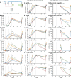
Figure 1
Figure 1. Study schedule and antibody binding data.
(A) Five rhesus macaques (RMs) were immunized 5 times (blue arrows above timeline) and peripheral blood taken 7 times (red arrows), rectal antibody sampled 4 times (green arrows), and the animals necropsied 2 weeks after the final immunization (black arrow). Individual macaques are represented by the same symbols in all figures. In plots BR, dotted vertical lines represent immunization time points. Plasma antibodies were tested for their ability to bind gp120AE.A244 in ELISA and block the binding of mAb A32 (B), soluble CD4 (sCD4) (C), mAb CH58 (D), mAb CH59 (E), and bnAb CH01 (F). Plasma antibody binding in ELISA (plotted as log10 AUC) was measured for gp120AE.A244 (G), gp120B.MN (H), gp120AE.92TH023 (I), Case A2 gp70 V1V2 (J), C.1086 V1V2 tags (K), and AE.A244 V1V2 tags (L). Rectal antibody was sampled using a surgical sponge inserted into the rectum and then eluted. Binding was measured and specific activity was calculated as specific activity = (antibody binding units × dilution factor)/measured rectal IgG in μg/ml for binding to gp120AE.A244 (M), gp120B.MN (N), gp120AE.92TH023 (O), Case A2 gp70 V1V2 (P), C.1086 V1V2 tags (Q), and AE.A244 V1V2 tags (R). RMs 22‑11, 46‑11, and 60‑11 boosted rectal antibody with the final immunization; those animals were selected for B cell repertoire analysis.
Figure 2
Figure 2. Functional activity of plasma/serum antibody.
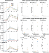
Serum antibody neutralization in the TZM-bl assay was measured for pseudoviruses AE.92TH023.6 (A), B.MN.3 (B), B.SF162.LS (C), B.BaL.26 (D), and C.MW965.26 (E). Plasma antibody-dependent cellular cytotoxicity (ADCC) activity was measured against gp120-coated targets for the AE.92TH023 (F, I, and L), AE.A244 (G, J, and M), and B.MN (H, K, and N). ADCC activity is shown as reciprocal ADCC titer (FH), peak activity in the assay (IK), and log AUC (LN). All animals had a rise in functional antibodies following vaccination. ID50, inhibitory dilution 50%.
Figure 3
Figure 3. Flow cytometric detection of antigen-specific B cells.
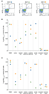
Peripheral blood mononuclear cells (PBMCs) were labeled to identify memory B cells, defined as CD3/14/16, CD20+, and surface IgD, and also with Env probes in 2 colors. (A) PBMCs labeled with gp120M.ConS in 2 colors are shown for the 3 rhesus macaques (RMs). (B) Cell samples from all sites obtained at necropsy were labeled with gp120M.ConS and shown as the frequency of memory B cells. Few antigen-specific B cells were observed in mesenteric lymph nodes (LNs) or terminal ileum.(C) Cell samples were also labeled with gp120AE.A244 and showed a similar pattern, except that the detected cell frequency was lower for RM 46‑11 (green triangles) in anterior pelvic LN, posterior pelvic LN, and periaortic LN. In mesenteric LN and terminal ileum, antigen-specific B cell frequencies were slightly higher for gp120AE.A244 than they were for gp120M.ConS.
Figure 4
Figure 4. Characteristics of isolated HIV-1 envelope–reactive mAbs.
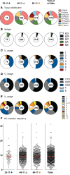
There were 1,493 mAbs isolated of which 1,242 were reactive with HIV‑1 antigens during screening. (A) For rhesus macaque (RM) 22‑11, mAbs were isolated only from peripheral blood mononuclear cells (PBMCs). For RMs 66‑11 and 60‑11, envelope-reactive (Env-reactive) mAbs were isolated from most tissue compartments. (B) The majority (95%) of isolated mAbs were of IgG isotype. (C) VH gene segment usage was similar among the RMs with the majority of mAbs using VH1, VH3, and VH4. Overall, 55% of mAbs used lambda chains. For kappa chain–using mAbs, the majority used Vκ1/1D or Vκ2/2D (D); for lambda chain–using mAbs, the majority used Vλ1, Vλ2, or Vλ3 (E). Each animal had some Env-reactive mAbs that used Vλ3~17, the lambda chain associated with genetically conserved V1V2 responses. (F) Heavy chain (HC) mutation frequencies were similar among Env-reactive antibodies for all 3 RMs; the overall mean mutation frequency was 9.3%.
Figure 5
Figure 5. Clonal lineage characteristics of isolated mAbs.
Analysis of isolated genes demonstrated that many of the mAbs were clonally related. (A) The majority of clonal lineages were comprised of 2 members; larger lineages were detected in each rhesus macaque (RM) up to 4 members for RM 22‑11 and up to 10 members for the other 2 animals. (B) For display of heavy chain complementarity-determining region 3 (HCDR3) length, each lineage was collapsed so that it was counted once; for each RM the median HCDR3 length was 16 amino acids. (C) Tissue distribution of 2‑member clonal lineages are shown with the site of origin displayed by color. Lineages consisting of antibodies that mapped to the V1V2 loop are identified as red circles. A, anterior; P, posterior; T, terminal; PBMC, peripheral blood mononuclear cell.
Figure 6
Figure 6. Tissue distribution of clonal lineages of 3 or more members from rhesus macaque 46‑11.
Clonal lineages are displayed in vertical stripes; below the line is shown the heavy chain complementarity-determining region 3 (HCDR3) length as an integer below and the VH gene used by the lineage. Lineages that used light chain lambda gene segment Vλ3~17 are indicated by a dagger (†) symbol. Lineages with members that mapped to the V1V2 loop are shown by a red vertical line. Individual mAbs are shown by horizontal tick marks; the length of the tick mark is proportional to heavy chain mutation frequency. Antibodies that were negative for HIV-1 envelope reactivity in screening are shown in light blue. Stripes corresponding to clonal lineages in Figures 8 and 9 are indicated. All clonal lineage mAbs were IgG. No clonal lineages had mAbs isolated from terminal ileum. LN, lymph node; PBMC, peripheral blood mononuclear cell.
Figure 7
Figure 7. Tissue distribution of clonal lineages of 3 or more members from rhesus macaque 60‑11.
As in Figure 6, clonal lineages are displayed in vertical stripes; below the line is shown the heavy chain complementarity-determining region 3 (HCDR3) length as an integer below and the VH gene used by the lineage. Clonal lineage mAbs were IgG except for 1 IgA (orange tick) and 1 IgM (green tick). Lineages that used light chain lambda gene segment Vλ3~17 are indicated by a dagger (†) symbol. Lineages with members that mapped to the V1V2 loop are shown by a red vertical line. Individual mAbs are shown by horizontal tick marks; the length of the tick mark is proportional to heavy chain mutation frequency. Antibodies that were negative for HIV-1 envelope reactivity in screening are shown in light blue. The stripe corresponding to lineage DH614 in Figures 8 and 9 is indicated. No clonal lineages had mAbs isolated from terminal ileum. LN, lymph node; PBMC, peripheral blood mononuclear cell.
Figure 8
Figure 8. Clonal lineages of isolated mAbs.
Phylograms of clonal lineages isolated from rhesus macaques (RMs) are shown with antibody data on each line to the right of the mAb name. (A) Mature lineage DH614 potently bound to gp120 proteins and V1V2 targets with the exception of RV144 breakthrough strain gp120AE.703357; the lineage members also neutralized AE.92TH023.6 and the mature antibodies bound to the surface of AE.CM235-infected target cells. Lineage characteristics: VH3~SC11/JH4, heavy chain (HC) complementarity-determining region 3 (HCDR3) length 13, mean HC mutation frequency 10.4%; Vλ3~17/Vλ2 CDR3 length 10, mean light chain (LC) mutation frequency 5.9%. (B) Mature lineage DH612 mAbs bound to gp120M.ConS and neutralized B.MN.3 but did not bind or neutralize clade AE targets. The mature mAbs bound to the surface of AE.CM235-infected target cells. Lineage characteristics: VH4~82/JH6, HCDR3 length 24, mean HC mutation frequency 8.4%; Vκ1-LC1c/Jκ2, CDR3 length 9, mean LC mutation frequency 9.2%. (C) Mature lineage DH613 mAbs potently bound gp120 proteins from clade AE and group M consensus but did not bind V1V2-specific targets. The mature antibodies potently neutralized both AE.92TH023.6 and B.MN.3; the mature antibodies weakly bound to AE.CM235-infected target cells. Lineage characteristics: VH3~SC11/JH4, HCDR3 length 18, mean HC mutation frequency 11.1%; Vκ1~23/Jκ1, CDR3 length 9, mean LC mutation frequency 4.4%. The anatomic site of origin appears after the mAb name: BL, peripheral blood; SP, spleen; IN, inguinal lymph node (LN); AP, anterior pelvic LN; PP, posterior pelvic LN; PA, periaortic LN; MT, mesenteric LN. Data are in μg/ml, color coded as shown in the legend for ELISA half maximal effective concentration (EC50) and neutralization inhibitory dilution 50% (ID50), or as % cell binding. MFI, mean fluorescence intensity; UCA, unmutated common ancestor.
Figure 9
Figure 9. Epitope mapping of mAbs and characterization of mAbs with long HCDR3 loops.
Phylograms for clonal lineages DH614 and DH621 shown with anatomic site of origin. Epitope mapping ELISA data shown as half maximal effective concentration (EC50) and color coded as in the legend. The sequence of the 22–amino acid V2 peptide is shown across the top of the grids (AC) and data below each amino acid are the observed EC50 for variant peptide constructs with substitutions at that position. (A) Mature lineage DH614 mAbs were most sensitive to changes at K169, H173, F176, and Y177. For this lineage, data for binding to infected cells are shown in Figure 8. (B) Mature lineage DH621 mAbs were most sensitive to changes at K168, Q170, K171, H173, K178, and D180. Lineage members bound the surface of AE.CM235-infected target cells. Lineage characteristics: VH5~117/JH4, heavy chain (HC) complementarity-determining region 3 (HCDR3) length 13, mean HC mutation frequency 5.2%; Vκ2~73/Jκ4, CDR3 length 9, mean light chain (LC) mutation frequency 4.7%. (C) V1V2-specific mAbs that did not use Vλ3~17 showed different patterns of sensitivity to amino acid substitutions. DH623 showed significantly reduced binding only for changes at H173. DH624 was not sensitive to H173 changes but was sensitive to changes at K178, L179, and D180. DH627 was sensitive to changes at H173, L175, and K178. All 3 mAbs bound the surface of AE.CM235-infected target cells. (D) Two mAbs that did use Vλ3~17 did not bind to a gp120 from RV144 breakthrough strain AE.703357, but did bind to proteins and V1V2 proteins reflective of the vaccine, and also bound to AE.CM235-infected target cells. (E) Tested long HCDR3 mAbs showed 3 patterns of binding to gp120 constructs. DH605 and DH622 showed reduced binding to gp120AE.A244 N259A/N301A/N332A. DH609 and DH611 were clonally related and showed reduced binding to gp120AE.A244 Δ371I/P363N and gp120C.YU2 D368R; DH607 did not bind clade AE proteins but was sensitive to gp120C.YU2 D368R. DH606, DH608, and DH610 potently bound all tested variants of gp120AE.A244 but differentially bound gp120C.YU2 variants. The anatomic site of origin appears after the mAb name: BL, peripheral blood; SP, spleen; IN, inguinal lymph node (LN); AP, anterior pelvic LN; PP, posterior pelvic LN; PA, periaortic LN; MT, mesenteric LN; ND, not done; MFI, mean fluorescence intensity; UCA, unmutated common ancestor.

Similar articles

Cited by

References

    1. Robb ML, et al. Risk behaviour and time as covariates for efficacy of the HIV vaccine regimen ALVAC-HIV (vCP1521) and AIDSVAX B/E: a post-hoc analysis of the Thai phase 3 efficacy trial RV 144. Lancet Infect Dis. 2012;12(7):531–537. doi: 10.1016/S1473-3099(12)70088-9. - DOI - PMC - PubMed
    1. Rerks-Ngarm S, et al. Vaccination with ALVAC and AIDSVAX to prevent HIV-1 infection in Thailand. N Engl J Med. 2009;361(23):2209–2220. doi: 10.1056/NEJMoa0908492. - DOI - PubMed
    1. Haynes BF, et al. Immune-correlates analysis of an HIV-1 vaccine efficacy trial. N Engl J Med. 2012;366(14):1275–1286. doi: 10.1056/NEJMoa1113425. - DOI - PMC - PubMed
    1. Tomaras GD, et al. Vaccine-induced plasma IgA specific for the C1 region of the HIV-1 envelope blocks binding and effector function of IgG. Proc Natl Acad Sci U S A. 2013;110(22):9019–9024. doi: 10.1073/pnas.1301456110. - DOI - PMC - PubMed
    1. Gottardo R, et al. Plasma IgG to linear epitopes in the V2 and V3 regions of HIV-1 gp120 correlate with a reduced risk of infection in the RV144 vaccine efficacy trial. PLoS One. 2013;8(9):e75665. doi: 10.1371/journal.pone.0075665. - DOI - PMC - PubMed

Publication types

MeSH terms