. 2017 Feb 1;83(4):e02991-16.
doi: 10.1128/AEM.02991-16. Print 2017 Feb 15.

Relationships and Evolution of Double-Stranded RNA Totiviruses of Yeasts Inferred from Analysis of L-A-2 and L-BC Variants in Wine Yeast Strain Populations

Affiliations

Relationships and Evolution of Double-Stranded RNA Totiviruses of Yeasts Inferred from Analysis of L-A-2 and L-BC Variants in Wine Yeast Strain Populations

Nieves Rodríguez-Cousiño et al. Appl Environ Microbiol. .

Abstract

Saccharomyces cerevisiae killer strains secrete a protein toxin active on nonkiller strains of the same (or other) yeast species. Different killer toxins, K1, K2, K28, and Klus, have been described. Each toxin is encoded by a medium-size (1.5- to 2.3-kb) M double-stranded RNA (dsRNA) located in the cytoplasm. M dsRNAs require L-A helper virus for maintenance. L-A belongs to the Totiviridae family, and its dsRNA genome of 4.6 kb codes for the major capsid protein Gag and a minor Gag-Pol protein, which form the virions that separately encapsidate L-A or the M satellites. Different L-A variants exist in nature; on average, 24% of their nucleotides are different. Previously, we reported that L-A-lus was specifically associated with Mlus, suggesting coevolution, and proposed a role of the toxin-encoding M dsRNAs in the appearance of new L-A variants. Here we confirm this by analyzing the helper virus in K2 killer wine strains, which we named L-A-2. L-A-2 is required for M2 maintenance, and neither L-A nor L-A-lus shows helper activity for M2 in the same genetic background. This requirement is overcome when coat proteins are provided in large amounts by a vector or in ski mutants. The genome of another totivirus, L-BC, frequently accompanying L-A in the same cells shows a lower degree of variation than does L-A (about 10% of nucleotides are different). Although L-BC has no helper activity for M dsRNAs, distinct L-BC variants are associated with a particular killer strain. The so-called L-BC-lus (in Klus strains) and L-BC-2 (in K2 strains) are analyzed.

Importance: Killer strains of S. cerevisiae secrete protein toxins that kill nonkiller yeasts. The "killer phenomenon" depends on two dsRNA viruses: L-A and M. M encodes the toxin, and L-A, the helper virus, provides the capsids for both viruses. Different killer toxins exist: K1, K2, K28, and Klus, encoded on different M viruses. Our data indicate that each M dsRNA depends on a specific helper virus; these helper viruses have nucleotide sequences that may be as much as 26% different, suggesting coevolution. In wine environments, K2 and Klus strains frequently coexist. We have previously characterized the association of Mlus and L-A-lus. Here we sequence and characterize L-A-2, the helper virus of M2, establishing the helper virus requirements of M2, which had not been completely elucidated. We also report the existence of two specific L-BC totiviruses in Klus and K2 strains with about 10% of their nucleotides different, suggesting different evolutionary histories from those of L-A viruses.

Keywords: L-A helper virus; double-stranded RNA virus; yeast killer toxins; yeast virus; yeast wine strains.


Figures

FIG 1
FIG 1
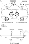
(A) Replication cycles of L-A virus and the killer toxin-encoding virus M1. L-A is transcribed by the transcriptase activity of Gag-Pol, yielding L-A positive strands (indicated by plus signs in parentheses) that are released from the virions into the cytoplasm and are either translated or encapsidated. Translation by ribosomes gives rise to Gag, the structural capsid protein, and Gag-Pol, the RNA polymerase. Gag-Pol interacts with L-A positive strands, triggering viral capsid assembly and encapsidation. The replicase activity of Gag-Pol inside the virions synthesizes the negative strand (indicated by a minus sign in parentheses) on the positive-strand template, forming the dsRNA genome. The replication cycle of M1 is similar to that of L-A and depends on L-A-encoded viral proteins Gag and Gag-Pol. Thus, M1 is a satellite virus of L-A. M1 positive strands are translated, giving rise to the preprotoxin, which is processed and secreted. (B) Genomic organization of L-A. The L-A positive strand and the two overlapping reading frames (ORF1 and ORF2) are indicated. Schematic representations of the secondary structures corresponding to the frameshifting region and to the cis signals required for encapsidation and replication in the 3′-end region of the L-A positive strand are shown. The sequences where oligonucleotides NR121 and NR122 anneal (indicated by arrows) are conserved in all L-A variants.
FIG 2
FIG 2
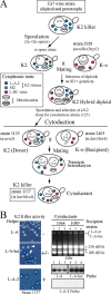
Construction of K2 strain 1137 and exclusion of K2 by L-A or L-A-lus. (A) Diagram of the experimental approach followed to introduce the K2 killer trait from wine strain Ca7 into laboratory strain 2405. The K2 strain Ca7 (diploid and prototroph) was induced to sporulate, and the spore clones were mated to laboratory strain 2928 (expressing Geneticin resistance [GenR] from a vector [23]). Hybrid diploids were selected on minimal medium (SD) with Geneticin. After sporulation, strain 1125 was selected (a ura3; K2). The cytoplasm of this K2 strain was then transferred by cytoduction into recipient strain 2405 (kar1 mutant defective in nuclear fusion and [rho0]). A transient heterokaryon was initially formed. After mitotic division, cytoductants carried the nucleus of the recipient strain and the mixed cytoplasms from the donor and recipient strains, and thus, they are K2 (strain 1137). Strain 1137 also carries L-BC virus (from strain 2928, since strain Ca7 is L-BC-o), and its mitochondria were inherited from the wine strain (information obtained from Fig. S5). The various cytoplasmic traits present in the cells are indicated as follows. K2 viruses L-A-2 and M2 are shown as dark-blue and light-blue hexagons, respectively; L-BC viruses are represented by red hexagons. Mitochondria from different parental strains are shown on a white or gray background. (B) K2 viruses are excluded by L-A or L-A-lus. L-A-2 and M2 viruses were transferred by cytoduction from strain 1125 into two strains isogenic with 2405 but containing L-A (strain 1064) or L-A-lus (strain 1089). (Left) (Top and center) K2 killer activity of isolated colonies from each strain, with the respective helper virus indicated. White arrows indicate colonies that have lost killer activity. (Bottom) K2 killer activity of strain 1137 carrying L-A-2 and M2, shown as a control. (Right) Total nucleic acids from three cytoductants from each recipient strain were separated in an agarose gel and were analyzed by Northern hybridization with probes specific for L-A (lanes 1 to 3) or L-A-lus (lanes 4 to 6). (Top) Ethidium bromide (EtBr)-stained gel. The band indicated by the asterisk corresponds to the 23S RNA narnavirus originally present in donor strain 1125. (Center) The two autoradiograms. (Bottom) The same samples (from lanes 1 to 6) annealed to the L-A-2-specific probe.
FIG 3
FIG 3
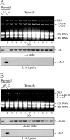
Exclusion of L-A-2 by L-A or L-A-lus. (A) Exclusion of L-A-2 by L-A. Strain 1163 (L-A-2) was mated with strain 1127 (L-A), and diploid colonies were selected. Total nucleic acids from the parental haploid strains (lanes 1 and 2) and from 10 independent diploid colonies (lanes 3 to 12) were separated in an agarose gel and were analyzed by Northern hybridization with specific probes. (Top) Ethidium bromide (EtBr)-stained gel. (Center and bottom) Autoradiograms of RNAs on blots detected with an L-A- or L-A-2-specific probe, respectively. (B) Exclusion of L-A-2 by L-A-lus. This experiment is similar to that for which results are shown in panel A, except that here the L-A-2-containing strain 1163 was mated with strain 1098, carrying L-A-lus. Ten independent diploid colonies were analyzed as described above, using probes against L-A-lus (center) or L-A-2 (bottom).
FIG 4
FIG 4
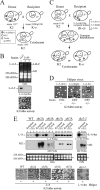
Helper activity of L-A variants. (A) Diagram of a cytoduction experiment to produce strain 1149, which maintains M2 virus by L-A proteins expressed from a vector. L-A-2 and M2 viruses (shaded and open hexagons, respectively) from strain 1137 were transferred to strain 2928 carrying plasmid p1290 (indicated by open circles in the nucleus). Although the cytoductants originally contained L-A-2 and M2, L-A-2 was eliminated by overexpression of viral proteins from the plasmid. (B) (Top) RNAs from two cytoductants (lanes 2 and 3) were analyzed in an agarose gel. RNAs from donor strain 1137 were used as a control (lane 1). Note that both cytoductants have lost L-A-2 but maintain large amounts of M2 dsRNA, visible as discrete bands in the ethidium bromide (EtBr)-stained gel (lanes 2 and 3). (Center) L-A-2 is detected by hybridization with an L-A-2-specific probe. (Bottom) K2 killer activity of isolated cytoductants. (C) Diagram of cytoduction experiments to introduce M2 virus from donor strain 1149 into three K-o recipient strains carrying L-A-lus or L-A helper viruses. The helper viruses (of any type) are indicated by shaded hexagons and M2 virus by open hexagons. (D) Killer assays of isolated cytoductants in each case. The recipient strain name is given below each image, and the respective helper virus is identified at the top. Only if an L-A variant has helper activity for M2 are the cytoductants K2 killers (+); otherwise they are nonkillers (−). (E) M2 maintenance by L-A or L-A-lus in ski mutants. M2 virions from strain 1161, which are maintained by viral proteins expressed from vector pI2L2, were introduced by cytoplasmic mixing into five strains carrying L-A: strain BY4741 (wild type [WT]) (lanes 1 to 3) or mutant derivative strains with deletions of SKI2 (strain Y05307) (lanes 4 to 6), SKI3 (strain Y05604) (lanes 7 to 9), SKI1 (strain Y04540) (lanes 10 to 12), or SKI7 (strain Y01852) (lanes 13 to 15). M2 virions from strain 1161 were also introduced into the L-A-lus-containing strain 1094, which carries the ski2-2 mutation (lanes 16 to 18). In each case, RNAs from the recipient strain and two independent cytoductants (Cytod.) were separated on an agarose gel and were analyzed by Northern hybridization with probes specific for L-A (or L-A-lus) and M2. All cytoductants carried 23S RNA (indicated by the asterisk), present in the donor strain but absent in the recipients. At the bottom, isolated colonies of independent cytoductants from each strain are analyzed for K2 killer activity.
FIG 5
FIG 5
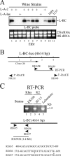
Different L-BC viruses in wine strains. (A) RNAs from a number of K2 or Klus wine strains (lanes 1 to 9) were hybridized with an L-BC probe made by runoff transcription from plasmid pRE442. Laboratory strains 1089 (Klus) and 1137 (K2) were also included (lanes 10 and 11). Ethidium bromide-stained rRNAs, used as loading controls, are shown below. The amounts of L-BC in lanes 5 and 9 are about 10 to 20% of the amounts of L-BC in other strains. (B) Strategy for sequencing L-BC-lus from strain EX198. Clone 20, obtained by random cDNA synthesis, is indicated. The rest of the sequence was obtained from RT-PCR clones or 3′ RACE. cDNA fragments were primed with the oligonucleotides indicated by the arrows. (C) RNAs from Klus wine strain EX198 (lanes 1 and 2), K2 strain 8F-13 (lanes 3 and 4), or laboratory strain BY4741 (lane 5) were used to amplify a fragment of 1.2 kb by RT-PCR with one of two pairs of oligonucleotides: RE635 plus RE636 (lanes 1 and 3) or RE635 plus RE637 (lanes 2, 4, and 5). The oligonucleotide sequences are given at the bottom. Note that the sequences of RE636 and RE637 differ only at two positions (marked by asterisks). RE636 was derived from L-BC in Klus strains, while RE637 comes from L-BC in laboratory strains.
FIG 6
FIG 6
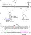
L-BC genomic organization and putative cis signals for frameshifting and replication. (A) Plus strand of L-BC dsRNA. Gag and Pol ORFs are represented by arrows. Pol is expressed as a Gag-Pol fusion protein by a −1 frameshift. Two cis signals, indicated as 1 and 2, are represented by schematic drawings above the plus strand. (B) Nucleotide sequences and predicted secondary structures of the two cis signals shown in panel A. For cis signal 1, the frameshifting region, with the slippery site in blue, is shown adjacent to a stem-loop structure. In L-BC-lus (and L-BC-2), the modification C1987U (indicated by the arrow) produces a change in the secondary-structure free energy. For cis signal 2, putative replication signals are identical in the three variants. (C) The nucleotide sequences of the 5′ and 3′ ends are shown with the start and stop codons for Gag and Pol, respectively. The putative replication signal is shown in pink.

Similar articles

Cited by

References

    1. Schmitt MJ, Breinig F. 2006. Yeast viral killer toxins: lethality and self-protection. Nat Rev Microbiol 4:212–221. doi:10.1038/nrmicro1347. - DOI - PubMed
    1. Schmitt MJ, Tipper DJ. 1995. Sequence of the M28 dsRNA: preprotoxin is processed to an α/β heterodimeric protein. Virology 213:341–351. doi:10.1006/viro.1995.0007. - DOI - PubMed
    1. Rodríguez-Cousiño N, Maqueda M, Ambrona J, Zamora E, Esteban R, Ramírez M. 2011. A new wine Saccharomyces cerevisiae killer toxin (Klus), encoded by a double-stranded RNA virus, with broad antifungal activity is evolutionarily related to a chromosomal host gene. Appl Environ Microbiol 77:1822–1832. doi:10.1128/AEM.02501-10. - DOI - PMC - PubMed
    1. Icho T, Wickner RB. 1989. The double-stranded RNA genome of yeast virus L-A encodes its own putative RNA polymerase by fusing two open reading frames. J Biol Chem 264:6716–6723. - PubMed
    1. Dinman JD, Icho T, Wickner RB. 1991. A −1 ribosomal frame shift in a double-stranded RNA virus of yeast forms a Gag–Pol fusion protein. Proc Natl Acad Sci U S A 88:174–178. doi:10.1073/pnas.88.1.174. - DOI - PMC - PubMed

MeSH terms

LinkOut - more resources