

Ophiobolin A Induces Autophagy and Activates the Mitochondrial Pathway of Apoptosis in Human Melanoma Cells
- PMID: 27936075
- PMCID: PMC5147944
- DOI: 10.1371/journal.pone.0167672
Ophiobolin A Induces Autophagy and Activates the Mitochondrial Pathway of Apoptosis in Human Melanoma Cells
Abstract
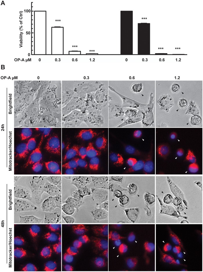
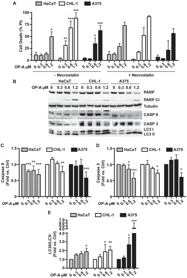
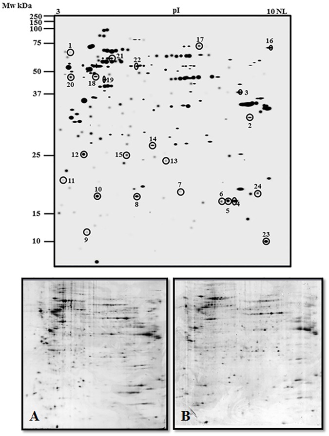
Ophiobolin A, a fungal toxin from Bipolaris species known to affect different cellular processes in plants, has recently been shown to have anti-cancer activity in mammalian cells. In the present study, we investigated the anti-proliferative effect of Ophiobolin A on human melanoma A375 and CHL-1 cell lines. This cellular model was chosen because of the incidence of melanoma malignant tumor on human population and its resistance to chemical treatments. Ophyobolin A strongly reduced cell viability of melanoma cells by affecting mitochondrial functionality. The toxin induced depolarization of mitochondrial membrane potential, reactive oxygen species production and mitochondrial network fragmentation, leading to autophagy induction and ultimately resulting in cell death by activation of the mitochondrial pathway of apoptosis. Finally, a comparative proteomic investigation on A375 cells allowed to identify several Ophiobolin A down-regulated proteins, which are involved in fundamental processes for cell homeostasis and viability.
Conflict of interest statement
The authors have declared that no competing interests exist.
Figures


Similar articles
-
Chaetocin induces apoptosis in human melanoma cells through the generation of reactive oxygen species and the intrinsic mitochondrial pathway, and exerts its anti-tumor activity in vivo.PLoS One. 2017 Apr 18;12(4):e0175950. doi: 10.1371/journal.pone.0175950. eCollection 2017. PLoS One. 2017. PMID: 28419143 Free PMC article.
-
Benzyl isothiocyanate (BITC) induces G2/M phase arrest and apoptosis in human melanoma A375.S2 cells through reactive oxygen species (ROS) and both mitochondria-dependent and death receptor-mediated multiple signaling pathways.J Agric Food Chem. 2012 Jan 18;60(2):665-75. doi: 10.1021/jf204193v. Epub 2012 Jan 6. J Agric Food Chem. 2012. PMID: 22148415
-
Phenethyl isothiocyanate triggers apoptosis in human malignant melanoma A375.S2 cells through reactive oxygen species and the mitochondria-dependent pathways.Hum Exp Toxicol. 2014 Mar;33(3):270-83. doi: 10.1177/0960327113491508. Epub 2013 Jun 11. Hum Exp Toxicol. 2014. PMID: 23760257
-
Chemistry and biology of ophiobolin A and its congeners.Bioorg Med Chem Lett. 2019 Apr 1;29(7):859-869. doi: 10.1016/j.bmcl.2019.02.007. Epub 2019 Feb 7. Bioorg Med Chem Lett. 2019. PMID: 30765189 Review.
-
Autophagy: In the cROSshairs of cancer.Biochem Pharmacol. 2017 Feb 15;126:13-22. doi: 10.1016/j.bcp.2016.10.006. Epub 2016 Oct 24. Biochem Pharmacol. 2017. PMID: 27789215 Free PMC article. Review.
Cited by
-
Physapruin A Induces Reactive Oxygen Species to Trigger Cytoprotective Autophagy of Breast Cancer Cells.Antioxidants (Basel). 2022 Jul 11;11(7):1352. doi: 10.3390/antiox11071352. Antioxidants (Basel). 2022. PMID: 35883843 Free PMC article.
-
PPARδ is a regulator of autophagy by its phosphorylation.Oncogene. 2020 Jun;39(25):4844-4853. doi: 10.1038/s41388-020-1329-x. Epub 2020 May 21. Oncogene. 2020. PMID: 32439863
-
Oregano Phytocomplex Induces Programmed Cell Death in Melanoma Lines via Mitochondria and DNA Damage.Foods. 2020 Oct 17;9(10):1486. doi: 10.3390/foods9101486. Foods. 2020. PMID: 33080917 Free PMC article.
-
Therapeutic Potential of Fungal Terpenes and Terpenoids: Application in Skin Diseases.Molecules. 2024 Mar 6;29(5):1183. doi: 10.3390/molecules29051183. Molecules. 2024. PMID: 38474692 Free PMC article. Review.
-
The Surprising Story of Fusicoccin: A Wilt-Inducing Phytotoxin, a Tool in Plant Physiology and a 14-3-3-Targeted Drug.Biomolecules. 2021 Sep 21;11(9):1393. doi: 10.3390/biom11091393. Biomolecules. 2021. PMID: 34572605 Free PMC article. Review.
References
-
- Au TK, Chick WS, Leung PC. The biology of ophiobolins. Life Sci. 2000;67: 733–742. - PubMed
-
- Fujiwara H, Matsunaga K, Kumagai H, Ishizuka M, Ohizumi Y. Ophiobolin A, a novel apoptosis-inducing agent from fungus strain f-7438. Pharm Pharmacol Commun. 2000;6: 427–431.
MeSH terms
Substances
Grants and funding
LinkOut - more resources
Full Text Sources
Other Literature Sources
Medical
Miscellaneous
