

Selective recruitment of non-classical monocytes promotes skeletal muscle repair
- PMID: 27930948
- PMCID: PMC5218730
- DOI: 10.1016/j.biomaterials.2016.11.021
Selective recruitment of non-classical monocytes promotes skeletal muscle repair
Abstract
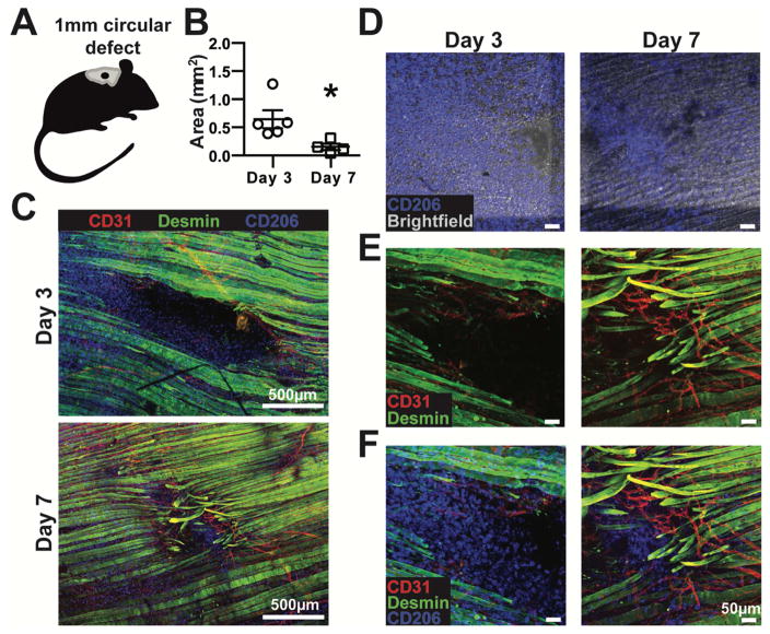
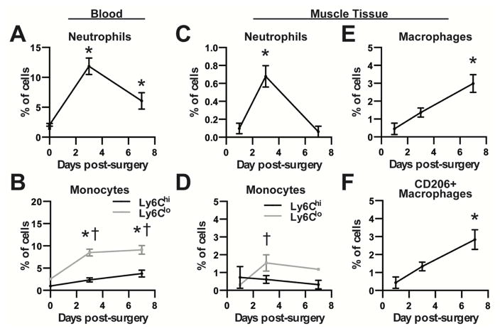
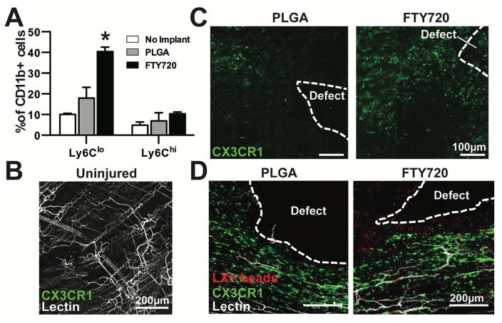
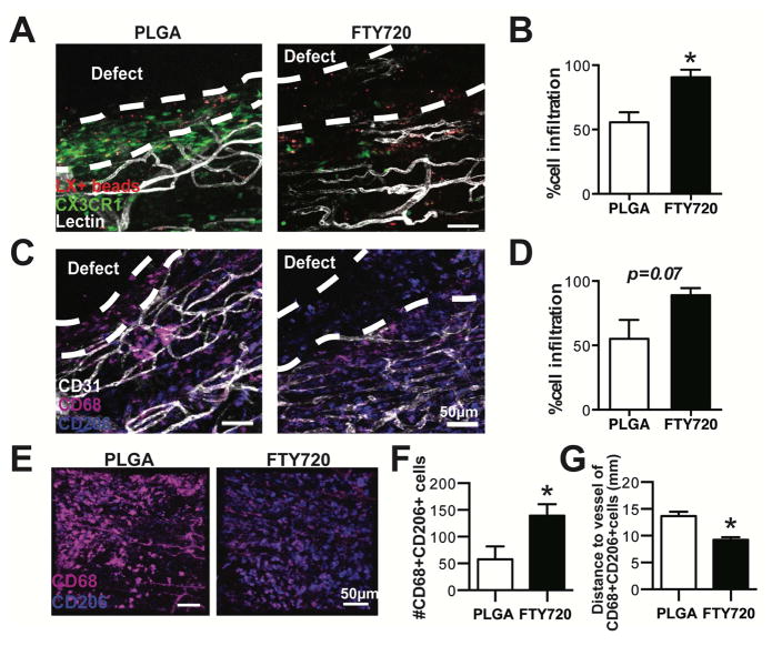
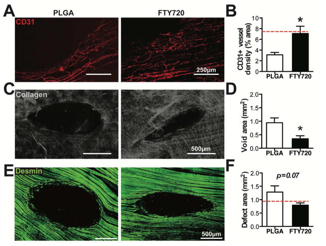
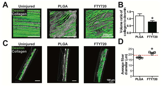
Regeneration of traumatic defects in skeletal muscle requires the synchronized behavior of multiple cells that participate in repair. The inflammatory cascade that is rapidly initiated after injury serves as a powerful node at which to guide the progression of healing and influence tissue repair. Here, we examine the role that myeloid cells play in the healing of traumatic skeletal muscle injury, and leverage their pro-regenerative functions using local delivery of the immunomodulatory small molecule FTY720. We demonstrate that increasing the frequency of non-classical monocytes in inflamed muscle coincides with increased numbers of CD206+ alternatively activated macrophages. Animals treated with immunomodulatory materials had greater defect closure and more vascularization in the acute phases of injury. In the later stages of repair, during which parenchymal tissue growth occurs, we observed improved regeneration of muscle fibers and decreased fibrotic tissue following localization of pro-regenerative inflammation. These results highlight non-classical monocytes as a novel therapeutic target to improve the regenerative outcome after traumatic skeletal muscle injury.
Keywords: Inflammation; Innate immunity; Macrophages; Monocytes; Regenerative medicine; Skeletal muscle.
Copyright © 2016 Elsevier Ltd. All rights reserved.
Figures

Similar articles
-
Non-classical monocytes are biased progenitors of wound healing macrophages during soft tissue injury.Sci Rep. 2017 Mar 27;7(1):447. doi: 10.1038/s41598-017-00477-1. Sci Rep. 2017. PMID: 28348370 Free PMC article.
-
Injury and subsequent regeneration of muscles for activation of local innate immunity to facilitate the development and relapse of autoimmune myositis in C57BL/6 mice.Arthritis Rheumatol. 2015 Apr;67(4):1107-16. doi: 10.1002/art.39017. Arthritis Rheumatol. 2015. PMID: 25580817
-
Calcium/Calmodulin-Dependent Protein Kinase IV (CaMKIV) Mediates Acute Skeletal Muscle Inflammatory Response.Inflammation. 2018 Feb;41(1):199-212. doi: 10.1007/s10753-017-0678-2. Inflammation. 2018. PMID: 28971270
-
Macrophage plasticity in skeletal muscle repair.Biomed Res Int. 2014;2014:560629. doi: 10.1155/2014/560629. Epub 2014 Apr 17. Biomed Res Int. 2014. PMID: 24860823 Free PMC article. Review.
-
Regeneration versus fibrosis in skeletal muscle.Curr Opin Rheumatol. 2011 Nov;23(6):568-73. doi: 10.1097/BOR.0b013e32834bac92. Curr Opin Rheumatol. 2011. PMID: 21934499 Review.
Cited by
-
Nanofiber-Based Delivery of Bioactive Lipids Promotes Pro-regenerative Inflammation and Enhances Muscle Fiber Growth After Volumetric Muscle Loss.Front Bioeng Biotechnol. 2021 Mar 19;9:650289. doi: 10.3389/fbioe.2021.650289. eCollection 2021. Front Bioeng Biotechnol. 2021. PMID: 33816455 Free PMC article.
-
Cells, scaffolds, and bioactive factors: Engineering strategies for improving regeneration following volumetric muscle loss.Biomaterials. 2021 Nov;278:121173. doi: 10.1016/j.biomaterials.2021.121173. Epub 2021 Oct 1. Biomaterials. 2021. PMID: 34619561 Free PMC article. Review.
-
Tissue engineering modalities in skeletal muscles: focus on angiogenesis and immunomodulation properties.Stem Cell Res Ther. 2023 Apr 15;14(1):90. doi: 10.1186/s13287-023-03310-x. Stem Cell Res Ther. 2023. PMID: 37061717 Free PMC article. Review.
-
The Hair Follicle: An Underutilized Source of Cells and Materials for Regenerative Medicine.ACS Biomater Sci Eng. 2018 Apr 9;4(4):1193-1207. doi: 10.1021/acsbiomaterials.7b00072. Epub 2017 Mar 21. ACS Biomater Sci Eng. 2018. PMID: 29682604 Free PMC article.
-
FTY720 in immuno-regenerative and wound healing technologies for muscle, epithelial and bone regeneration.Front Physiol. 2023 May 12;14:1148932. doi: 10.3389/fphys.2023.1148932. eCollection 2023. Front Physiol. 2023. PMID: 37250137 Free PMC article. Review.
References
-
- Summan M, Warren GL, Mercer RR, Chapman R, Hulderman T, Van Rooijen N, Simeonova PP. Macrophages and skeletal muscle regeneration: a clodronate-containing liposome depletion study. American Journal of Physiology - Regulatory, Integrative and Comparative Physiology. 2006;290(6):R1488–R1495. - PubMed
-
- Willenborg S, Lucas T, van Loo G, Knipper JA, Krieg T, Haase I, Brachvogel B, Hammerschmidt M, Nagy A, Ferrara N, Pasparakis M, Eming SA. CCR2 recruits an inflammatory macrophage subpopulation critical for angiogenesis in tissue repair. Blood. 2012;120(3):613–25. - PubMed
Publication types
MeSH terms
Substances
Grants and funding
LinkOut - more resources
Full Text Sources
Other Literature Sources
Medical
