. 2016 Dec 8;12(12):e1006469.
doi: 10.1371/journal.pgen.1006469. eCollection 2016 Dec.

Whole-Organism Developmental Expression Profiling Identifies RAB-28 as a Novel Ciliary GTPase Associated with the BBSome and Intraflagellar Transport

Affiliations

Whole-Organism Developmental Expression Profiling Identifies RAB-28 as a Novel Ciliary GTPase Associated with the BBSome and Intraflagellar Transport

Victor L Jensen et al. PLoS Genet. .

Abstract

Primary cilia are specialised sensory and developmental signalling devices extending from the surface of most eukaryotic cells. Defects in these organelles cause inherited human disorders (ciliopathies) such as retinitis pigmentosa and Bardet-Biedl syndrome (BBS), frequently affecting many physiological and developmental processes across multiple organs. Cilium formation, maintenance and function depend on intracellular transport systems such as intraflagellar transport (IFT), which is driven by kinesin-2 and IFT-dynein motors and regulated by the Bardet-Biedl syndrome (BBS) cargo-adaptor protein complex, or BBSome. To identify new cilium-associated genes, we employed the nematode C. elegans, where ciliogenesis occurs within a short timespan during late embryogenesis when most sensory neurons differentiate. Using whole-organism RNA-Seq libraries, we discovered a signature expression profile highly enriched for transcripts of known ciliary proteins, including FAM-161 (FAM161A orthologue), CCDC-104 (CCDC104), and RPI-1 (RP1/RP1L1), which we confirm are cilium-localised in worms. From a list of 185 candidate ciliary genes, we uncover orthologues of human MAP9, YAP, CCDC149, and RAB28 as conserved cilium-associated components. Further analyses of C. elegans RAB-28, recently associated with autosomal-recessive cone-rod dystrophy, reveal that this small GTPase is exclusively expressed in ciliated neurons where it dynamically associates with IFT trains. Whereas inactive GDP-bound RAB-28 displays no IFT movement and diffuse localisation, GTP-bound (activated) RAB-28 concentrates at the periciliary membrane in a BBSome-dependent manner and undergoes bidirectional IFT. Functional analyses reveal that whilst cilium structure, sensory function and IFT are seemingly normal in a rab-28 null allele, overexpression of predicted GDP or GTP locked variants of RAB-28 perturbs cilium and sensory pore morphogenesis and function. Collectively, our findings present a new approach for identifying ciliary proteins, and unveil RAB28, a GTPase most closely related to the BBS protein RABL4/IFT27, as an IFT-associated cargo with BBSome-dependent cell autonomous and non-autonomous functions at the ciliary base.


Conflict of interest statement

The authors have declared that no competing interests exist.

Figures
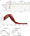
Fig 1
Fig 1. Whole-organism expression profiling identifies a signature pattern enriched for known ciliary genes.
(A) Timeline (in minutes) of C. elegans cell births according to tissues. Small shapes in each row represent the birth of a cell of the corresponding tissue type. Embryonic development, hatching and post-embryonic development are highlighted above the x-axis with the worm stages illustrated below. Most ciliated neurons are born in late embryogenesis with ciliogenesis occurring shortly after these cells are born. Worm illustrations modified from WormAtlas (www.wormatlas.org). Q/G/K/P/H/V/T/M refer to different embryonic cell lineages (http://www.wormatlas.org/celllineages.html). (B) Normalised expression levels for all ‘bait’ cilium-associated genes (red) and the top 276 target genes (black, p-value < 5e-7) over the developmental series of analysed RNA-Seq libraries. Expression peaks for all genes during late embryo or larval stage 1, which matches closely the birth of the ciliated neurons shown in (A). This cilia-specific gene expression pattern shows much lower levels of expression from larval stage 2 to young adult. (C) Box and whisker plots of the normalised expression across the whole-organism developmental RNA-Seq libraries for the chemosensory and ciliary gene sets. Expression of the ciliary set immediately precedes that of the chemosensory set. Peak expression for ciliary genes is at the late embryo-L1 larval stage, whereas expression peaks at L1-L2 larval stages for chemosensory genes. EE; early embryo. LE; late embryo. L1; larval stage 1. L2; larval stage 2. L3; larval stage 3. L4; larval stage 4. YA; young adult.
Fig 2
Fig 2. Candidate cilium-associated proteins localise to ciliary structures Representative images of amphid (head) and phasmid (tail) cilia from N2 wild type worms co-expressing GFP-tagged ‘translational’ reporters for candidate ciliary genes and tdTomato-tagged XBX-1 (IFT protein that localises to the ciliary base and axoneme).
All reporters driven by the endogenous gene promoter, except for YAP-1 and RAB-28, where a bbs-8 gene promoter sequence was used (see Fig 3A for a GFP::RAB-28 reporter driven by the endogenous promoter). GFP tags are on the N- or C-terminus, as indicated above the panels. PCMC (periciliary membrane compartment); distal dendrite (den) swelling enriched for endocytosis-associated proteins and vesicles that regulate ciliary membrane homeostasis [19]. The transition zone (TZ) extends from basal body (BB) and functions as a ciliary gate that regulates protein entry and exit to and from cilia [6]. Middle (MS) and distal (DS) segments; characterised by a circular array of 9 doublet (A and B tubules) and 9 singlet (A tubules) microtubules, respectively (not shown). Note that for GFP::CCDC-149, signals are observed at the amphid/phasmid basal bodies (arrows) and as punctae (asterisks) along the dendrite of the OLQ ciliated neuron running parallel to the amphid neurons. Scale bars; 5 μm.
Fig 3
Fig 3. RAB-28 undergoes IFT.
(A, B) Representative images of phasmid cilia (left) from N2 wild type and che-11(e1810) worms expressing GFP::RAB-28, together with corresponding kymographs and kymograph schematics derived from time-lapse imaging. Distribution plot (and mean values) in B shows kymography-determined anterograde and retrograde GFP::RAB-28 velocities from wild type worms. MS; middle segment, DS; distal segment, PC; periciliary membrane compartment, DD; distal dendrite. Scale bars; 3 μm (phasmid image; and horizontal bar on kymographs); 3 seconds (vertical bar on kymographs). (C) Fluorescence recovery after photobleaching (FRAP) plots for GFP::RAB-28 in the phasmid neurons of N2 and che-11(e1810) mutant worms. GFP::RAB-28 also undergoes free diffusion in wild-type (N2) and che-11 IFT mutant animals. Ciliary GFP signals bleached at time 0. Intensity measurements normalised to pre-bleach levels. Curves derived from 3 separate FRAP experiments. Error bars; SEM. Images taken from a representative FRAP experiment in N2 wild type worms. s; seconds. Scale bar; 2 μm.
Fig 4
Fig 4. BBSome-dependent recruitment of activated RAB-28 to the periciliary membrane.
(A, B) Representative images of amphid and phasmid cilia from N2 wild type (A) and bbs-8(nx77) mutant (B) worms expressing GFP-tagged RAB-28(WT), RAB-28(GDP) and RAB-28(GTP) reporters. All reporters are driven by the endogenous rab-28 gene promoter. Kymographs and kymograph schematics derived from time-lapse imaging of GFP signals in phasmid cilia. Phenotypes summarised in cartoons. Large phasmid images are placed on black backgrounds. m; middle segment, d; distal segment. pcmc; periciliary membrane compartment (also denoted by arrow). Scale bars; 2 μm and 5 μm (low magnification phasmid images).
Fig 5
Fig 5. Worms overexpressing RAB-28(GTP) or RAB-28(GDP) display overlapping and distinct defects in sensory pore formation and function.
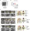
(A) Dye filling (DiI) of amphid neurons in non-transgenic (control) or transgenic wild type (N2) worms expressing GFP::RAB-28(WT), GFP::RAB-28(GTP) or GFP::RAB-28(GDP). All constructs driven by the endogenous rab-28 gene promoter and expressed to similar levels in transgenic animals (all injected at 5 ng/ul). For each amphid pore, the number of dye-filling neurons was scored. Each dataset represents mean ± standard deviation (error bars) from 3 independent experiments (at least 40 amphid pores scored for each strain per experiment). (B) Roaming scores for non-transgenic (control) and transgenic wild type (N2) worms expressing GFP-tagged RAB-28 variants. Scores normalised to non-transgenic N2 worms. For each strain, >45 worms were scored. *p<0.01 (unpaired Student t-test vs non-transgenic N2 controls). (C) Transmission electron microscopy images of the amphid pore from serial cross-section of wild type (N2) and N2 worms expressing GFP-tagged RAB-28(GTP) or RAB-28(GDP). Low magnification images (top rows) show the enlarged amphid channel (yellow outline and asterisk) in RAB-28(GTP)-expressing worms and dense matrix filled vesicles (mfv) in the amphid sheath cell of RAB-28(GDP) expressing worms (white arrows). High magnification images (bottom rows) display close ups of the amphid pore and ciliary axonemes, showing the distal segments (DS), middle segments (MS), transition zones (TZ) and periciliary membrane compartments (PCMC). Note the misplaced (disconnected) channel axoneme (white arrowhead) that fails to enter the channel of worms expressing RAB-28(GDP). Images representative of at least 4 analysed pores for each strain. Cartoons show the amphid channel in cross section and longitudinal orientations (only 3 of the 10 axonemes shown for simplicity in longitudinal cartoon), and indicate observed phenotypes. Numbers above images indicate the position of the section relative to the most anterior section (at ‘0’); section positions also indicated in cartoon. Scale bars; 1 μm (top rows); 200 nm (large images in bottom rows); 100 nm (small images in bottom rows).
Fig 6
Fig 6. Model of RAB-28 ciliary associations in C. elegans.
Shown is a cartoon of the amphid sensory pore. The ciliary axonemes (only one shown for simplicity) invaginate through the sheath cell process into a matrix-filled channel. The process of the socket cell forms a doughnut-like ending that establishes the distal part of the channel, by sitting on top of the sheath cell process. Binding of GTP to RAB-28 promotes its targeting to the periciliary membrane (PCM) via interactions with the ciliary base-associated BBSome complex. RAB-28(GTP)-BBSome assemblies associate with the IFT machinery at the ciliary base and enter the cilium via IFT-directed movement. Activated RAB-28 is proposed to serve cell non-autonomous functions by regulating the release (or cell surface expression) of a neuronal signal that controls sheath cell channel morphogenesis. In worms overexpressing RAB-28(GTP) or RAB-28(GDP), potentially opposing amphid channel size phenotypes are observed (enlarged vs small), together with modest cilium structure and sensory behaviour abnormalities (not shown in above model; see Fig 5 for details).

Similar articles

Cited by

References

    1. Goetz SC, Anderson KV. The primary cilium: a signalling centre during vertebrate development. Nat Rev Genet. 2010;11: 331–344. 10.1038/nrg2774 - DOI - PMC - PubMed
    1. Satir P, Pedersen LB, Christensen ST. The primary cilium at a glance. J Cell Sci. 2010;123: 499–503. 10.1242/jcs.050377 - DOI - PMC - PubMed
    1. Waters AM, Beales PL. Ciliopathies: an expanding disease spectrum. Pediatr Nephrol Berl Ger. 2011;26: 1039–1056. - PMC - PubMed
    1. Novas R, Cardenas-Rodriguez M, Irigoín F, Badano JL. Bardet-Biedl syndrome: Is it only cilia dysfunction? FEBS Lett. 2015;589: 3479–3491. 10.1016/j.febslet.2015.07.031 - DOI - PubMed
    1. Blacque OE, Sanders AA. Compartments within a compartment: What C. elegans can tell us about ciliary subdomain composition, biogenesis, function, and disease. Organogenesis. 2014;10: 126–137. 10.4161/org.28830 - DOI - PMC - PubMed

MeSH terms

Grants and funding

MRL acknowledges funding from the Canadian Institutes of Health Research (CIHR; grant MOP-82870) and a senior scholarship award from Michael Smith Foundation for Health Research (MSFHR). RDM acknowledges funding from the Natural Sciences and Engineering Council of Canada (NSERC; 435398-2013) and holds a CIHR New Investigator Award. VLJ holds postdoctoral fellowships from MSFHR and KRESCENT, and TAT is the recipient of a Banting Postdoctoral Fellowship. OEB acknowledges principal investigator funding from Science Foundation Ireland (11/PI/1037), and SC holds an Irish Research Council Government of Ireland postgraduate award (GOIPG/2014/683). The funders had no role in study design, data collection and analysis, decision to publish, or preparation of the manuscript.

LinkOut - more resources