. 2017 Jan 3;127(1):335-348.
doi: 10.1172/JCI88353. Epub 2016 Nov 28.

The H3K9 dimethyltransferases EHMT1/2 protect against pathological cardiac hypertrophy

The H3K9 dimethyltransferases EHMT1/2 protect against pathological cardiac hypertrophy

Bernard Thienpont et al. J Clin Invest. .

Abstract

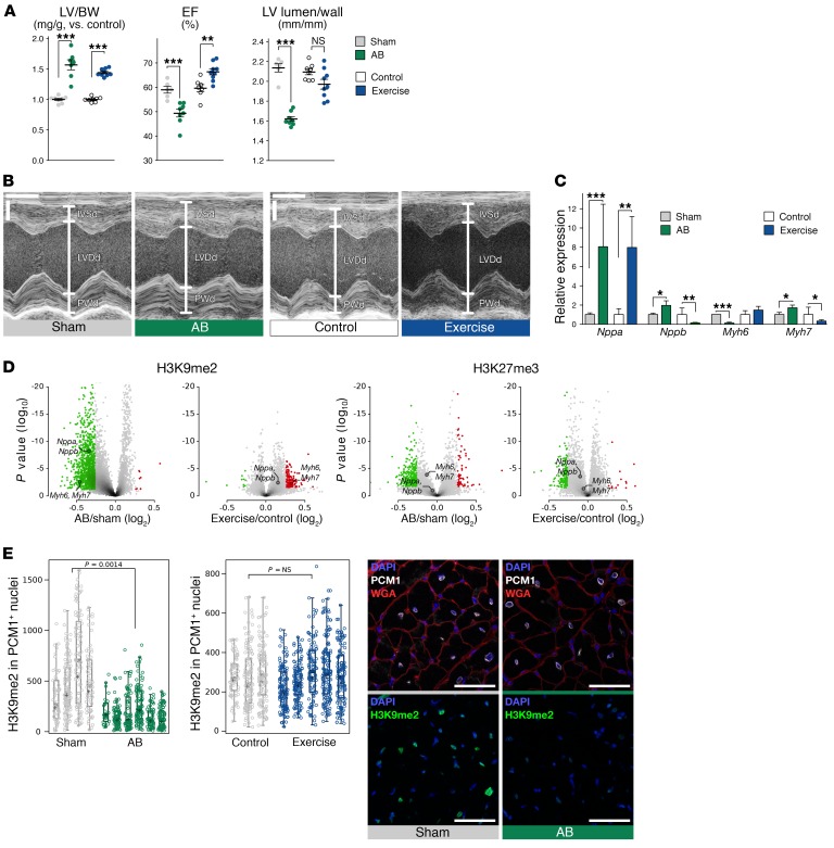
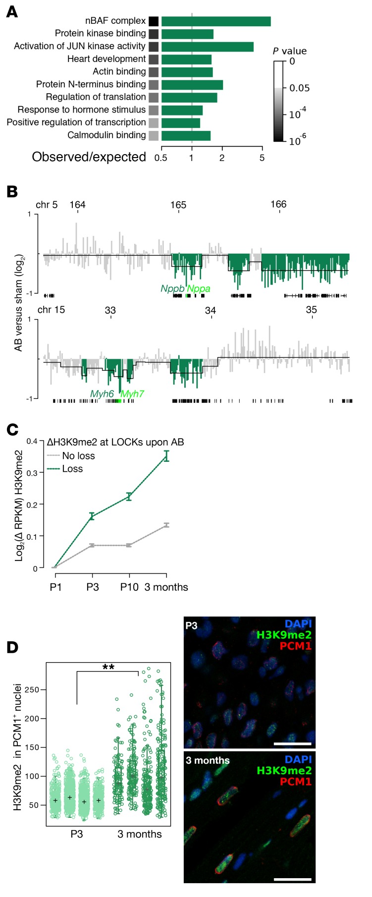
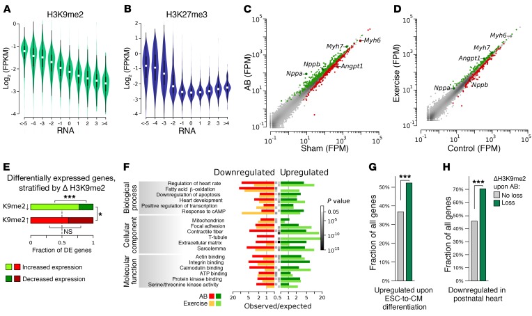
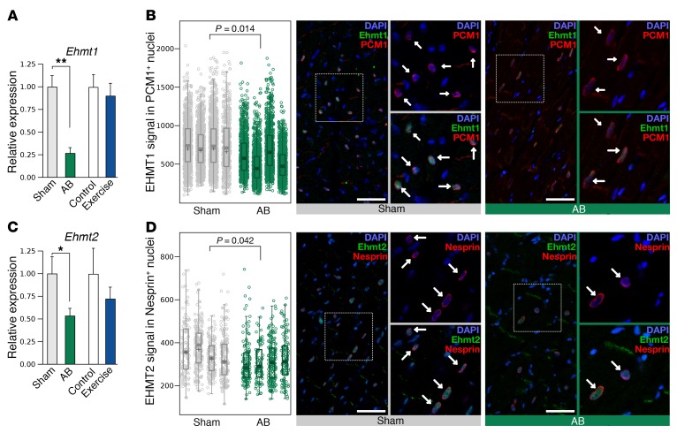
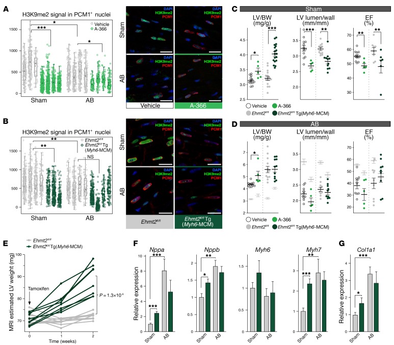
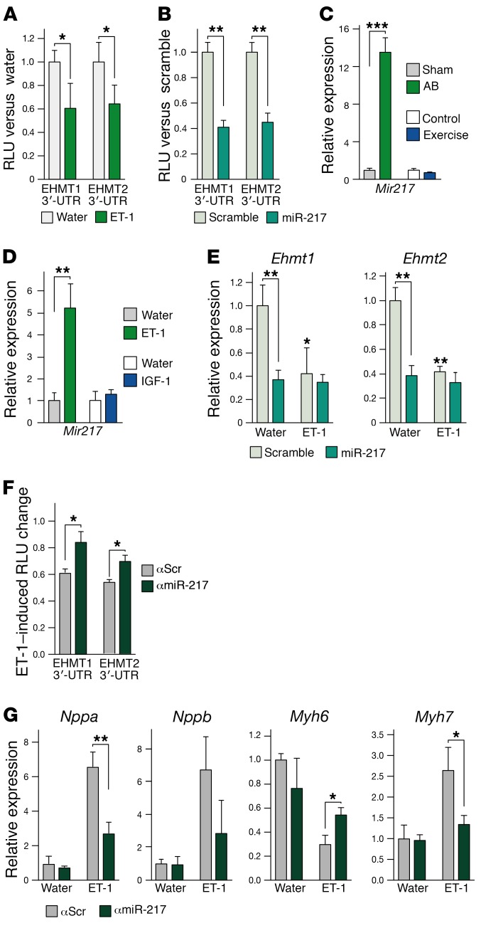
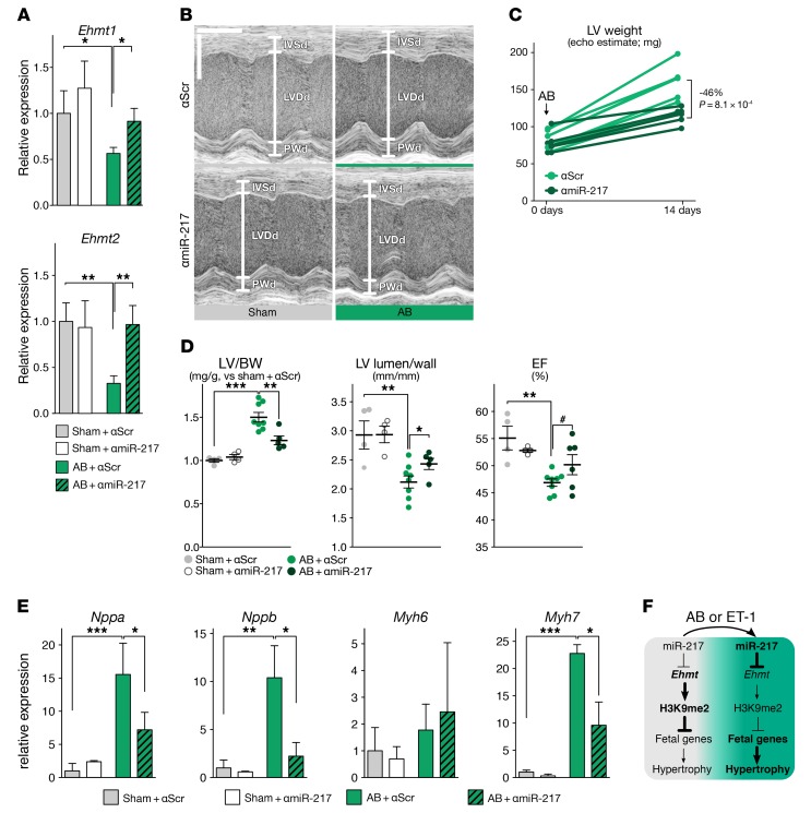
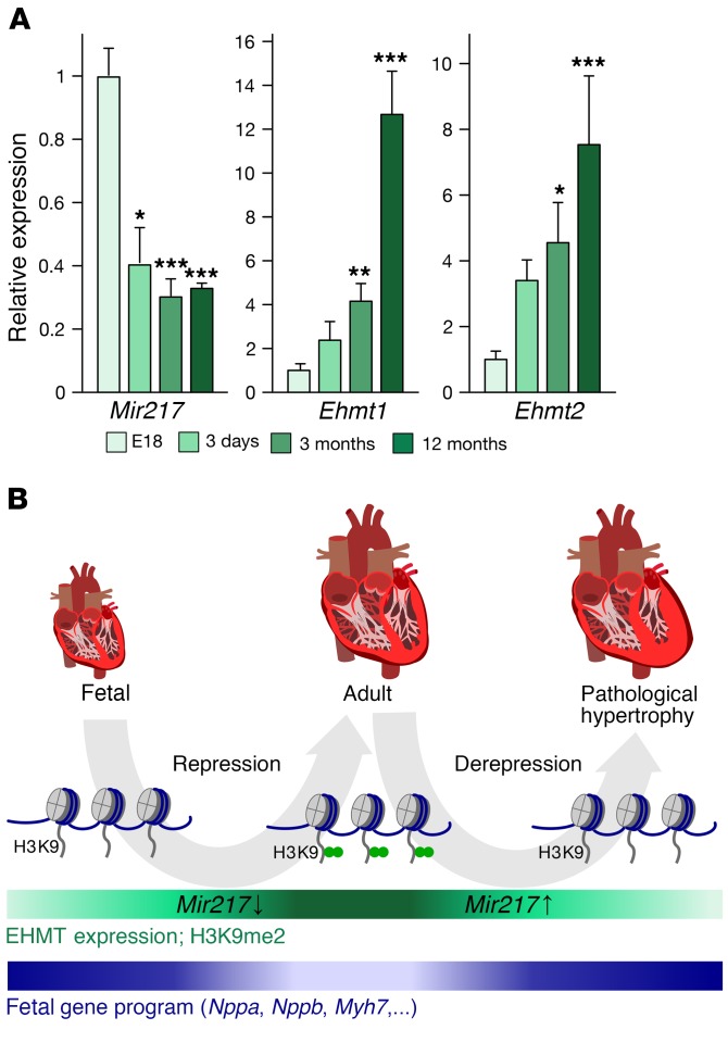
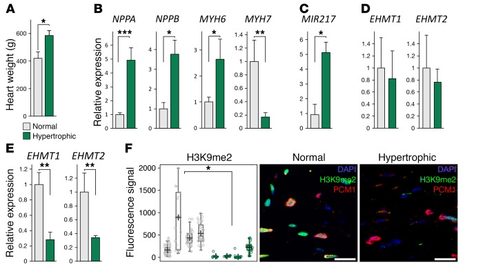
Cardiac hypertrophic growth in response to pathological cues is associated with reexpression of fetal genes and decreased cardiac function and is often a precursor to heart failure. In contrast, physiologically induced hypertrophy is adaptive, resulting in improved cardiac function. The processes that selectively induce these hypertrophic states are poorly understood. Here, we have profiled 2 repressive epigenetic marks, H3K9me2 and H3K27me3, which are involved in stable cellular differentiation, specifically in cardiomyocytes from physiologically and pathologically hypertrophied rat hearts, and correlated these marks with their associated transcriptomes. This analysis revealed the pervasive loss of euchromatic H3K9me2 as a conserved feature of pathological hypertrophy that was associated with reexpression of fetal genes. In hypertrophy, H3K9me2 was reduced following a miR-217-mediated decrease in expression of the H3K9 dimethyltransferases EHMT1 and EHMT2 (EHMT1/2). miR-217-mediated, genetic, or pharmacological inactivation of EHMT1/2 was sufficient to promote pathological hypertrophy and fetal gene reexpression, while suppression of this pathway protected against pathological hypertrophy both in vitro and in mice. Thus, we have established a conserved mechanism involving a departure of the cardiomyocyte epigenome from its adult cellular identity to a reprogrammed state that is accompanied by reexpression of fetal genes and pathological hypertrophy. These results suggest that targeting miR-217 and EHMT1/2 to prevent H3K9 methylation loss is a viable therapeutic approach for the treatment of heart disease.


Conflict of interest statement

W. Reik is a consultant and shareholder of Cambridge Epigenetix.

Figures

Figure 1
Figure 1. H3K9me2 is decreased in CMs upon AB in rats.
(A) Graphs show LV/BW ratio, normalized to sham or control (mg/g); EF percentage; and ratio of LV lumen diameter to myocardial wall thickness at end-diastole. n = 7, 9, 6, and 8 animals for control, exercise, sham, and AB, respectively. (B) M-mode echocardiogram showing, at end-diastole, the LV diameter (LVDd) and thickness of the posterior wall (PWd) and of the interventricular septum (IVSd). Horizontal scale bars: 0.1 second; vertical scale bars: 2 mm. (C) qRT-PCR showing expression of Nppa, Nppb, Myh6, and Myh7 in the LV. (D) Volcano plot of differential markings by H3K9me2 and H3K27me3 of, respectively, LOCKs and BLOCs in PCM1+ nuclei upon pathological (left) and physiological (right) hypertrophy, as determined by ChIP-seq. Red and green dots represent regions enriched and depleted upon hypertrophy (FDR <10%; >20% change). (E) Plots show H3K9me2 signal in PCM1+ nuclei, determined by immunofluorescence analysis of the LV from AB, sham-operated, exercise, and control animals (n = 4, 5, 3, and 5, respectively). Representative confocal immunofluorescence images show staining for wheat germ agglutinin (WGA, red) and PCM1 (white), and H3K9me2 (green) in the LV. Scale bars: 50 μm. Error bars indicate the SEM. n = 5 (C) and 4 (D) replicates. *P < 0.05, **P < 0.01, and ***P < 0.001, by Student’s t test (A and C) and nested ANOVA (E).
Figure 2
Figure 2. H3K9me2 is acquired during cardiomyocyte maturation and lost at fetal genes upon AB.
(A) Selected ontology terms enriched among genes in regions altered in H3K9me2 upon AB. (B) H3K9me2 enrichment at loci on chromosomes (Chr) 5 and 15 in PCM1+ nuclei. Enrichment is displayed per 10-kb bin as log2(ratio of AB vs. sham). Green denotes significantly altered LOCKs. (C) Changes in H3K9me2 during CM maturation at LOCKs showed loss or no loss (green or gray) upon AB. (D) Quantification and representative images of immunofluorescence staining of H3K9me2 (green) in CMs (PCM1, red) in neonatal (3-day-old) and adult (3-month-old) mice (n = 4 each). Hypertrophy was induced for 6 weeks. Error bars indicate the SEM. n = 4 (B and D) and 2 (C) replicates. **P < 0.01, by nested ANOVA (D).
Figure 3
Figure 3. Hypertrophy-associated transcriptomic changes in flow-sorted rat CMs.
(A and B) Violin plots illustrating the gene-wise relation between RNA expression and H3K9me2 (A) or H3K27me3 (B). Data were quantified per gene and are represented as log2((fragments + 1) per kb per million) (FPKM). (C and D) Gene expression, represented as fragments per million (FPM), in PCM1+ nuclei sorted from hypertrophied and control hearts. Highlighted are the genes that were significantly upregulated (green) and downregulated (red) (FDR <10%) after 6 weeks of hypertrophy induction, with characteristic genes indicated. (E) Fractions of differentially expressed genes (FDR<10%, difference >20%) in LOCKs with H3K9me2 loss or gain upon AB. (F) Selected ontological terms significantly enriched among genes significantly up- or downregulated in PCM1+ nuclei upon exercise and AB. The gray box before each bar represents the P value of enrichment; there are no boxes for P values greater than 0.05. (G and H) Characterization of genes displaying loss of H3K9me2 upon AB. (G) Graph represents the fraction of genes that were upregulated during the in vitro differentiation of ESCs into CMs, as determined by Wamstad and colleagues (18). (H) Graph shows the fraction of genes that were downregulated in the adult versus newborn heart (19). Data for G and H were from the mouse genome. For comparisons with the rat genome, analyses were limited to genes having 1-to-1 paralogs between both species. n = 4. *P < 0.05 and ***P < 0.001, by χ2 test.
Figure 4
Figure 4. EHMT1/2 expression is decreased following AB in rats.
(A and C) qRT-PCR of Ehmt1 (A) and Ehmt2 (C) mRNA expression in sorted PCM1+ nuclei isolated from rats subjected to AB or exercise for 6 weeks. Error bars represent the mean ± SEM of 4 (sham, AB) or 7 (control, exercise) biological replicates. *P < 0.05 and **P < 0.01, by Student’s t test. (B and D) (left) Jitter plot illustrating EHMT1 and EHMT2 expression (n = 4 for all conditions) in PCM1+ and nesprin+ cardiomyocytes from hearts as in A. P values were calculated using nested ANOVA. Representative images of immunofluorescence staining for PCM1 (red) and EHMT1 (green) (B) and nesprin (red) and EHMT2 (green) (D) in LV sections from rats that underwent sham operation or AB. The dashed white box indicates the location of the zoomed images (zoom, ×2) to the right of each panel. Scale bars: 50 μm. Nuclei were counterstained with DAPI (blue), and CM nuclei are indicated by arrows.
Figure 5
Figure 5. Suppression of EHMTs induces hypertrophy in vitro.
(A and B) Protein synthesis (A) and qRT-PCR for Nppa, Nppb, Myh6, and Myh7 (B) in primary NRVMs exposed for 48 hours to ET-1 or vehicle (water) and in response to EHMT1/2 inhibition (A-366). (C) Immunofluorescence staining for α-actinin (white) and ANF (green) in NRVMs treated with or without ET-1 and/or A-366. Nuclei were counterstained with DAPI (blue). Scale bars: 20 μm. (D) Immunofluorescence signal intensities of perinuclear ANF (shown is 1 representative experiment from 4 repeats). (E and F) Protein synthesis analysis by 3H-leucine incorporation (E) and Nppa, Nppb, Myh6, and Myh7 expression (F) in NRVMs transduced with empty or EHMT2 adenovirus 8 hours prior to agonist addition (ET-1 or water). Cells were assayed 48 hours after agonist addition. Note that the hypertrophic response appears dampened in transduced NRVMs compared with nontransduced NRVMs in A and B. Error bars in A, B, E, and F represent the mean ± SEM. n = 5 biological replicates. *P < 0.05,**P < 0.01, and ***P < 0.001, by Student’s t test.
Figure 6
Figure 6. Pharmacological or genetic inactivation of EHMTs induces pathological hypertrophy.
(A and B) H3K9me2 signal in PCM1+ nuclei and representative immunofluorescence images for PCM1 (red) and H3K9me2 (green) in sections from LVs of sham- and AB-operated mice (A) after 10 days with A-366 or vehicle administration through s.c. mini-osmotic pumps (n = 3 for sham vehicle and 4 for other treatments) and (B) from Ehmt2fl/fl and Ehmt2fl/fl Tg(Myh6-MCM) mice [n = 3 for sham-treated Ehmt2fl/fl Tg(Myh6-MCM) mice and n = 4 for all other conditions], 2 weeks after tamoxifen injection. Scale bars: 20 μm. Nuclei are stained with DAPI (blue). (C and D) Alterations in LV weight, shape, and EF for sham-operated mice (C) (n = 12, 5, 11, and 11 mice for the indicated conditions) and AB-operated mice (D) (n = 10, 5, 10, and 10 mice for the indicated conditions), after 10 days of administration of A-366 or vehicle and for Ehmt2fl/fl and Ehmt2fl/fl Tg(Myh6-MCM) mice, 2 weeks after tamoxifen injection. Vertical dashed line indicates experiments conducted separately to inactivate EHMT1/2 by chemical or genetic means. To enable comparison within experiments, averaged data for sham-operated animals from C were added to the data in D (grayed averages ± SEM). (E) Evolution of LV weight following tamoxifen injection into mice of the indicated genotypes, as estimated by MRI at weeks 0, 1, and 2 [n = 8 for Ehmt2fl/fl mice and n = 9 for Ehmt2fl/fl Tg(Myh6-MCM) mice]. (F and G) qRT-PCR showing expression levels of Nppa, Nppb, Myh6, Myh7 (F), and collagen, type I, α 1 (Col1a1) (G) in the LVs of Ehmt2fl/fl and Ehmt2fl/fl Tg(Myh6-MCM) mice, 2 weeks after tamoxifen injection and after sham or AB operations. Error bars indicate the SEM. n = 5 replicates (F and G). *P < 0.05, **P < 0.01, and ***P < 0.001, by nested ANOVA (A and B) and Student’s t test (CG).
Figure 7
Figure 7. Downregulation of Ehmt1/2 by miR-217 is required for hypertrophy induction in vitro.
(A and B) RLU of luciferase constructs harboring the 3′-UTR of EHMT1 or EHMT2, transfected into NRVMs treated with water or ET-1 (A), or into HEK293 cells transfected with scrambled or miR-217 mimic (B). (C and D) Expression of miR-217 in rat CM nuclei sorted from sham-operated, AB-operated, control, or exercised hearts (C), and in NRVMs treated for 48 hours with ET-1, IGF-1, or water (D). (E) Expression of Ehmt1 and Ehmt2 in NRVMs transfected with scrambled or miR-217 mimic for 48 hours and treated for 48 hours with ET-1 or water. (F) RLU of luciferase constructs harboring the 3′-UTR of Ehmt1 or Ehmt2, transfected into NRVMs treated with control (anti-scramble; αScr) or miR-217 antagomirs (anti–miR-217; αmiR-217). Shown are the RLU in response to 48 hours of exposure to ET-1 relative to water-treated controls. (G) Expression of Nppa, Nppb, Myh6, and Myh7 in NRVMs treated with control or miR-217 antagomirs and exposed for 48 hours to water or ET-1. *P < 0.05, **P < 0.01, and ***P < 0.001, by Student’s t test. Error bars represent the mean ± SEM of 5 independent experiments.
Figure 8
Figure 8. Downregulation of Ehmt1/2 by miR-217 is required for hypertrophy induction in vivo.
(A) Effect of miR-217 antagomir or scramble treatment upon expression of Ehmt1 and Ehmt2 in hearts from sham- or AB-operated mice 2 weeks after surgery. (B) M-mode echocardiogram from parasternal location of mice treated as in A. Indicated are, at end-diastole, the LV diameter (LVDd) and the thickness of the posterior wall (PWd) and of the interventricular septum (IVSd). Horizontal scale: 0.1 second; vertical scale: 2 mm. (C) LV mass as estimated by echocardiography (echo) 1 day after AB (before administration of antagomirs) and 2 weeks after AB (n = 5 for miR-217– and 8 for scramble-injected animals). (D) LV/BW ratio, LV lumen/wall diameter ratio, and EF of hearts treated as in A (n = 4, 4, 8, 5 for the indicated conditions). (E) Expression of Nppa, Nppb, Myh6, and Myh7 in hearts treated as in A. (F) Schematic of miR-217 regulation of Ehmt expression and pathological hypertrophy. ***P < 0.001, **P < 0.01, *P < 0.05, and #P = 0.07, by Student’s t test. Error bars in A and E represent the mean ± SEM of 4 (sham conditions), 8 (AB plus αScramble), or 6 (AB plus αmiR-217) mice.
Figure 9
Figure 9. Gain of Ehmt1/2 expression and loss of miR-217 during CM maturation.
(A) qRT-PCR showing expression of Ehmt1, Ehmt2, and Mir217 in CMs isolated from rats at the indicated ages, relative to E18. Error bars represent the mean ± SEM of 5 biological replicates. ***P < 0.001, **P < 0.01, and *P < 0.05, by Student’s t test. (B) Proposed role of EHMT-induced repression of the fetal gene program in suppressing pathological hypertrophic remodeling of the heart.
Figure 10
Figure 10. Loss of EHMT1/2 expression and H3K9me2 in human hypertrophic hearts.
(A) Weight of normal and hypertrophic human hearts. (BE) Expression of NPPA, NPPB, MYH6, and MYH7 (B), MIR217 (C), and EHMT1 and EHMT2 nascent transcripts (D) and mature mRNAs (E) in PCM1+ nuclei in LVs of normal and hypertrophic human hearts. (F) Quantification of H3K9me2 signal in human CMs. Representative images of H3K9me2 staining (green) in PCM1+ CM nuclei (red) in normal and hypertrophic human hearts. Scale bars: 20 μm. Error bars represent the mean ± SEM of 5 (AE) and 4 (F) biological replicates. *P < 0.05, **P < 0.01, and ***P < 0.001, by Student’s t test (AE) or nested ANOVA (F).

Similar articles

Cited by

References

    1. Mathers CD, Loncar D. Projections of global mortality and burden of disease from 2002 to 2030. PLoS Med. 2006;3(11):e442. doi: 10.1371/journal.pmed.0030442. - DOI - PMC - PubMed
    1. Heineke J, Molkentin JD. Regulation of cardiac hypertrophy by intracellular signalling pathways. Nat Rev Mol Cell Biol. 2006;7(8):589–600. doi: 10.1038/nrm1983. - DOI - PubMed
    1. Soonpaa MH, Field LJ. Survey of studies examining mammalian cardiomyocyte DNA synthesis. Circ Res. 1998;83(1):15–26. doi: 10.1161/01.RES.83.1.15. - DOI - PubMed
    1. Bergmann O, et al. Dynamics of Cell Generation and Turnover in the Human Heart. Cell. 2015;161(7):1566–1575. doi: 10.1016/j.cell.2015.05.026. - DOI - PubMed
    1. Bergmann O, et al. Evidence for cardiomyocyte renewal in humans. Science. 2009;324(5923):98–102. doi: 10.1126/science.1164680. - DOI - PMC - PubMed

Publication types