. 2016 Nov 17;7(11):e2474.
doi: 10.1038/cddis.2016.371.

De-ubiquitinating enzyme, USP11, promotes transforming growth factor β-1 signaling through stabilization of transforming growth factor β receptor II

Affiliations

De-ubiquitinating enzyme, USP11, promotes transforming growth factor β-1 signaling through stabilization of transforming growth factor β receptor II

A M Jacko et al. Cell Death Dis. .

Abstract

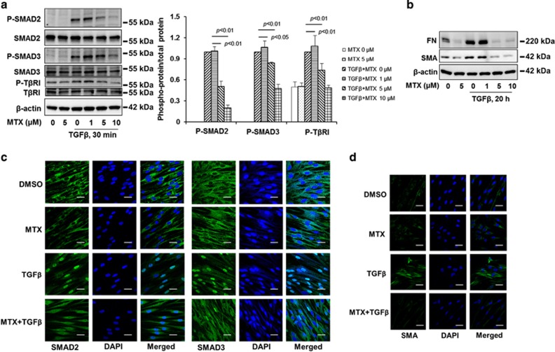
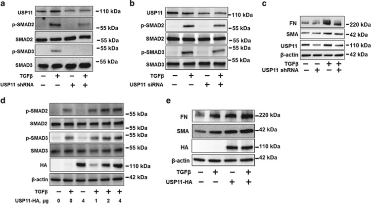
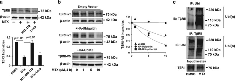
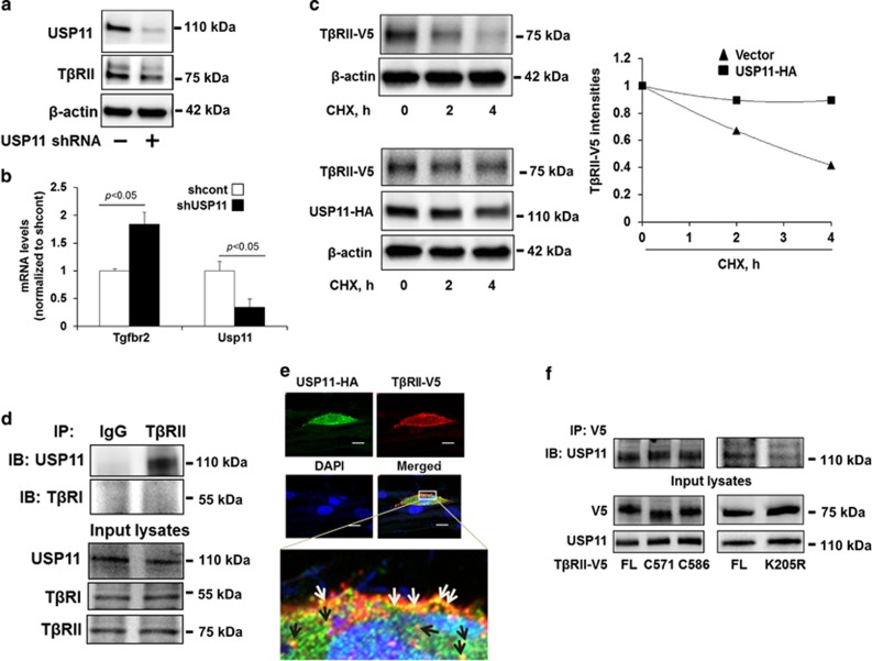
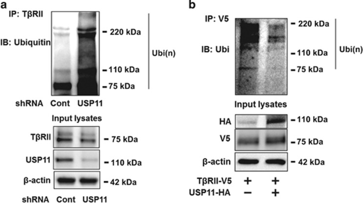
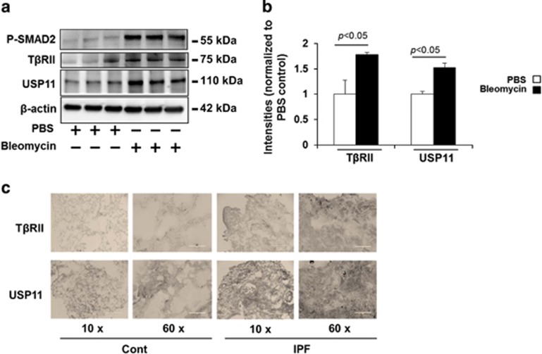
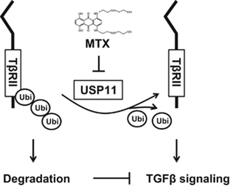
The transforming growth factor β-1 (TGFβ-1) signaling pathway plays a central role in the pathogenesis of pulmonary fibrosis. Two TGFβ-1 receptors, TβRI and TβRII, mediate this pathway. TβRI protein stability, as mediated by the ubiquitin/de-ubiquitination system, has been well studied; however, the molecular regulation of TβRII still remains unclear. Here we reveal that a de-ubiquitinating enzyme, USP11, promotes TGFβ-1 signaling through de-ubiquitination and stabilization of TβRII. We elucidate the role that mitoxantrone (MTX), an USP11 inhibitor, has in the attenuation of TGFβ-1 signaling. Inhibition or downregulation of USP11 results in increases in TβRII ubiquitination and reduction of TβRII stability. Subsequently, TGFβ-1 signaling is greatly attenuated, as shown by the decreases in phosphorylation of SMAD2/3 levels as well as that of fibronectin (FN) and smooth muscle actin (SMA). Overexpression of USP11 reduces TβRII ubiquitination and increases TβRII stabilization, thereby elevating phosphorylation of SMAD2/3 and the ultimate expression of FN and SMA. Further, elevated expression of USP11 and TβRII were detected in lung tissues from bleomycin-challenged mice and IPF patients. Therefore, USP11 may contribute to the pathogenesis of pulmonary fibrosis by stabilization of TβRII and promotion of TGFβ-1 signaling. This study provides mechanistic evidence for development of USP11 inhibitors as potential antifibrotic drugs for pulmonary fibrosis.


Figures

Figure 1
Figure 1
MTX attenuates TGFβ-1 signaling in human lung fibroblast. (a) MRC5 cells were treated with increasing doses of MTX (0, 1, 5, 10 μM) for 1 h, and then cells were treated with TGFβ-1 (2 ng/ml) for 30 min; the phosphorylated and total forms of SMAD2, SMAD3, and TβRI were then analyzed by western blotting. Western blotting images were cropped to improve the conciseness of the data; samples derived from the same experiment and the blots were processed in parallel. Representative of experiments performed at least three independent times. Intensities of blots were measured. The ratio of phosphorylated/total protein was analyzed by the ImageJ software. (b) MRC5 cells were treated with increasing doses of MTX (0, 1, 5, 10 μM) for 1 h, and then cells were treated with TGFβ-1 (2 ng/ml) for 20 h. FN and SMA levels were analyzed by western blotting. Western blotting images were cropped to improve the conciseness of the data; samples derived from the same experiment and the blots were processed in parallel. Representative of experiments performed at least three independent times. (c) MRC5 cells grown on glass-bottom dishes were treated with dimethyl sulfoxide (DMSO) or MTX (5 μM) for 1 h, and then cells were treated with TGFβ-1 (2 ng/ml) for 1 h. Localization of SMAD2 or SMAD3 (green) in the cells were detected by immunostaining with SMAD2 and SMAD3 antibodies. DAPI (4,6-diamidino-2-phenylindole) was used for nuclei staining (blue). Bars, 10 μM. Representative images were shown. (d) MRC5 cells grown on glass-bottom dishes were treated with DMSO or MTX (5 μM) for 1 h, and then cells were treated with TGFβ-1 (2 ng/ml) for 20 h. Localization of SMA (green) in the cells were detected by immunostaining with an SMA antibody. DAPI was used for nuclei staining (blue). Bars, 10 μm. Representative images are shown
Figure 2
Figure 2
USP11 regulates TGFβ-1 signaling pathway. (a) MRC5 cells were infected with cont shRNA (−) or USP11 shRNA lentivirus for 3 days as described in the Materials and Methods section, and then cells were treated with TGFβ-1 (2 ng/ml) for 30 min. USP11 and phosphorylated and total SMAD2/3 levels were analyzed by western blotting. (b) MRC5 cells were transfected with cont siRNA (−) or USP11 siRNA for 3 days, and then cells were treated with TGFβ-1 (2 ng/ml) for 30 min. USP11, total, and phosphorylated SMAD2/3 levels were analyzed by western blotting. (c) MRC5 cells were infected with cont shRNA (−) or USP11 shRNA lentivirus for 3 days, and then cells were treated with TGFβ-1 (2 ng/ml) for 20 h. USP11, FN, SMA, and β-actin protein levels were analyzed by western blotting. (d) MRC5 cells were transfected with USP11-HA plasmids (0–4 μg) for 2 days, and then cells were treated with TGFβ-1 (2 ng/ml) for 30 min. USP11-HA, phosphorylated and total SMAD2/3, and β-actin levels were analyzed by western blotting. (e) MRC5 cells were transfected with USP11-HA plasmids (2 μg) for 2 days, and then cells were treated with TGFβ-1 (2 ng/ml) for 20 h. USP11-HA, FN, SMA, and β-actin protein levels were analyzed by western blotting. Western blotting images were cropped to improve the conciseness of the data; samples derived from the same experiment and the blots were processed in parallel. Representative of experiments performed at least three independent times
Figure 3
Figure 3
MTX reduces TβRII levels in human lung fibroblast cells. (a) MRC5 cells were treated with 5 uM MTX for 0–24 h. TβRII and β-actin levels were analyzed by western blotting. Intensities of TβRII were analyzed by the ImageJ software. *P<0.01 compared with 0 h. (b) MRC5 cells were treated with MTX (0–5 μM) for 4 h. TβRII and β-actin levels were analyzed by western blotting. Intensities of TβRII were analyzed by the ImageJ software. *P<0.01 compared with 0 μM. (c) MRC5 cells were transfected with TβRII-V5 plasmid for 48 h, and then cells were treated with MTX (0–20 μM) for 4 h. TβRII-V5 and β-actin levels were examined by western blotting. Intensities of TβRII-V5 were analyzed by the ImageJ software. *P<0.01 compared with 0 μM. (d) MRC5 cells were treated with 5 μM MTX for 0–24 h. TβRI and β-actin levels were analyzed by western blotting. Intensities of TβRI were analyzed by the ImageJ software. Western blotting images were cropped to improve the conciseness of the data; samples derived from the same experiment and the blots were processed in parallel. Representative of experiments performed at least three independent times. (e) MRC5 cells grown on glass-bottom dishes were transfected with TβRII-V5 plasmid for 48 h, and then cells were treated with MTX (10 μM) for 4 h. Localization of TβRII-V5 (green) in the cells were detected by immunostaining with V5 tag. DAPI (4,6-diamidino-2-phenylindole) was used for nuclei staining (blue). Bars, 50 μm. Representative images were shown. Pixels of TβRII-V5 on the plasma membrane were quantified with the NIS-Elements software. *P<0.01 compared with dimethyl sulfoxide (DMSO)
Figure 4
Figure 4
MTX promotes TβRII ubiquitination and degradation in the proteasome. (a) MRC5 cells were treated with MG-132 (20 μM) or leupeptin (100 μM) for 1 h prior to MTX treatment (5 μM, 2 h). TβRII and β-actin levels were analyzed by western blotting. Intensities of TβRII were analyzed by the ImageJ software. (b) MRC5 cells were co-transfected with TβRII-V5 and with either empty vector, HA-ubiquitin, or HA-ubiquitin without lysine (HA-UbiK0) plasmids, cells were then treated with increasing concentrations of MTX (0–10 μM) for 4 h. TβRII-V5 and β-actin levels were analyzed by western blotting. Intensities of TβRII-V5 were analyzed by the ImageJ software and then compared between the three groups. (c) MRC5 cells were treated with 5 μM MTX for 1 h, and then cell lysates were subjected to immunoprecipitation with an ubiquitin antibody or a TβRII antibody, followed by TβRII or ubiquitin immunoblotting. Input lysates were analyzed by TβRII immunoblotting. Western blotting images were cropped to improve the conciseness of the data; samples derived from the same experiment and the blots were processed in parallel. Representative of experiments performed at least three independent times
Figure 5
Figure 5
USP11 regulates TβRII stability in human lung fibroblast cells. (a) MRC5 cells were infected with cont shRNA (−) or USP11 shRNA lentivirus for 3 days, and then USP11, TβRII, and β-actin levels were analyzed by western blotting. (b) MRC5 cells were infected with cont shRNA (−) or USP11 shRNA lentivirus for 3 days, and then total RNA was extracted. Tgfbr2 and Usp11 gene expression were examined by RT-real time PCR. (c) MRC5 cells were co-transfected with TβRII-V5 and empty vector or USP11-HA plasmids for 48 h, and then cells were treated with CHX (20 μg/ml) for 0–4 h. TβRII-V5, USP11-HA, and β-actin levels were analyzed by western blotting. Intensities of TβRII-V5 were analyzed by the ImageJ software and then compared between the two groups. (d) MRC5 cell lysates were subjected to immunoprecipitation with IgG or a TβRII antibody, followed by USP11 and TβRI immunoblotting. Input lysates were analyzed by USP11, TβRI, and TβRII immunoblotting. (e) MRC5 cells grown on glass-bottom dishes were co-transfected with USP11-HA and TβRII-V5 plasmids for 48 h. Localization of USP11-HA (green) and TβRII-V5 (red) in MRC5 cells were examined by immunostaining. Nuclei were stained by DAPI (4,6-diamidino-2-phenylindole; blue). USP11-HA and TβRII-V5 co-localization on the plasma membrane are indicated by white arrows; two protein co-localization in the cytoplasm are indicated by black arrows. Bars, 50 μm. Representative images are shown. (f) MRC5 cells were transfected with plasmids encoding TβRII-V5 full-length (FL), C571, C586 deletion mutants, or K205R mutants for 48 h. Cell lysates were subjected to immunoprecipitation with a V5 antibody, followed by USP11 immunoblotting. Input lysates were analyzed by USP11 and V5 immunoblotting. Western blotting images were cropped to improve the conciseness of the data; samples derived from the same experiment and the blots were processed in parallel. Representative of experiments performed at least three independent times
Figure 6
Figure 6
USP11 de-ubiquitinates TβRII. (a) MRC5 cells were infected with cont shRNA or USP11 shRNA lentivirus for 3 days. Cell lysates were subjected to immunoprecipitation with a TβRII antibody, followed by ubiquitin immunoblotting. Input lysates were analyzed by TβRII, USP11, and β-actin immunoblotting. (b) MRC5 cells were co-transfected with TβRII-V5 and empty vector or USP11-HA plasmids for 48 h, and then cell lysates were subjected to immunoprecipitation with a V5 antibody, followed by ubiquitin immunoblotting. Input lysates were analyzed by HA, V5, and β-actin immunoblotting. Western blotting images were cropped to improve the conciseness of the data; samples derived from the same experiment and the blots were processed in parallel. Representative of experiments performed at least three independent times
Figure 7
Figure 7
TβRII and USP11 are increased in the lungs from bleomycin-induced fibrosis model and IPF patients (a) C57BL/6 mice were challenged with intranasal injection of bleomycin for 3 weeks. P-SMAD2, TβRII, USP11, and β-actin levels were analyzed by western blotting. (b) Intensities of TβRII and USP11 were analyzed by the ImageJ software. (c) Human normal and IPF lung tissues were fixed and immunostained with TβRII and USP11 antibodies. Scale bars in × 10 image, 400 μm; scale bars in × 60 images: 50 μm. Representative images (from three per each group) are shown
Figure 8
Figure 8
USP11 de-ubiquitinates and stabilizes TβRII. TβRII stability is regulated by its poly-ubiquitination. Destabilization of TβRII attenuates TGFβ-1 signaling. USP11 stabilizes TβRII through de-ubiquitination of TβRII. MTX, an inhibitor of USP11, promotes ubiquitination and degradation of TβRII, thus mitigating TGFβ-1 signaling

Similar articles

Cited by

References

    1. Wrana JL, Attisano L, Carcamo J, Zentella A, Doody J, Laiho M et al. TGF beta signals through a heteromeric protein kinase receptor complex. Cell 1992; 71: 1003–1014. - PubMed
    1. Wrana JL, Carcamo J, Attisano L, Cheifetz S, Zentella A, Lopez-Casillas F et al. The type II TGF-beta receptor signals diverse responses in cooperation with the type I receptor. Cold Spring Harb Symp Quant Biol 1992; 57: 81–86. - PubMed
    1. Dennler S, Goumans MJ, ten Dijke P. Transforming growth factor beta signal transduction. J Leukoc Biol 2002; 71: 731–740. - PubMed
    1. Heldin CH, Miyazono K, ten Dijke P. TGF-beta signalling from cell membrane to nucleus through SMAD proteins. Nature 1997; 390: 465–471. - PubMed
    1. Kawabata M, Miyazono K. Signal transduction of the TGF-beta superfamily by Smad proteins. J Biochem 1999; 125: 9–16. - PubMed

MeSH terms