. 2017 Jan 3;91(2):e01774-16.
doi: 10.1128/JVI.01774-16. Print 2017 Jan 15.

A Transformation-Defective Polyomavirus Middle T Antigen with a Novel Defect in PI3 Kinase Signaling

Affiliations

A Transformation-Defective Polyomavirus Middle T Antigen with a Novel Defect in PI3 Kinase Signaling

Deborah Denis et al. J Virol. .

Abstract

Middle T antigen (MT), the principal oncoprotein of murine polyomavirus, transforms by association with cellular proteins. Protein phosphatase 2A (PP2A), YAP, Src family tyrosine kinases, Shc, phosphatidylinositol 3-kinase (PI3K), and phospholipase C-γ1 (PLCγ1) have all been implicated in MT transformation. Mutant dl1015, with deletion of residues 338 to 347 in the C-terminal region, has been an enigma, because the basis for its transformation defect has not been apparent. This work probes the dl1015 region of MT. Because the region is proline rich, the hypothesis that it targets Src homology domain 3 (SH3) domains was tested, but mutation of the putative SH3 binding motif did not affect transformation. During this work, two point mutants, W348R and E349K, were identified as transformation defective. Extensive analysis of the E349K mutant is described here. Similar to wild-type MT, the E349K mutant associates with PP2A, YAP, tyrosine kinases, Shc, PI3 kinase, and PLCγ1. The E349K mutant was examined to determine the mechanism for its transformation defect. Assays of cell localization and membrane targeting showed no obvious difference in localization. Src association was normal as assayed by in vitro kinase and MT phosphopeptide mapping. Shc activation was confirmed by its tyrosine phosphorylation. Association of type 1 PI3K with MT was demonstrated by coimmunoprecipitation, showing both PI3K subunits and in vitro activity. Nonetheless, expression of the mutants failed to lead to the activation of two known downstream targets of PI3K, Akt and Rac-1. Strikingly, despite normal association of the E349K mutant with PI3K, cells expressing the mutant failed to elevate phosphatidylinositol (3,4,5)-trisphosphate (PIP3) in mutant-expressing cells. These results indicate a novel unsuspected aspect to PI3K control.

Importance: The gene coding for middle T antigen (MT) is the murine polyomavirus oncogene most responsible for tumor formation. Its study has a history of uncovering novel aspects of mammalian cell regulation. The importance of PI3K activity and tyrosine phosphorylation are two examples of insights coming from MT. This study describes new mutants unable to transform like the wild type that point to novel regulation of PI3K signaling. Previous mutants were defective in PI3K because they failed to bind the enzyme and bring the activity to the membrane. These mutants recruit PI3K activity like the wild type, but fail to elevate the cellular level of PIP3, the product used to signal downstream of PI3K. As a result, they fail to activate either Akt or Rac1, explaining the transformation defect.

Keywords: Akt; PI3 kinase; Rac; middle T antigen; polyomavirus; transformation.


Figures

FIG 1
FIG 1
(A) Murine polyomavirus MT showing associated proteins. (B) dl1015 is defective in transformation. Rat-1 fibroblasts were transfected with control vector DNA or DNA expressing either wild-type or dl1015 MT. After 2 weeks, cells were fixed and stained with crystal violet. (C) Wild-type (WT) and mutant MT sequences from residues 336 to 350. (D) MT interactions with the N-terminal SH3 of GRB2. Anti-MT immunoprecipitates from control cells or cells expressing wild-type, E349K, or 2XP MT were labeled in vitro with 32P. MT was then reisolated and incubated with either GST-agarose or Grb2 N-SH3-GST-agarose. Washed precipitates were analyzed by PAGE and autoradiography. (E) SH3 association does not correlate with transformation. Transformation experiments performed as described for panel B compared wild-type MT to dl1015 or 2XP (P338A P341A). Expression is shown by MT blotting of equal amounts of cell protein from each cell type.
FIG 2
FIG 2
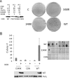
E349K mutant is defective in transformation. (A) Focus formation assays in Rat1 cells show an E349K defect in transformation. Cells were transfected with the empty vector control (CON) or vectors expressing wild-type, P347A, W348R, or E349K MT. MT expression was assessed by Western blotting. Focus formation assays were carried out as described in the legend to Fig. 1B. (B) E349K MT is defective in soft agar assays of transformation. NIH/3T3 cells were exposed to doxycycline for 24 h to induce expression of EGFP, WT MT, or E349K MT, after which the cells were seeded into soft agar in the continued presence of doxycycline. Colonies were counted after 4 weeks. Results are shown on the left. Representative images of individual NIH/3T3 colonies are shown on the right. Control Western blotting of NIH/3T3 cell extract was conducted to detect MT. PLCγ1 served as a loading control.
FIG 3
FIG 3
E349K MT associations with cellular proteins. (Left panel) Inducible NIH/3T3 cells were induced to express EGFP (CON), WT MT, or E349K MT by exposure to doxycycline for 24 h. Western blotting of MT immunoprecipitates (IP) shows WT binding of PP2A, Src, YAP, Shc, and PI3K (p85) to E349K MT. Actin was used as a loading control. (Right panel) 293 cells were induced to express EGFP, WT MT, or E349K MT by exposure to doxycycline for 6 h. Cell extracts were made and MT immunoprecipitated. After SDS-PAGE, blots were probed for p110 subunits of PI3K, PLCγ1, and MT.
FIG 4
FIG 4
E349K MT associates normally with Src family PTKs. (A) Equal protein amounts of extracts from control cells or stable cell lines expressing either wild-type or mutant MT were immunoprecipitated with an MT-specific antibody. An in vitro kinase assay was performed on washed immunoprecipitates. 32P-labeled proteins were resolved by SDS-PAGE. The gel was treated with alkaline to remove serine and threonine phosphate backgrounds. The image was developed using a Molecular Dynamics PhosphorImager screen. (B) Partial proteolytic digests of E349K MT resulted in the expected phosphopeptides. WT and E349K MT labeled in vitro with 32P were resolved in the first dimension in 7.5% acrylamide cylinders. After the appropriate regions of the cylinders were cut out, digestion was carried out with S. aureus V8 on a second-dimension 12.5% acrylamide gel. After alkaline treatment, that gel was dried and exposed to a Molecular Dynamics PhosphorImager screen. Full-length wild-type and E349K MTs are indicated by the arrowheads. A known shorter MT fragment is indicated by solid lines. The phosphopeptides are indicated by molecular weight.
FIG 5
FIG 5
E349K MT association with PI3 kinase. (A) E349K MT has associated PI3 kinase activity. Washed MT immunoprecipitates from cell lines expressing wild-type or E349K MT were analyzed in an in vitro lipid kinase assay. As described in Materials and Methods, phosphatidylinositol was used as the substrate. Phosphorylated lipids were extracted in CHCl3-methanol (1:1) and spotted onto TLC plates. The plates were developed by chromatography in n-propanol–2 N acetic acid (65:35), dried, and exposed to a Molecular Dynamics PhosphorImager screen. (B) PI3 kinase p85 associated with E349K MT is phosphorylated normally. Washed wild-type or E349K MT immunoprecipitates were labeled in an in vitro tyrosine kinase reaction. Complexes were disrupted with SDS and p85 isolated by immunoprecipitation. p85 was analyzed by two-dimensional chymotryptic mapping as described in the legend to Fig. 4.
FIG 6
FIG 6
E349K MT activates Shc. (A) Expression of WT and mutant MTs leads to the phosphorylation of Shc. Cell lines expressing either WT or mutant middle T antigen (MT) or normal NIH/3T3 cells were grown under serum-starved conditions for 24 h. Equal amounts of extract were immunoprecipitated with an antibody directed against pTyr (4G10). The washed immunoprecipitates were resolved on a 10% acrylamide gel, along with cell extracts for blotting with total Shc or MT. (B) Expression of WT and mutant MTs leads to the phosphorylation of Shc and Erk. NIH/3T3 cells were induced to express EGFP, WT MT, or E349K MT upon exposure to doxycycline for 24 h, the last 16 h of which took place without serum. Western blotting of whole-cell extracts shows wild-type activation of Shc (Y317) and Erk1/2 (pErk, T202 Y204).
FIG 7
FIG 7
The wild-type MT, but not dl1015, W348R, or E349K MT, activates Akt. (A) Control NIH/3T3 cells or lines stably expressing either wild-type or mutant MT were grown under serum-starved conditions for 24 h. Extracts were resolved by PAGE and blotted for pan-serine 473 phosphorylated Akt, total Akt, or MT. (B) E349K MT is defective in both S473 and T308 Akt phosphorylation. Control cells or cells expressing wild-type or E349K MT were treated with doxycycline for 24 h, of which the last 16 h were serum free. Extracts were made and Western blotting was carried out after SDS-PAGE using phosphospecific antibodies for pT308 and pS473 as well as antibody for total Akt and MT. (C) E349K MT does not prevent Akt activation. 3T3 cells stably expressing EGFP (control [CON]) or E349K MT were infected with empty vector (EV) pBABE retrovirus or pBABE retrovirus encoding constitutively activatable Akt2 or Akt3 and selected in puromycin. Cells were induced to express EGFP or E349K MT by exposure to doxycycline for 24 h, the last 16 h of which took place in the absence of serum. Blotting of cell extracts was carried with antibody recognizing S473 phosphorylation, total Akt, PI3K p85 subunit or MT. (D) Activated Akt3 partially restores E349K MT transformation. Cells with or without mutant Akt expression were induced to express EGFP (CON), WT MT, or E349K MT by exposure to doxycycline for 24 h, after which the cells were seeded into soft agar in the continued presence of doxycycline. Colonies were counted after 4 weeks.
FIG 8
FIG 8
The wild-type MT, but not dl1015, W348R, or E349K MT, activates Rac. Cell lines expressing either WT or mutant middle T antigen (MT) or control NIH/3T3s were grown under serum-starved conditions for 24 h. Equal amounts of extract were incubated with a GST-PAK-Rac1-binding domain fusion, bound to glutathione (GSH) beads. The washed GST pulldowns were resolved on a 10% acrylamide gel, transferred to nitrocellulose, then immunoblotted with an antibody directed against Rac1. For total Rac1 and MT, equal protein concentrations of cell extract were immunoblotted for either Rac1 or MT.
FIG 9
FIG 9
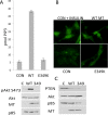
E349K cells lack PIP3. (A) 3T3 cells were exposed to doxycycline for 24 h to induce expression of EGFP (CON), wild-type, or E349K MT. The last 16 h of the induction took place in the absence of serum. Lipids were extracted, and PIP3 was measured by ELISA (top). Control Western blotting of duplicate samples shows MT in cell extracts and confirms that E349K MT was not activating Akt (bottom). (B) Control cells and those expressing either wild-type or E349K MT were grown under serum-starved conditions for 24 h. A subset of the controls were treated with insulin (100 ng/ml) for 30 min. Cells were then stained with antibody to PIP3 (Echelon). (C) PTEN levels are unaffected by MT. Induction of MT was carried out as in panel A. Cell extracts were analyzed for PTEN by Western blotting.
FIG 10
FIG 10
E349K MT and membrane localization. (A) E349K is associated with membranes: NIH/3T3 cells were induced to express EGFP (C) or wild-type or E349K MT by exposure to doxycycline for 24 h. Hypotonic cell fractionation followed by centrifugation was conducted to isolate the membrane fraction. After SDS-PAGE, Western blotting was carried out to detect MT in whole-cell lysates or the membrane fraction. The relative amount of whole-cell lysate was 1/15 that of the purified membranes. (B) Using H-RAS targeting of E349K MT to membranes fails to restore transformation. Rat1 cells were transfected with either wild-type, wild type-CAAX, E349K, or E349K-CAAX MT. Foci were stained as described in the legend to Fig. 1B.

Similar articles

Cited by

References

    1. Dawe CJ, Freund R, Mandel G, Ballmer-Hofer K, Talmage DA, Benjamin TL. 1987. Variations in polyoma virus genotype in relation to tumor induction in mice. Characterization of wild type strains with widely differing tumor profiles. Am J Pathol 127:243–261. - PMC - PubMed
    1. Eddy B. 1969. Polyoma virus. Virol Monogr 7:1–114.
    1. Fluck MM, Haslam SZ. 1996. Mammary tumors induced by polyomavirus. Breast Cancer Res Treat 39:45–56. doi:10.1007/BF01806077. - DOI - PubMed
    1. Schaffhausen BS, Roberts TM. 2009. Lessons from polyoma middle T antigen on signaling and transformation: a DNA tumor virus contribution to the war on cancer. Virology 384:304–316. doi:10.1016/j.virol.2008.09.042. - DOI - PMC - PubMed
    1. Fluck MM, Schaffhausen BS. 2009. Lessons in signaling and tumorigenesis from polyomavirus middle T antigen. Microbiol Mol Biol Rev 73:542–563. doi:10.1128/MMBR.00009-09. - DOI - PMC - PubMed

Substances

LinkOut - more resources