. 2016 Nov 11;2(11):e1601865.
doi: 10.1126/sciadv.1601865. eCollection 2016 Nov.

Overcoming a nucleosomal barrier to replication

Affiliations

Overcoming a nucleosomal barrier to replication

Han-Wen Chang et al. Sci Adv. .

Abstract

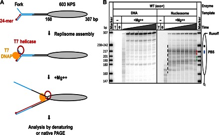
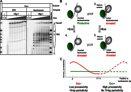
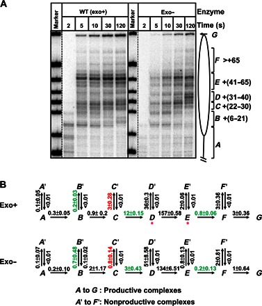
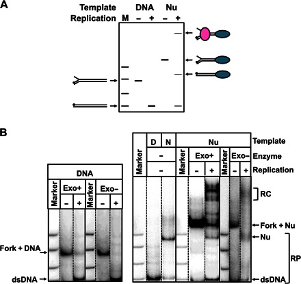
Efficient overcoming and accurate maintenance of chromatin structure and associated histone marks during DNA replication are essential for normal functioning of the daughter cells. However, the molecular mechanisms of replication through chromatin are unknown. We have studied traversal of uniquely positioned mononucleosomes by T7 replisome in vitro. Nucleosomes present a strong, sequence-dependent barrier for replication, with particularly strong pausing of DNA polymerase at the +(31-40) and +(41-65) regions of the nucleosomal DNA. The exonuclease activity of T7 DNA polymerase increases the overall rate of progression of the replisome through a nucleosome, likely by resolving nonproductive complexes. The presence of nucleosome-free DNA upstream of the replication fork facilitates the progression of DNA polymerase through the nucleosome. After replication, at least 50% of the nucleosomes assume an alternative conformation, maintaining their original positions on the DNA. Our data suggest a previously unpublished mechanism for nucleosome maintenance during replication, likely involving transient formation of an intranucleosomal DNA loop.

Keywords: Replication; chromatin; mechanism; nucleosome.


Figures

Fig. 1
Fig. 1. Nucleosomes cause strong pausing of the T7 replisome.
(A) Experimental approach for analysis of replication through chromatin. Each template contains the fork DNA structure, linker DNA, and the strong 603 NPS. The fork DNA structure (in blue) is ligated to the nucleosomal template (gray oval). After ligation, the P32-end–labeled 24-mer DNA primer (red arrow) is annealed to the template DNA strand. Next, DNAP, which was preassembled with E. coli trx and T7 helicase (red ring), is added to the reaction and forms a replisome in the presence of all dNTPs. The reaction is then activated by the addition of Mg++. Replication products are analyzed by denaturing or native PAGE. (B) Analysis of labeled products after replication of 603 DNA (histone-free or organized in a nucleosome) for different time intervals (0, 2, 5, 10, 30, 60, 120, 240, and 480 s) by denaturing PAGE. The locations of the nucleosome (oval), the nucleosome dyad (square), the PBS (blue line), and the runoff transcript and nucleosome-specific pausing (black dashed line) are shown. T, DNA template only; 0, reaction before addition of Mg++. Note that the nucleosomal pausing regions +(31–40) and +(41–65) are indicated by green and red dashed lines, respectively. Markers are pBR322 DNA–Msp I digest marker (New England Biolabs). The sizes of marker DNA fragments are indicated on the left side.
Fig. 2
Fig. 2. Exonuclease activity increases the efficiency of replication through the nucleosome.
(A) Analysis of labeled products after replication of 603 DNA or nucleosomal templates by the exo T7 replisome for different time intervals (0, 2, 5, 10, 30, 60, 120, 240, and 480 s) by denaturing PAGE. Note the ~10-bp periodic pausing pattern (indicated by black dots; also see fig. S3) detected after replication for short time periods that is not present after replication by the exo+ T7 replisome (Fig. 1B). (B) Proposed mechanism explaining the 10-bp periodic nucleosomal pausing patterns. It is proposed that discrete 10-bp DNA regions of nucleosomal DNA are uncoiled from the octamer stepwise, after the T7 replisome encounters DNA-histone interactions. As the T7 replisome proceeds along uncoiled DNA (complex 1), it arrests after encountering DNA-histone interactions (complex 2) and possible backtracking (complex 3). When the replisome is backtracked, the DNA may also recoil to bind back to the octamer, which leads to peeling of the primer end from the template, resulting in a more stable arrest. To proceed further, DNA has to be uncoiled from the octamer (complex 4) and the replisome has to recover from the backtracked state. The recovery is likely facilitated by the exonuclease activity that can excise the peeled primer end and regenerate the functionally active, fully annealed primer template. The exo enzyme must wait for DNA uncoiling to move forward, resulting in a lower efficiency and 10-bp periodicity of replication through chromatin. Nucleosomal DNA and histone octamer are shown in blue and green. DNA polymerase is in gray. The arrow indicates the direction of replisome progression. (C) The expected efficiency of arrest of exo and exo+ replisomes (red and green lines, respectively) in a nucleosome.
Fig. 3
Fig. 3. Kinetic analysis of replication through a nucleosome by exo+ and exo replisomes.
(A) 603 nucleosomes were replicated by exo+ and exo replisomes for indicated time intervals. End-labeled DNA was analyzed by denaturing PAGE. The intranucleosomal pauses and runoff (from A to G) were quantified using a Phosphorimager and the OptiQuant software. (B) The quantified data were analyzed using an elongation model that produces a good fit of the experimental data to the calculated curves (fig. S4). The fitting curves (fig. S4) and kinetic parameters were obtained using the KinTek Explorer software. All rate constants were averages from three independent experiments. Rate constants that are more than threefold different between the two forms of DNAPs are marked by green and red colors (for positive and negative effects on processivity of the exo+ replisome, respectively). Note that the exo+ replisome has a higher overall rate of replication through the nucleosome than exo; the rates of replication through the +(31–40) and +(41–65) regions have the largest differences.
Fig. 4
Fig. 4. Nucleosomes survive after replication by the T7 replisome.
(A) The diagram shows mobility of the substrates and the products of replication in native gel. The nucleosome is shown as blue, and DNAP are shown as pink ovals. The labeled DNA end is indicated by a black circle. M, pBR322–Msp I digest. (B) Analysis of labeled templates after replication by the exo+/ T7 replisome of the 603 DNA or nucleosome for 240 s by native PAGE. Some RCs are stalled in the nucleosome. The nucleosomes (N) assembled on the 307-bp dsDNA (D) are the expected replication products (RP). Marker is pBR322–Msp I digest. Nucleosomes correspond to ~45 and ~65% of the replication product (average of three experiments) in the case of the exo+ and exo T7 replisome, respectively.
Fig. 5
Fig. 5. Nucleosomes remain at the original position on the DNA after replication by the exo+ T7 replisome.
(A) Positions of sites for restriction enzymes on the nucleosomal template. (B) Analysis of nucleosome fate using restriction enzyme sensitivity assay (B, Bss SI; M, Msl I; Ca, Cac8 I; Cl: Cla I). The PAGE-purified nucleosomes after replication, 307-bp dsDNA, and the nucleosomes assembled on the 307-bp dsDNA were incubated in the presence of an excess of indicated restriction enzymes and analyzed by native PAGE. DNA fragment resistant to digestion by Cla I (likely due to dissociation of nucleosomal DNA, resistant to the enzyme, during the electrophoresis) is indicated by asterisks.
Fig. 6
Fig. 6. The length of the spacer DNA dictates the efficiency of replication through the nucleosome.
(A) Design of the templates containing linker DNA between the replication fork and the nucleosome of different lengths. (B and C) Efficiency of replication of nucleosomal templates is directly proportional to the length of the linker DNA. The templates of different lengths were replicated by the exo+ (B) or exo (C) T7 replisome for 8 min, as described in Fig. 1. Runoff products of replication by the T7 replisome of the nucleosome templates were quantified after separation by denaturing PAGE and normalized to the amount of runoff products detected after replication of the DNA templates. The fraction of all RCs capable of overcoming the nucleosomal barrier is shown. Average values from three (B) or two experiments (C) with SDs are shown.
Fig. 7
Fig. 7. Proposed model of chromatin reassembly during or after replication.
The original histones are nearly quantitatively recovered on leading strand (after survival and translocation) and lagging strand (after translocation). Three pathways of nucleosome reformation after replication are proposed: (i) original histone octamers survive at the original positions on DNA, (ii) original histone octamers are transferred within the 400-bp region upstream of the replisome, or (iii) de novo histone assembly after replication.

Similar articles

Cited by

References

    1. Struhl K., Segal E., Determinants of nucleosome positioning. Nat. Struct. Mol. Biol. 20, 267–273 (2013). - PMC - PubMed
    1. Annunziato A. T., The fork in the road: Histone partitioning during DNA replication. Genes 6, 353–371 (2015). - PMC - PubMed
    1. Das C., Tyler J. K., Histone exchange and histone modifications during transcription and aging. Biochim. Biophys. Acta 1819, 332–342 (2013). - PMC - PubMed
    1. Ramachandran S., Henikoff S., Replicating nucleosomes. Sci. Adv. 1, e1500587 (2015). - PMC - PubMed
    1. Whitehouse I., Smith D. J., Chromatin dynamics at the replication fork: There’s more to life than histones. Curr. Opin. Genet. Dev. 23, 140–146 (2013). - PMC - PubMed

LinkOut - more resources