. 2016 Nov 11;17(1):469.
doi: 10.1186/s12891-016-1326-y.

Anti-fibrotic action of pirfenidone in Dupuytren's disease-derived fibroblasts

Affiliations

Anti-fibrotic action of pirfenidone in Dupuytren's disease-derived fibroblasts

Chaoming Zhou et al. BMC Musculoskelet Disord. .

Abstract

Background: Dupuytren's disease (DD) is a complex fibro-proliferative disorder of the hand that is often progressive and eventually can cause contractures of the affected fingers. Transforming growth factor beta (TGF-β1) has been implicated as a key stimulator of myofibroblast activity and fascial contraction in DD. Pirfenidone (PFD) is an active small molecule shown to inhibit TGF-β1-mediated action in other fibrotic disorders. This study investigates the efficacy of PFD in vitro in inhibiting TGF-β1-mediated cellular functions leading to Dupuytren's fibrosis.

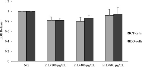
Methods: Fibroblasts harvested from (DD) and carpal tunnel (CT)- tissues were treated with or without TGF-β1 and/or PFD and were subjected to cell migration, cell proliferation and cell contraction assays. ELISA; western blots and real time RT-PCR assays were performed to determine the levels of fibronectin; p-Smad2/Smad3; alpha-smooth muscle actin (α-SMA), α2 chain of type I collagen and α1 chain of type III collagen respectively.

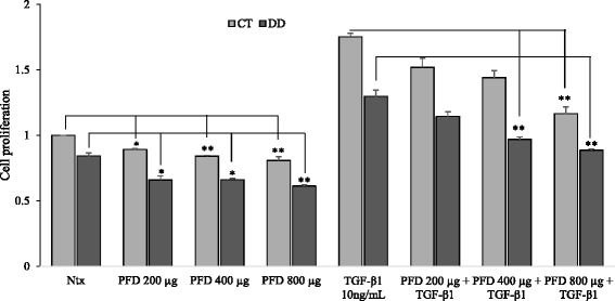
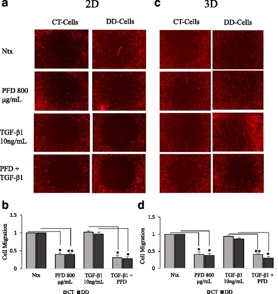
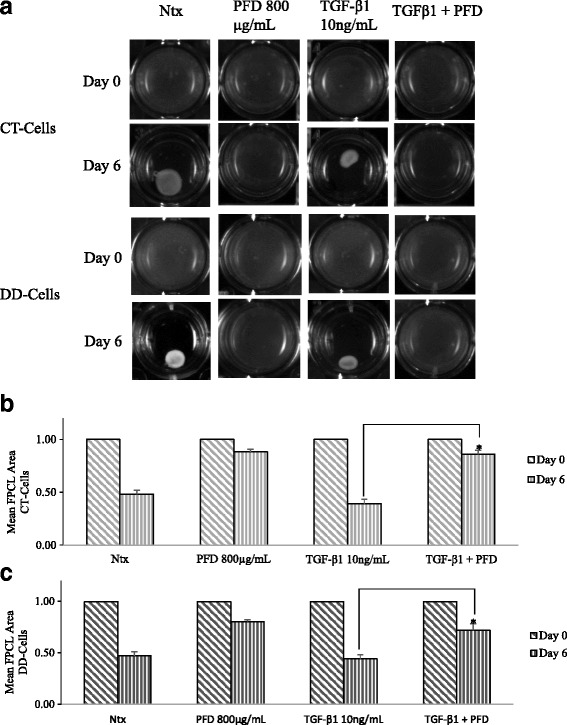
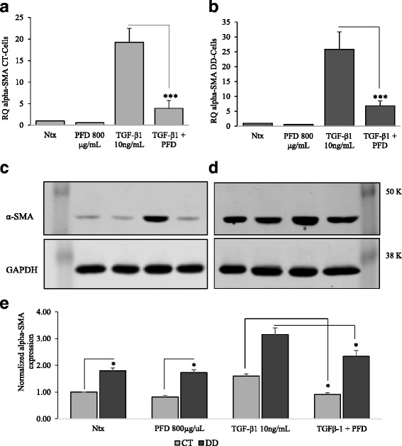
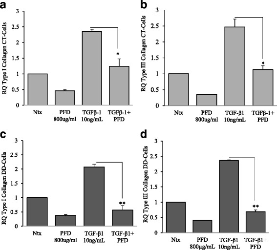
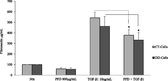
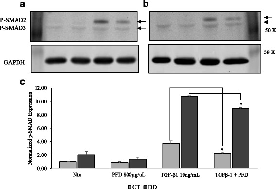
Results: Our results show that PFD effectively inhibits TGF-β1-induced cell migration, proliferation and cell contractile properties of both CT- and DD-derived fibroblasts. TGF-β1-induced α-SMA mRNA and protein levels were inhibited at the higher concentration of PFD (800 μg/ml). Interestingly, TGF-β1 induction of type I and type III collagens and fibronectin was inhibited by PFD in both CT- and DD- derived fibroblasts, but the effect was more prominent in DD cells. PFD down-regulated TGF-β1-induced phosphorylation of Smad2/Smad3, a key factor in the TGF-β1 signaling pathway.

Conclusion: Taken together these results suggest the PFD can potentially prevent TGF-β1-induced fibroblast to myofibroblast transformation and inhibit ECM production mainly Type I- and Type III- collagen and fibronectin in DD-derived fibroblasts. Further in-vivo studies with PFD may lead to a novel therapeutic application in preventing the progression or recurrence of Dupuytren's disease.

Keywords: Alpha-SMA; Carpal tunnel; Cell contraction; Cell migration; Collagen; Dupuytren’s contracture; Palmar fascia fibrosis; Smad2/Smad3.


Figures

Fig. 1
Fig. 1
Cell proliferative ability was diminished after the addition of Pirfenidone. (a) CT- and (b) DD-derived fibroblasts derived from four different patient samples (N = 4/group) were exposed to varying concentrations of PFD (0, 200, 400, 800 μg/ml) and TGF-β1 (10 ng/ml). Cells were then subjected to MTT assay to determine the effect of PFD on basal and TGF-β1-induced cell proliferation. Cell proliferation is displayed as absorbance values of untreated CT-derived fibroblasts normalized to 1 and compared with other treatments. Data are shown as mean ± SEM of the averages of triplicate reads for each culture derived from the four different patient samples both for CT- and DD-derived fibroblasts. Statistical analysis was performed using one-way ANOVA. *p < 0.05; **p < 0.01
Fig. 2
Fig. 2
Cell viability was not affected by the presence of Pirfenidone. CT- and DD-derived fibroblasts derived from three different patient samples (N = 3/group) were incubated with different concentrations of PFD (0, 200, 400, 800 μg/ml), and the extracellular LDH release into the culture medium was measured as described in Materials and Methods. Data are shown as mean ± SEM of the averages of triplicate reads for each culture derived from the four different patient samples both for CT- and DD-derived fibroblasts. Ntx = no treatment (neither PFD nor TGF-β1 added control). Statistical analysis was performed using one-way ANOVA. *p < 0.05; **p < 0.01
Fig. 3
Fig. 3
Pirfenidone suppressed basal and TGF-β1 induced cell migration. CT- and DD-derived fibroblasts derived from three different pateint samples (N = 3/group) were maintained in low-serum medium for 24 h, then were left untreated (Ntx) or exposed to PFD (800 μg/ml) in the presence or absence of TGF-β1 (10 ng/ml) to perform the 2D (a & b) and OrisTM 3D (c & d) cell migration assays. Shown here are representative images of three different experiments from three different cultures (N = 3/group) of CT- and DD-derived fibroblasts done for 2D (a) and 3D (c) migration assays in triplicate. NIH- image analysis was used to assess the number of cells that entered the detection zones after various treatments. Data are shown as mean ± SEM of the averages of triplicate reads for each culture derived from the three different patient samples both for CT- and DD- derived fibroblasts. Statistical analysis was performed using one-way ANOVA. *p < 0.05; **p < 0.01, ***p < 0.001
Fig. 4
Fig. 4
Cellular contractile ability was diminished in the presence of Pirfenidone. CT- and DD-derived fibroblasts derived from four different patient samples (N = 4/group) were left untreated (Ntx) or exposed to PFD (800 μg/ml) in the presence or absence of TGF-β1 (10 ng/ml) to perform the stressed fibroblast populated collagen lattice (sFPCL) assay. Collagen lattices cultured for 24 h were detached from the surface of the well, and the digital images of the floating lattices were captured at different time points (day 0- day 6). a Shown here are representative images of four independent experiments performed in duplicate. b & c Data were obtained using Adobe Photoshop to analyze photographic images; data are shown as the area of the contracted collagen lattice normalized to the average area of contraction seen in untreated cells, set as a baseline value of 1. Each data point represents the mean ± SEM of the averages of triplicate reads for each culture derived from the four different patient samples both for CT- and DD-derived fibroblasts. Statistical significance was determined using one-way ANOVA. *p < 0.05; **p < 0.01
Fig. 5
Fig. 5
Inhibition of α-SMA levels was evident after addition of Pirfenidone. CT- and DD-derived fibroblasts derived from three different patient samples (N = 3/group) of were left untreated (Ntx) or were stimulated with PFD (800 μg/ml) in the presence or absence of TGF-β1 (10 ng/ml) in α-MEM medium containing 0.1 % dialyzed FBS. Twenty-four hours later cell lysates were collected to determine the mRNA and protein expression of α-SMA by real-time RT-PCR (a & b) and Western blot analyses (c & d). Real-time RT-PCR experiments were done on three independent cultures of each of the cell types. Values are mean ± SEM of three independent studies performed in triplicate. One-way ANOVA test was used to determine the statistical significance. *p < 0.05, **p < 0.01. Western blot analyses shown here are representative images of experiments performed using three independent primary cultures of CT- and DD-cord derived fibroblasts. Proteins obtained from different patients were processed in parallel to confirm the changes that are observed are consistent. Protein accumulation was quantified by densitometry using GAPDH as a loading control (e)
Fig. 6
Fig. 6
Pirfenidone effectively inhibited TGF-β1-induction of α-2 chain of Type I and α-1 chain of Type III collagen. CT- and DD-derived fibroblasts derived from three independent patient samples (N = 3/group) were left untreated (Ntx) or exposed to PFD (800 μg/ml) in the presence or absence of TGF-β1 for 24 h. Twenty-four hours later RNA was extracted and mRNA expression levels of type I and type III collagen of CT- (a & b) and DD-derived fibroblasts (c & d) were determined using real-time RT-PCR analysis. Shown is the mean ± SEM of n = 3 of experimental replicates from three different culture of CT- and DD-fibroblasts, each performed in triplicate. *p < 0.05, **p < 0.01
Fig. 7
Fig. 7
Pirfenidone substantially reduced TGF-β1 induction of fibronectin expression. CT- and DD-cord derived fibroblasts grown in growth medium overnight were treated with medium containing 0.1 % dialyzed FBS for 24 h. Following which, cells were treated with 800 μg/mL of PFD with or without 10 ng/ml TGF-β1 for 24 h. After the treatment, secreted proteins in the growth medium were collected to determine the fibronectin expression using Human Fibronectin SimpleStep ELISA Kit. CT- and DD-derived fibroblasts derived from three different patient samples (N = 3/group) were used to perform the experiment three times in triplicate. Statistical significance was determined by One-way ANOVA. *p < 0.05
Fig. 8
Fig. 8
Pirfenidone inhibits TGF-β1-induced phosphorylation of Smad2/Smad3. CT-and DD-cord-derived fibroblasts derived from three different patient samples (N = 3/group) were maintained in MEM- α medium containing 0.1 % dialyzed FBS for 24 h. After 24 h, cells were either treated or untreated with PFD (800 μg/ml) in the presence or absence of TGF-β1 (10 ng/ml) for additional 24 h. Cell lysates collected from CT- (a & b) and DD- derived fibroblasts were used to examine the expression of phosphorylated Smad2/Smad3 by Western blot analysis. Proteins obtained from different patients were processed in parallel to confirm the changes that are observed are consistent. Densitometry results are reported as the ratio of phosphorylated Smad2/Smad3 protein to GAPDH expression (c). Shown here is the representation of Western blot experiments performed using three different culture each of CT- and DD-derived fibroblasts essentially with identical results. *p < 0.05

Similar articles

Cited by

References

    1. Rayan GM. Dupuytren disease: anatomy, pathology, presentation, and treatment. J Bone Joint Surg Am. 2007;89:189–98. doi: 10.2106/00004623-200701000-00026. - DOI - PubMed
    1. Shih B, Bayat A. Scientific understanding and clinical management of Dupuytren disease. Nat Rev Rheumatol. 2010;6:715–26. doi: 10.1038/nrrheum.2010.180. - DOI - PubMed
    1. Michou L, Lermusiaux JL, Teyssedou JP, Bardin T, Beaudreuil J, et al. Genetics of Dupuytren’s disease. Joint Bone Spine. 2012;79:7–12. doi: 10.1016/j.jbspin.2011.05.027. - DOI - PubMed
    1. Arkkila PE, Kantola IM, Viikari JS. Dupuytren’s disease: association with chronic diabetic complications. J Rheumatol. 1997;24(1):153–9. - PubMed
    1. Mikkelsen OA. Dupuytren’s disease-initial symptoms, age of onset and spontaneous course. Hand. 1977;9:11–5. doi: 10.1016/S0072-968X(77)80023-5. - DOI - PubMed

Publication types

MeSH terms