. 2016 Dec 20;7(51):85109-85123.
doi: 10.18632/oncotarget.13198.

RanBPM (RanBP9) regulates mouse c-Kit receptor level and is essential for normal development of bone marrow progenitor cells

Affiliations

RanBPM (RanBP9) regulates mouse c-Kit receptor level and is essential for normal development of bone marrow progenitor cells

Sandrine Puverel et al. Oncotarget. .

Abstract

c-Kit is a tyrosine kinase receptor important for gametogenesis, hematopoiesis, melanogenesis and mast cell biology. Dysregulation of c-Kit function is oncogenic and its expression in the stem cell niche of a number of tissues has underlined its relevance for regenerative medicine and hematopoietic stem cell biology. Yet, very little is known about the mechanisms that control c-Kit protein levels. Here we show that the RanBPM/RanBP9 scaffold protein binds to c-Kit and is necessary for normal c-Kit protein expression in the mouse testis and subset lineages of the hematopoietic system. RanBPM deletion causes a reduction in c-Kit protein but not its mRNA suggesting a posttranslational mechanism. This regulation is specific to the c-Kit receptor since RanBPM reduction does not affect other membrane proteins examined. Importantly, in both mouse hematopoietic system and testis, RanBPM deficiency causes defects consistent with c-Kit loss of expression suggesting that RanBPM is an important regulator of c-Kit function. The finding that this regulatory mechanism is also present in human cells expressing endogenous RanBPM and c-Kit suggests a potential new strategy to target oncogenic c-Kit in malignancies.

Keywords: RanBP9; c-Kit signaling; hematopoietic system; spermatogenesis; stem cells.


Conflict of interest statement

CONFLICTS OF INTEREST

The authors declare no competing financial interest.

Figures
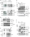
Figure 1
Figure 1. RanBPM interacts with c-Kit through its SPRY domain
RanBPM associates with c-Kit in vitro (A) and in vivo (B, C). A. protein lysates from HEK293 cells transfected with RanBPM-HA and c-Kit cDNAs and stimulated or not with 100 ng/mL SCF for 30' were subjected to immunoprecipitation (IP) with the indicated antibodies and analyzed by Western blot. A negative control IP was performed using a goat serum isotype (IP: Ct). B, C. c-Kit IP RanBPM (B) and RanBPM IP c-kit (C) in mouse testis. Testis lysates from 3 month-old wild type mice were immunoprecipitated with two different c-kit antibodies (B) or an anti-RanBPM antibody (C), control IgG serum isotypes or only beads as negative controls and blotted with the indicated antibodies. D. RanBPM interacts with c-Kit through its SPRY domain. HEK293 cells were transfected with vectors expressing a full-length HA-tagged control or c-DNA RANBPM deletion mutants lacking specific RanBPM domains as indicated in Supplementary Figure S2 [48] and subjected to IP experiments with an c-kit antibody and Western analysis with the indicated antibodies. E. RanBPM interacts with the tyrosine kinase domain of c-kit. HEK293 cells transfected with vectors expressing a full-length or c-kit c-DNA mutants lacking specific c-Kit intracellular domains as indicated in Supplementary Figure S2 were subjected to IP experiments with an anti-c-Kit antibody and Western analysis with the indicated antibodies. Inputs represent 10% of the lysates used for the IP experiments.
Figure 2
Figure 2. RanBPM affects c-Kit protein levels and signaling in the testis of young RanBPM−/− mice
A. c-Kit protein levels are decreased in the testis of young RanBPM−/− mice. C-Kit protein was pulled-down using wheat germ agglutinin (WGA)-conjugated beads from postnatal day 9 (P9) testis lysates of WT or RanBPM−/− mice (WGA lanes). Immunoblots (IB) were performed using anti-c-Kit and anti-RanBPM antibodies. MVH level in input lysates (right panels) was used as a marker of germ cells whereas β-actin was used as a loading control. B. quantification of c-Kit and MVH levels are shown as the mean ±SEM from three independent experiment as in A. C. c-Kit PI3K signaling is reduced in the testis of young RanBPM−/− mice. Cells dissociated from P9 WT and RanBPM−/− testes were stimulated or not with 100 ng/ml SCF for 15 minutes and, pAkt (both S473 and T308) and pErk levels were analyzed by Western blot. β-actin served as loading control. D. quantification of pAkt (S473) and pErk levels are shown as the mean ±SEM from three independent experiment as in C. E. c-Kit mRNA levels are unchanged in the testis of young RanBPM−/− mice. Q-PCR analysis was performed using P9 WT and RanBPM−/− testes and data are shown as the mean ±SEM from three independent experiments. Both MVH and Dazl were used as markers of germ cells. Molecular weight markers (MW) are indicated in kilo Dalton (kDa). * p<0.01.
Figure 3
Figure 3. Silencing of RanBPM in EML cells causes a decrease in c-Kit levels and reduced cell growth
A. sh-RNA for RanBPM is specific. Non-infected MEFs, MEFs infected with lentiviral vectors expressing sh-Control1 or sh-RanBPM1 RNAs and MEFs isolated from RanBPM−/− mice as control were analyzed by Western blot using an anti-RanBPM antibody. β-actin was used as a loading control. B, C. RanBPM-silenced EML cells have reduced cells in S phase. Representative flow cytometry graphs from EML cells infected with either sh-Control or sh-RanBPM constructs incubated with BrdU at day 3 after lentiviral infection and subsequent labeling with anti-BrdU antibodies and 7AAD (B). C. Bar graphs showing the percentage of cells in each phase of the cell cycle from cytometry analysis in B. D. growth curves showing reduced proliferation of EML cells infected with a sh-RanBPM lentiviral construct compared to non-infected or sh-Control infected cells. E. c-Kit protein levels are decreased in EML cells with silenced RanBPM. Protein lysates from non-infected EML cells and EML cells infected with 2 different sh-RanBPM or sh-Control constructs were analyzed by Western blot for c-Kit and RanBPM levels at day 3. GAPDH (bottom, left panel) and β-actin (bottom, right panel) served as loading controls. F. Bar graph showing quantification of RanBPM and c-Kit levels as the mean ±SEM from three independent experiments as in E. * p<0.05, **p<0.01.
Figure 4
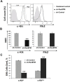
Figure 4. RanBPM silencing in EML cells causes specific downregulation of c-Kit
A. Flow cytometric analysis of EML cells transduced with RanBPM-specific or control shRNA lentivirus showing the mean fluorescence intensity (MFI) of c-Kit (left panel) and Flk2 (right panel). Note the specific MFI reduction of c-Kit and not Flk2 in sh-RanBPM EML cells compared to sh-Control cells. B. quantification of the averages of c-kit and Flk2 MFI of EML cells transduced with Control or RanBPM shRNA from 3 independent experiments as in A. C. percentage of live EML cells with or without RanBPM knockdown expressing high or low levels of c-Kit in gated live cells. Data are presented as the mean ± S.E.M. and *** = P<0.001. C-kit intensity level was set as depicted in panel A (Low and High). Note the dramatic reduction in EML cells with high c-Kit membrane level after sh-RanBPM transduction.
Figure 5
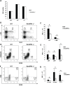
Figure 5. Hematopoietic progenitor cell populations are significantly reduced in RanBPM−/− mice
A. Absolute number of platelets (PLT), white (WBC) and red (RBC) blood cells in control and RanBPM−/− mice (number/uL). B-G. Analysis of hematopoietic stem and progenitor cell populations in the bone marrow of WT and RanBPM−/− mice. Representative flow cytometric analyses of Lineage-negative cells (B), LK cells (D) and LSK cells (F) from the bone marrow of WT and RanBPM−/− mice. The gates used to distinguish the different populations together with the specific total number of cells are displayed. C, E and G: quantification of gated cells respectively from B, D and F; note, in all graphs, the significant reduction in all lineages total number± SEM (n = 3 mice per genotype) of different c-kit progenitor cells. * p<0.05. Cells were incubated with antibodies that recognize lineage specific cell surface antigens (Mac-1, Gr-1, B220, Ter119, CD4, CD8, and IL-7Rα) and antibodies to distinguish HSPC including PE-conjugated c-Kit, APC-conjugated Sca-1, FITC–conjugated CD34, PE-Cy5-conjugated Flk2 (Flt3), and PE-Cy7-conjugated FcγRII/III. LK, lineage-negative c-kit+/Sca1 (LK) progenitors; LSK, lineage negative c-Kit+/Sca1+; MPP, multi-potent progenitors; CMP, common myeloid progenitors; GMP, granulocyte macrophage progenitors; MEP, megakaryocyte erythrocyte progenitors.
Figure 6
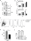
Figure 6. RanBPM regulation of c-kit level influences human cell lines growth
A. c-Kit binds to RanBPM in the M07e human myeloid leukemia cell line. M07e and K562 cell lysates were immunoprecipitated with an anti-c-Kit antibody and blotted with the indicated antibodies. Note the presence of RanBPM pulled down by c-Kit in the M07e cells. Inputs represent 10% of the protein lysates used for the IP experiment. MW, molecular weight markers in kDa. B. Total cellularity of M07e and K562 cultures following transduction with sh-Control or sh-RanBPM lentiviruses. Cells were transduced on day 0, and puromycin treated (1 ug/mL) on day 3 and continuously thereafter. Error bars indicate total cell number +/− SEM. C. Representative dot plot showing forward and side scatter gating for live cells. Bold lines indicate the live gate, excluding dead and dying cells (left panel). In central and right panels are the representative flow cytometry histograms showing c-Kit expression (indicated by anti-c-Kit-PE fluorescence) on live-gated M07e or K562 cells. D. mean fluorescence intensity (MFI) of c-Kit-PE-stained cells for live-gated M07e cells. Cells were transduced with either sh-Control or sh-RanBPM lentiviruses as described above. Data are from cultures analyzed at day 10. Error bars represent mean fluorescence intensity +/− SEM. **** P<0.0001. E. Western blot analysis of protein lysates from the K562 and M07e cells at day 10 from transduction with the sh-Control or sh-RanBPM RNA as in B-D. Blots were hybridized with c-Kit, RanBPM and GAPDH specific antibodies as indicated. Note the effectiveness of the Sh-RanBPM-specific RNA in downregulating RanBPM in both K562 and M07e cells and the reduction in c-Kit expression in the M07e cells.

Similar articles

Cited by

References

    1. Morrison-Graham K, Takahashi Y. Steel factor and c-kit receptor: from mutants to a growth factor system. BioEssays. 1993;15:77–83. - PubMed
    1. Lennartsson J, Ronnstrand L. Stem cell factor receptor/c-Kit: from basic science to clinical implications. Physiological reviews. 2012;92:1619–1649. - PubMed
    1. Rojas-Sutterlin S, Lecuyer E, Hoang T. Kit and Scl regulation of hematopoietic stem cells. Curr Opin Hematol. 2014;21:256–264. - PubMed
    1. Kimura Y, Ding B, Imai N, Nolan DJ, Butler JM, Rafii S. c-Kit-mediated functional positioning of stem cells to their niches is essential for maintenance and regeneration of adult hematopoiesis. PloS one. 2011;6:e26918. - PMC - PubMed
    1. Shin JY, Hu W, Naramura M, Park CY. High c-Kit expression identifies hematopoietic stem cells with impaired self-renewal and megakaryocytic bias. The Journal of experimental medicine. 2014;211:217–231. - PMC - PubMed

MeSH terms

LinkOut - more resources