

Chitin and Chitosan: Production and Application of Versatile Biomedical Nanomaterials
- PMID: 27819009
- PMCID: PMC5094803
Chitin and Chitosan: Production and Application of Versatile Biomedical Nanomaterials
Abstract
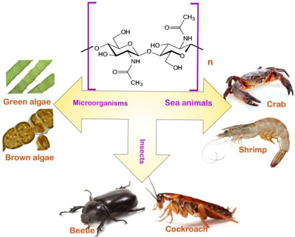
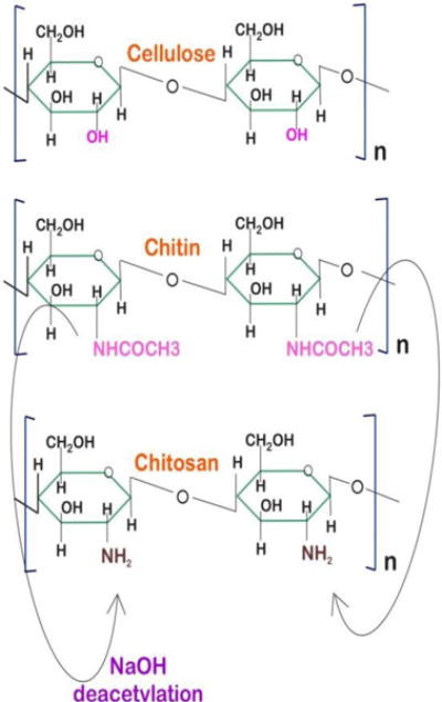
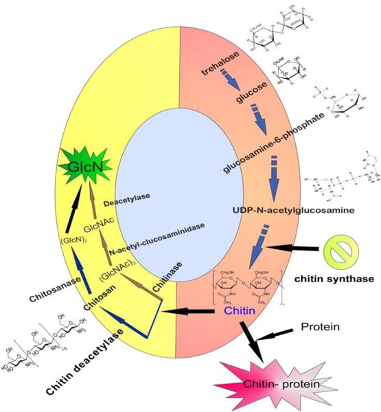
Chitin is the most abundant aminopolysaccharide polymer occurring in nature, and is the building material that gives strength to the exoskeletons of crustaceans, insects, and the cell walls of fungi. Through enzymatic or chemical deacetylation, chitin can be converted to its most well-known derivative, chitosan. The main natural sources of chitin are shrimp and crab shells, which are an abundant byproduct of the food-processing industry, that provides large quantities of this biopolymer to be used in biomedical applications. In living chitin-synthesizing organisms, the synthesis and degradation of chitin require strict enzymatic control to maintain homeostasis. Chitin synthase, the pivotal enzyme in the chitin synthesis pathway, uses UDP-N-acetylglucosamine (UDPGlcNAc), produce the chitin polymer, whereas, chitinase enzymes degrade chitin. Bacteria are considered as the major mediators of chitin degradation in nature. Chitin and chitosan, owing to their unique biochemical properties such as biocompatibility, biodegradability, non-toxicity, ability to form films, etc, have found many promising biomedical applications. Nanotechnology has also increasingly applied chitin and chitosan-based materials in its most recent achievements. Chitin and chitosan have been widely employed to fabricate polymer scaffolds. Moreover, the use of chitosan to produce designed-nanocarriers and to enable microencapsulation techniques is under increasing investigation for the delivery of drugs, biologics and vaccines. Each application is likely to require uniquely designed chitosan-based nano/micro-particles with specific dimensions and cargo-release characteristics. The ability to reproducibly manufacture chitosan nano/microparticles that can encapsulate protein cargos with high loading efficiencies remains a challenge. Chitosan can be successfully used in solution, as hydrogels and/or nano/microparticles, and (with different degrees of deacetylation) an endless array of derivatives with customized biochemical properties can be prepared. As a result, chitosan is one of the most well-studied biomaterials. The purpose of this review is to survey the biosynthesis and isolation, and summarize nanotechnology applications of chitin and chitosan ranging from tissue engineering, wound dressings, antimicrobial agents, antiaging cosmetics, and vaccine adjuvants.
Keywords: Chitin; biomedical nanotechnology; chitosan; drug delivery; nanoparticle; synthetic nanofiber; vaccine adjuvant.
Figures
Similar articles
-
Extraction, chemical modification and characterization of chitin and chitosan.Int J Biol Macromol. 2018 Dec;120(Pt A):1181-1189. doi: 10.1016/j.ijbiomac.2018.08.139. Epub 2018 Aug 30. Int J Biol Macromol. 2018. PMID: 30172808 Review.
-
Chitins and chitosans as immunoadjuvants and non-allergenic drug carriers.Mar Drugs. 2010 Feb 21;8(2):292-312. doi: 10.3390/md8020292. Mar Drugs. 2010. PMID: 20390107 Free PMC article. Review.
-
Chitin, Characteristic, Sources, and Biomedical Application.Curr Pharm Biotechnol. 2020;21(14):1433-1443. doi: 10.2174/1389201021666200605104939. Curr Pharm Biotechnol. 2020. PMID: 32503407 Review.
-
Chitosan: Applications in Drug Delivery System.Mini Rev Med Chem. 2023;23(2):187-191. doi: 10.2174/1389557522666220609102010. Mini Rev Med Chem. 2023. PMID: 35692143 Review.
-
Chitin--the undisputed biomolecule of great potential.Crit Rev Food Sci Nutr. 2003;43(1):61-87. doi: 10.1080/10408690390826455. Crit Rev Food Sci Nutr. 2003. PMID: 12587986 Review.
Cited by
-
Improving Polysaccharide-Based Chitin/Chitosan-Aerogel Materials by Learning from Genetics and Molecular Biology.Materials (Basel). 2022 Jan 28;15(3):1041. doi: 10.3390/ma15031041. Materials (Basel). 2022. PMID: 35160985 Free PMC article. Review.
-
Coating of chitosan on poly D,L-lactic-co-glycolic acid thymoquinone nanoparticles enhances the anti-tumor activity in triple-negative breast cancer.Front Chem. 2023 Feb 8;11:1044953. doi: 10.3389/fchem.2023.1044953. eCollection 2023. Front Chem. 2023. PMID: 36846852 Free PMC article.
-
Efficient tribological properties of azomethine-functionalized chitosan as a bio-lubricant additive in paraffin oil: experimental and theoretical analysis.RSC Adv. 2020 Sep 10;10(55):33401-33416. doi: 10.1039/d0ra07011d. eCollection 2020 Sep 7. RSC Adv. 2020. PMID: 35515070 Free PMC article.
-
Microbial chitinases: properties, current state and biotechnological applications.World J Microbiol Biotechnol. 2019 Sep 6;35(9):144. doi: 10.1007/s11274-019-2721-y. World J Microbiol Biotechnol. 2019. PMID: 31493195 Review.
-
Improved Metal Cation Optosensing Membranes through the Incorporation of Sulphated Polysaccharides.Molecules. 2022 Aug 7;27(15):5026. doi: 10.3390/molecules27155026. Molecules. 2022. PMID: 35956976 Free PMC article.
References
-
- Jayakumar R, Prabaharan M, Sudheesh Kumar PT, Nair SV, Tamura H. Biomaterials based on chitin and chitosan in wound dressing applications. Biotechnology advances. 2011;29(3):322–37. - PubMed
-
- Rinaudo M. Chitin and chitosan: Properties and applications. Elsevier Ltd; 2006.
-
- Merzendorfer H. Insect chitin synthases: a review. Journal of comparative physiology B, Biochemical, systemic, and environmental physiology. 2006;176(1):1–15. - PubMed
Grants and funding
LinkOut - more resources
Full Text Sources
Other Literature Sources
Research Materials