

Keeping it in check: chronic viral infection and antiviral immunity in the brain
- PMID: 27811921
- PMCID: PMC5477650
- DOI: 10.1038/nrn.2016.140
Keeping it in check: chronic viral infection and antiviral immunity in the brain
Abstract
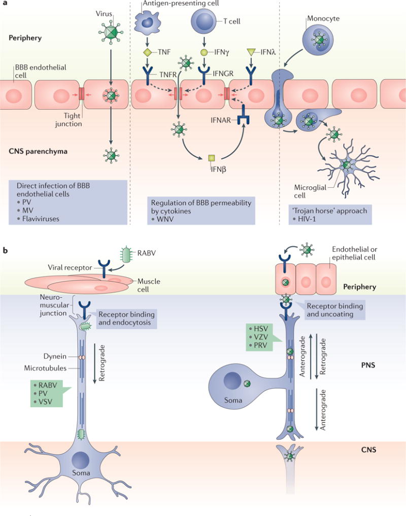
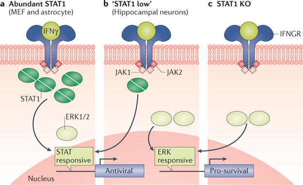
It is becoming clear that the manner by which the immune response resolves or contains infection by a pathogen varies according to the tissue that is affected. Unlike many peripheral cell types, CNS neurons are generally non-renewable. Thus, the cytolytic and inflammatory strategies that are effective in controlling infections in the periphery could be damaging if deployed in the CNS. Perhaps for this reason, the immune response to some CNS viral infections favours maintenance of neuronal integrity and non-neurolytic viral control. This modified immune response - when combined with the unique anatomy and physiology of the CNS - provides an ideal environment for the maintenance of viral genomes, including those of RNA viruses. Therefore, it is possible that such viruses can reactivate long after initial viral exposure, contributing to CNS disease.
Conflict of interest statement
The authors declare no competing interests.
Figures
Similar articles
-
Alpha-Synuclein Expression Restricts RNA Viral Infections in the Brain.J Virol. 2015 Dec 30;90(6):2767-82. doi: 10.1128/JVI.02949-15. J Virol. 2015. PMID: 26719256 Free PMC article.
-
Immune-Mediated Control of a Dormant Neurotropic RNA Virus Infection.J Virol. 2019 Aug 28;93(18):e00241-19. doi: 10.1128/JVI.00241-19. Print 2019 Sep 15. J Virol. 2019. PMID: 31270232 Free PMC article.
-
Immune responses to RNA-virus infections of the CNS.Nat Rev Immunol. 2003 Jun;3(6):493-502. doi: 10.1038/nri1105. Nat Rev Immunol. 2003. PMID: 12776209 Free PMC article. Review.
-
Mechanisms of innate immune evasion in re-emerging RNA viruses.Curr Opin Virol. 2015 Jun;12:26-37. doi: 10.1016/j.coviro.2015.02.005. Epub 2015 Mar 9. Curr Opin Virol. 2015. PMID: 25765605 Free PMC article. Review.
-
Neuroinflammation During RNA Viral Infections.Annu Rev Immunol. 2019 Apr 26;37:73-95. doi: 10.1146/annurev-immunol-042718-041417. Annu Rev Immunol. 2019. PMID: 31026414 Free PMC article. Review.
Cited by
-
The Role of Herpes Simplex Virus Type 1 Infection in Demyelination of the Central Nervous System.Int J Mol Sci. 2020 Jul 16;21(14):5026. doi: 10.3390/ijms21145026. Int J Mol Sci. 2020. PMID: 32708697 Free PMC article. Review.
-
Neuronal Ablation of Alpha/Beta Interferon (IFN-α/β) Signaling Exacerbates Central Nervous System Viral Dissemination and Impairs IFN-γ Responsiveness in Microglia/Macrophages.J Virol. 2020 Sep 29;94(20):e00422-20. doi: 10.1128/JVI.00422-20. Print 2020 Sep 29. J Virol. 2020. PMID: 32796063 Free PMC article.
-
Long-term persistence of infectious Zika virus: Inflammation and behavioral sequela in mice.PLoS Pathog. 2020 Dec 10;16(12):e1008689. doi: 10.1371/journal.ppat.1008689. eCollection 2020 Dec. PLoS Pathog. 2020. PMID: 33301527 Free PMC article.
-
Antigen non-specific CD8+ T cells accelerate cognitive decline in aged mice following respiratory coronavirus infection.bioRxiv [Preprint]. 2024 Jan 3:2024.01.02.573675. doi: 10.1101/2024.01.02.573675. bioRxiv. 2024. PMID: 38260669 Free PMC article. Preprint.
-
Antiviral Functions of Type I and Type III Interferons in the Olfactory Epithelium.Biomolecules. 2023 Dec 8;13(12):1762. doi: 10.3390/biom13121762. Biomolecules. 2023. PMID: 38136633 Free PMC article.
References
Publication types
MeSH terms
Grants and funding
LinkOut - more resources
Full Text Sources
Other Literature Sources
