. 2017 Jan 3;91(2):e01504-16.
doi: 10.1128/JVI.01504-16. Print 2017 Jan 15.

Evolution and Cryo-electron Microscopy Capsid Structure of a North American Bat Adenovirus and Its Relationship to Other Mastadenoviruses

Affiliations

Evolution and Cryo-electron Microscopy Capsid Structure of a North American Bat Adenovirus and Its Relationship to Other Mastadenoviruses

Nicole Hackenbrack et al. J Virol. .

Abstract

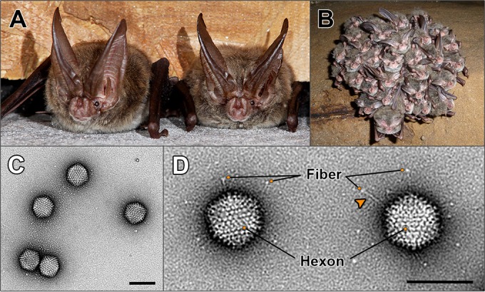
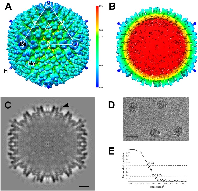
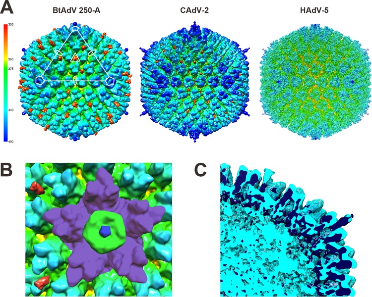
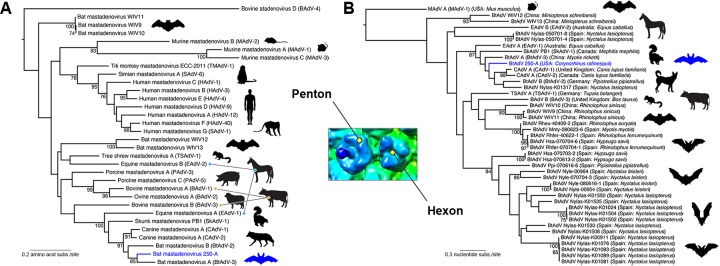
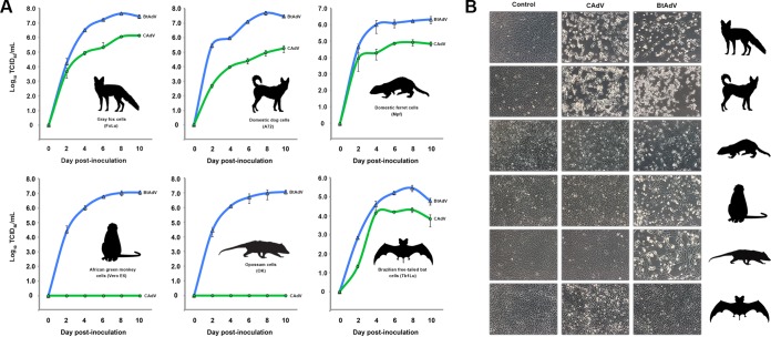
Since the first description of adenoviruses in bats in 2006, a number of micro- and megabat species in Europe, Africa, and Asia have been shown to carry a wide diversity of adenoviruses. Here, we report on the evolutionary, biological, and structural characterization of a novel bat adenovirus (BtAdV) recovered from a Rafinesque's big-eared bat (Corynorhinus rafinesquii) in Kentucky, USA, which is the first adenovirus isolated from North American bats. This virus (BtAdV 250-A) exhibits a close phylogenetic relationship with Canine mastadenovirus A (CAdV A), as previously observed with other BtAdVs. To further investigate the relationships between BtAdVs and CAdVs, we conducted mass spectrometric analysis and single-particle cryo-electron microscopy reconstructions of the BtAdV 250-A capsid and also analyzed the in vitro host ranges of both viruses. Our results demonstrate that BtAdV 250-A represents a new mastadenovirus species that, in contrast to CAdV, has a unique capsid morphology that contains more prominent extensions of protein IX and can replicate efficiently in a phylogenetically diverse range of species. These findings, in addition to the recognition that both the genetic diversity of BtAdVs and the number of different bat species from disparate geographic regions infected with BtAdVs appears to be extensive, tentatively suggest that bats may have served as a potential reservoir for the cross-species transfer of adenoviruses to other hosts, as theorized for CAdV.

Importance: Although many adenoviruses are host specific and likely codiverged with their hosts over millions of years, other adenoviruses appear to have emerged through successful cross-species transmission events on more recent time scales. The wide geographic distribution and genetic diversity of adenoviruses in bats and their close phylogenetic relationship to Canine mastadenovirus A (CAdV A) has raised important questions about how CAdV A, and possibly other mammalian adenoviruses, may have emerged. Although most adenoviruses tend to cause limited disease in their natural hosts, CAdV A is unusual in that it may cause high morbidity and sometimes fatal infections in immunocompetent hosts and is thus an important pathogen of carnivores. Here, we performed a comparative evolutionary and structural study of representative bat and canine adenoviruses to better understand the relationship between these two viral groups.

Keywords: bat adenovirus; canine adenovirus; cross-species transmission; cryo-electron microscopy; host range; mastadenovirus; virus evolution.


Figures

FIG 1
FIG 1
Host species from which bat adenovirus (BtAdV) 250-A was isolated and morphology of purified virions. (A) Two adult Rafinesque's big-eared bats (Corynorhinus rafinesquii) roosting in a building in Mammoth Cave National Park (MCNP), Kentucky, the site of isolation of BtAdV 250-A (photo courtesy of Tom Uhlman). (B) Rafinesque's big-eared bats hibernating in a cave in MCNP. Note the densely packed clusters formed by the bats during winter hibernation (photo courtesy of Steven Thomas, National Park Service). (C) Transmission electron micrograph of BtAdV 250-A virions negatively stained with uranyl formate, showing particles of icosahedral symmetry ∼90 nm in diameter (scale bar, 100 nm). (D) High-magnification view (52,476×) of BtAdV 250-A virions clearly showing the long flexible fiber proteins with their C-terminal knob domain visible (adjacent to the orange circles). Note an apparent kink (arrowhead) in the fiber protein, suggesting a potential hinge region imparting flexibility. Also highlighted is the hexon protein, which comprises the bulk of the adenovirus capsid, where 12 hexons form each of the 20 icosahedral facets of the particle (i.e., 240 copies per virion) (scale bar, 100 nm).
FIG 2
FIG 2
Genomic organization of BtAdV 250-A. (A) The 31,481-nt genome of BtAdV 250-A is shown. The inverted terminal repeats (ITRs) found at the ends of the genome are highlighted in royal blue, with genes transcribed from left to right (from the top DNA strand) shown above the blue-gray solid line, and those transcribed from right to left (from the bottom DNA strand) are indicated below. Genes that are believed to be spliced are indicated by two genetic elements that are connected by adjoining lines in a “V” or “V” pattern. (B) The 31 ORFs identified in the BtAdV 250-A genome that share homology to other BtAdVs and canine mastadenoviruses (CAdVs) are shown. All 13 tentative structural proteins are highlighted with shading: (i) the three major capsid proteins—II (hexon), III (penton), and IV (fiber)—found on the particle surface (blue), (ii) four minor capsid/cement proteins—IIIa, VI, VIII, and IX—found on or just beneath the particle surface (green), and (iii) six core proteins—IVa2, V, VII, X, TP, and the adenovirus protease (AVP)—found internally (gray). The percent amino acid identity of the BtAdV 250-A proteins to other viruses was determined using the following GenBank accession numbers: BtAdV 250-A (KX871230), BtAdV A (NC_016895), BtAdV B (JN252129), CAdV A (CAdV-1) (AC_000003), and CAdV A (CAdV-2) (AC_000020). The virus sharing the highest degree of amino acid identity to BtAdV 250-A for each protein is underlined. A lowercase “p” preceding a protein's name (i.e., pTP, pIIIa, pVI, pVII, pVIII, and pX) indicates a precursor form of the protein that is subsequently cleaved by the AVP.
FIG 3
FIG 3
Mass spectrometry of purified virions of BtAdV 250-A. Purified virus was analyzed by SDS-PAGE, and individual protein bands were excised and subjected to nano-HPLC-MS/MS. Peptide sequencing identified the three major capsid proteins (hexon, fiber, and penton), the four minor capsid/cement proteins (IIIa, VI, VIII, and IX), and two of the six core proteins (V and VII). Note that the lower band in the central group of triplet bands was a mixture of both fiber and penton proteins (estimated molecular masses of 56.1 and 54.5 kDa, respectively). Peptides corresponding to proteins VII, VIII, and IX were detected in a band migrating at approximately ≤10-kDa (not visible after separation of the V-IIIa-fiber-penton cluster).
FIG 4
FIG 4
Three-dimensional reconstruction of the BtAdV 250-A capsid. (A) The surface-rendered BtAdV map is colored according to radius (key) to illustrate the topology of the capsid. An asymmetric unit is highlighted (white) with the axes of icosahedral symmetry denoted by an oval (2-fold), triangle (3-fold), and a pentagon (5-fold). Only a short portion of the fiber (Fi) protein, shown extending from the penton (Pe) base at the 5-fold axis, could be reconstructed due to its flexible nature (see full-length protein in Fig. 1D). Hexon (He) proteins are also denoted. (B) A cross section of the BtAdV particle, color-coded by radius, reveals a disordered genome (red) and the internal structures of the hexons and pentons that comprise the capsid shell. (C) The central cross-section of BtAdV shows external capsid densities are distinct, but less well defined internally. Weak density connects protein IX (arrowhead) to the capsid. Scale bar, 10 nm. (D) Image of BtAdV capsids, embedded in vitreous ice, that were used for the reconstruction. Scale bar, 100 nm. (E) The Fourier shell correlation is shown as a function of resolution indicating 17.9 and 13.7 Å, where the curve crosses 0.5 and 0.143, respectively.
FIG 5
FIG 5
BtAdV 250-A has a capsid morphology similar to CAdV-2 and a prominent protein IX extending from its surface. (A) The surface-rendered maps of BtAdV 250-A, CAdV A (serotype CAdV-2) (EMDB 1462), and Human mastadenovirus C (HAdV C; serotype HAdV-5) (EMDB 5172) are colored according to radius (key) to illustrate similar topologies. In addition to the radial coloring, protein IX was highlighted in crimson in BtAdV and CAdV to demonstrate its location and prominence in the capsid. Note that protein IX in BtAdV protrudes well beyond the hexon proteins. (B) Magnification of the icosahedral 5-fold vertex of BtAdV. The five peripentonal hexons (purple), which surround a single tulip-shaped penton base (green) with its central projecting fiber stalk (royal blue), are visible. The C-terminal surface exposed spikes of two protein IX triskelions (crimson) are shown on the left. (C) The central section of the superimposed structures of BtAdV 250-A (light blue) and CAdV-2 (dark blue) reveal overlapping internal structural similarities between the viruses.
FIG 6
FIG 6
Maximum-likelihood phylogenetic analysis of BtAdV 250-A with other mastadenoviruses. For both the penton and the hexon trees, bootstrap values (>70%) are shown for key nodes. The structural location of the penton and hexon proteins in the BtAdV capsid are shown in the central inset. BtAdV 250-A is highlighted by a blue branch and bat in each tree for reference. (A) Phylogenetic tree of the amino acid sequences of the penton protein for member species of the genus Mastadenovirus (i.e., from a diversity of mammalian taxa). The tree is midpoint rooted for clarity only, and horizontal branch lengths are scaled according to the number of amino acid substitutions per site. GenBank accession numbers for the sequences used in the penton tree were as follows: BAdV-1 (YP_094036), BAdV-2 (NP_597922), BAdV-3 (AEW91335), BAdV-4 (NP_077393), BtAdV-2 (YP_004782104), BtAdV-3 (YP_005271186), BtAdV 250-A (KX871230), BtAdV WIV9 (KT698853), BtAdV WIV10 (KT698854), BtAdV WIV11 (KT698855), BtAdV WIV12 (KT698856), BtAdV WIV13 (KT698852), CAdV-1 (NP_044193), CAdV-2 (AP_000617), EAdV-1 (AEP16413), EAdV-2 (YP_009162351), HAdV-1 (AP_000507), HAdV-3 (YP_002213774), HAdV-4 (AAW33302), HAdV-9 (YP_001974428), HAdV-12 (NP_040920), HAdV-40 (NP_040857), MAdV-1 (NP_015541), MAdV-2 (YP_004123742), MAdV-3 (YP_002822211), PAdV-3 (YP_009205), PAdV-5 (NP_108662), SAdV-1 (YP_213970), SAdV-6 (AFG19591), SkAdV-1 (YP_009162592), TMAdV (YP_007518317), and TSAdV-1 (YP_068064). (B) Evolutionary relationships between BtAdV 250-A and BtAdVs from Europe and Asia were assessed using nucleotide sequences of the hexon gene. Closely related viruses (CAdV-1, CAdV-2, SkAdV-1, EAdV-1, EAdV-2, TSAdV-1, and BAdV-3) as determined from the penton phylogeny in panel A, were also included in the analysis. The tree is rooted using the more divergent sequence of Murine mastadenovirus A (MAdV A), and horizontal branch lengths are scaled according to the number of nucleotide substitutions per site. GenBank accession numbers for the sequences used in the hexon tree were as follows: BAdV-3 (AF030154), BtAdV-2 (JN252129), BtAdV-3 (NC_016895), BtAdV 250-A (KX871230), BtAdV WIV9 (KT698853), BtAdV WIV10 (KT698854), BtAdV WIV11 (KT698855), BtAdV WIV12 (KT698856), BtAdV WIV13 (KT698852), CAdV-1 (AC_000003), CAdV-2 (AC_000020), EAdV-1 (JN418926), EAdV-2 (KT160425), MAdV-1 (M81889), SkAdV-1 (KP238322), TSAdV-1 (AF258784), and BtAdV population set HM856327 to HM856353 (courtesy of I. Casas, S. Vazquez, J. Juste, A. Falcon, C. Aznar, C. Ibanez, F. Pozo, G. Ruiz, J. M. Berciano, I. Garin, J. Aihartza, P. Perez-Brena, and J. E. Echevarria, National Centre of Microbiology, Madrid, Spain).
FIG 7
FIG 7
Comparative in vitro host ranges of BtAdV and CAdV. (A) Gray fox (FoLu), domestic dog (A72), domestic ferret (Mpf), African green monkey (Vero E6), Virginia opossum (OK), and Brazilian free-tailed bat (Tb1Lu) cells were infected with either BtAdV 250-A or CAdV-1 (isolate 24-05), and viral titers (log10 TCID50/ml) were measured at 2-day intervals over a 10-day period. BtAdV 250-A growth curves are shown in blue, whereas CAdV 24-05 growth curves are shown in green. Note that although CAdV was host restricted to cells derived from members of the order Carnivora (fox, dog, and ferret) and bats, BtAdV 250-A replicated efficiently in cells derived from a phylogenetically diverse group of mammals, including primates, as well as a marsupial species (opossum). The data shown are from multistep single growth curve experiments performed in triplicate, with error bars indicating the standard deviations. (B) Cell morphology of the cell lines used in panel A at day 6 postinfection with either BtAdV 250-A or CAdV 24-05. Note the induction of cytopathic effects by BtAdV in all of the cell lines tested beside bat (Tb1Lu) cells.

Similar articles

Cited by

References

    1. Harrach B, Benkö M, Both GW, Brown M, Davison AJ, Echavarría M, Hess M, Jones MS, Kajon A, Lehmkuhl HD, Mautner V, Mittal SK, Wadell G. 2012. Family Adenoviridae, p 125–141. In King AMQ, Adams MJ, Carstens EB, Lefkowitz EJ (ed), Virus taxonomy: ninth report of the International Committee on Taxonomy of Viruses. Elsevier Academic Press, San Diego, CA.
    1. Davison AJ, Benko M, Harrach B. 2003. Genetic content and evolution of adenoviruses. J Gen Virol 84:2895–2908. doi:10.1099/vir.0.19497-0. - DOI - PubMed
    1. Benkö M, Harrach B. 2003. Molecular evolution of adenoviruses. Curr Top Microbiol Immunol 272:3–35. - PubMed
    1. Harrach B. 2000. Reptile adenoviruses in cattle? Acta Vet Hung 48:485–490. doi:10.1556/004.48.2000.4.11. - DOI - PubMed
    1. Chen EC, Yagi S, Kelly KR, Mendoza SP, Tarara RP, Canfield DR, Maninger N, Rosenthal A, Spinner A, Bales KL, Schnurr DP, Lerche NW, Chiu CY. 2011. Cross-species transmission of a novel adenovirus associated with a fulminant pneumonia outbreak in a new world monkey colony. PLoS Pathog 7:e1002155. doi:10.1371/journal.ppat.1002155. - DOI - PMC - PubMed

MeSH terms